0972
Quantifiable study of magnetic resonance super resolution reconstruction in Placenta Accreta Spectrum using Image Quality Metrics1School of Biomedical Engineering and Imaging Sciences (BMEIS), Kings College London, London, United Kingdom, 2Elizabeth Garrett Anderson Institute for Women's Health, University College London, London, United Kingdom, 3Women's Health Division, University College London, London, United Kingdom, 4Depart. Medical Physics and Biomedical Engineering, University College London, London, United Kingdom, 5Centre for Medical Imaging, University College London, London, United Kingdom, 6WEISS (Wellcome /EPSRC Centre for Medical Imaging, University College London, London, United Kingdom
Synopsis
Magnetic Resonance Imaging is increasingly used for assessment of placental complications1. Here we apply MRI to examine Placenta Accreta Spectrum (PAS) a condition where the placenta is abnormally adherent or invasive. Super-Resolution Reconstruction (SRR) provides a high-resolution 3D reconstruction from 2D MRI slices to improve the visibility of structures for future clinical use2. Image Quality metrics allows quantitative evaluation of the SRR images to compare with the original 2D images. These metrics are found to be statistical significant, providing an objective assessment of the SRR images.
Introduction
Magnetic Resonance Imaging (MRI) of the placenta is used clinically to diagnose and manage pregnancies affected by placental complications1.Placental Attachment Spectrum(PAS) is a major obstetric complication where the placenta is abnormally adherent or invasive, resulting in major postpartum haemorrhage with the need for hysterectomy.Correctly assessing the degree of placental invasion is important for surgical management.MRI can provide quantitative measures relating to the tissue properties and function as well as the visualisation of the maternal and fetal blood vessels2.Interpreting MRI data can be complex as movement of both the mother and fetus can lead to image artefacts and distort features. Super-Resolution Reconstruction(SRR) can be used to remove these errors and improve image visualisation through reconstructing a high-resolution image from multiple, overlapping low-resolution images3,4.SRR is increasingly being used to visualise and evaluate the fetal brain and neck particularly structural anomalies, with both structures considered to be rigid organs5.The application of SRR to the placenta is still being explored due to the non-rigid nature of this organ.MRI of PAS is commonly performed in late preterm gestation prior to Caesarean section when there is less movement in utero allowing for improved image quality.Qualitative methods for the assessment of MRI use ratings of the region of interest by from clinical experts to evaluate whether the reconstructions improve the visualisation.Quantitative methods however provide an understanding of the degree of benefit between the 2D MRI and the 3D reconstructions.Image Quality Metrics measure parameters such as contrast, resolution, noise, artefacts and distortion6.Methods
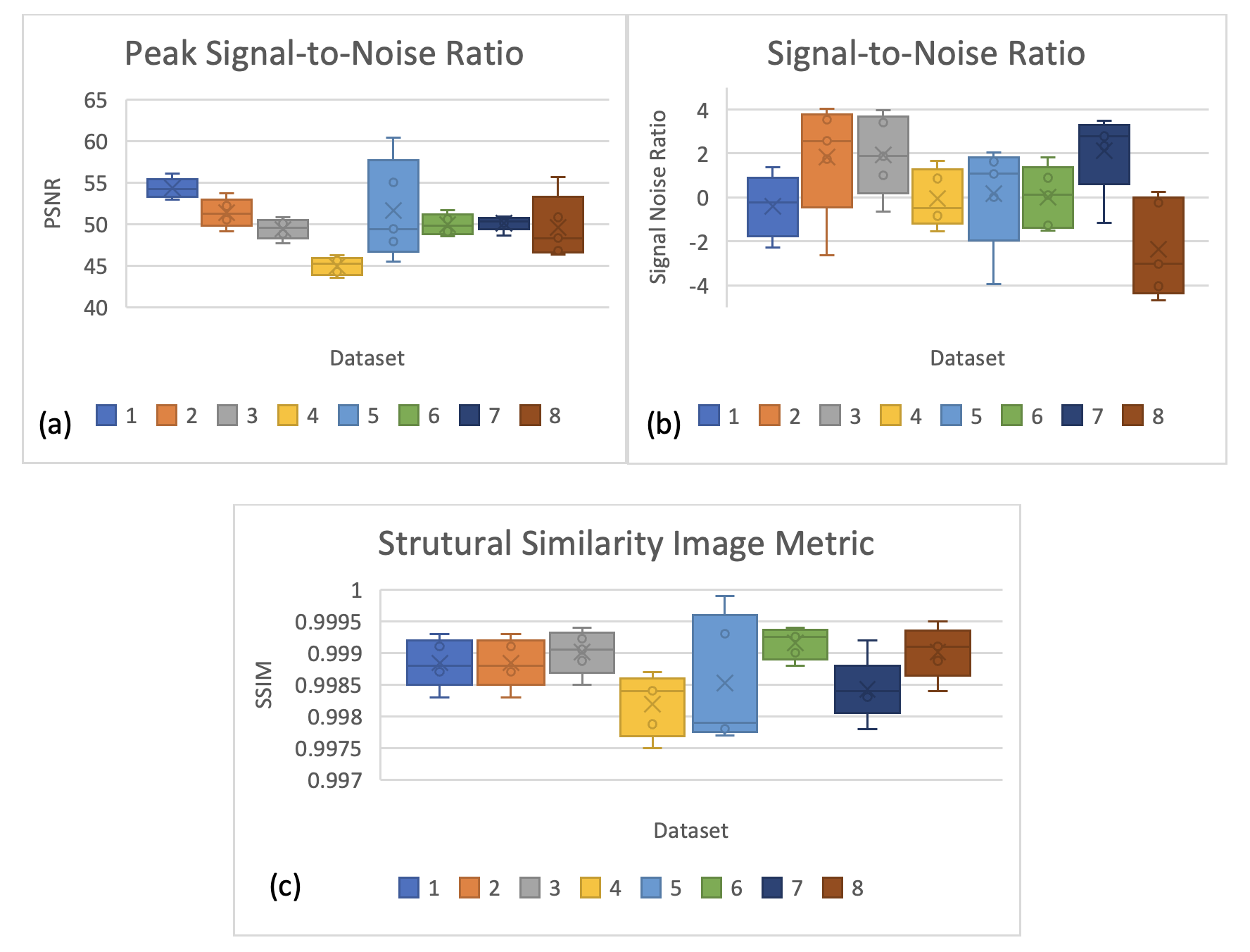
We imaged eight women with suspected Placenta Accreta Spectrum Disorders by MRI, at late-gestation.Imaging was performed on 1.5T Siemens Symphony, with images acquired in three orthogonal planes with TR/TE resolution and 50 slices.A 3D super resolution reconstruction was completed for all of the datasets4, after which the SRR files being converted to 2D by exporting as DICOM files and cropping the original MRIs to the same dimensions as the new SRR.This allowed calculations of the Peak Signal to Noise Ratio(PSNR), the Signal to Noise Ratio(SNR), Structural Similarity Image Metric(SSIM) and Entropy to be completed.The Peak Signal-to-Noise Ratio(PSNR) is an expression of the ratio between the maximum values of signal and the power of the distorting noise that affects the quality of the image, it is calculated as:7$$PSNR=20log_{10}\left(\frac{MAX_f}{√MSE}\right)$$
The Signal-to-Noise Ratio of an MR image is measured by taking the mean of a high-intensity region of interest and dividing by the standard deviation of the region of noise outside of the imaged object7.Structural Similarity Image metric (SSIM) measures the local structural similarity, by using a correlation between the quality and the perception of the human visual system.Instead of using traditional error summation methods, the SSIM models image distortion and contrast distortion7.Entropy is a statistical measure of the randomness that can be used to characterise the texture of the input image. It is a quantitative measure of the information transmitted in the image. It is defined as:
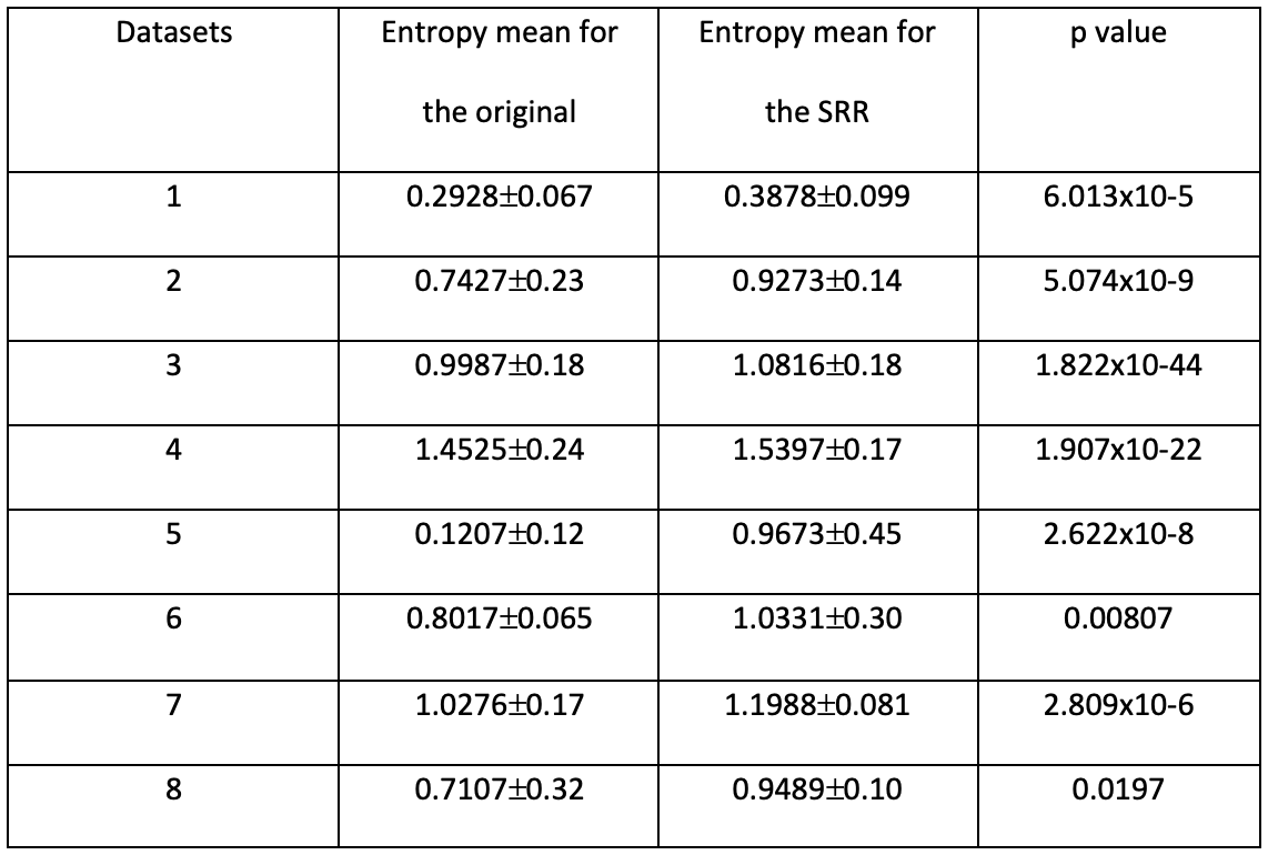
$$Entropy=-∑_{i=1}^np∙log_{2}p $$Where p contains the normalised histogram counts6,9,10.Statistical significance was taken as a p value of <0.05 using a 2 tailed t-test.
Results
At delivery patient’s 2,3,4,6,7,8 were PAS, 1 was placenta previa and patient 5 is yet to deliver.Figure 1 shows the PSNR, SNR and SSIM between the images of the datasets where the SRR are the denominators.
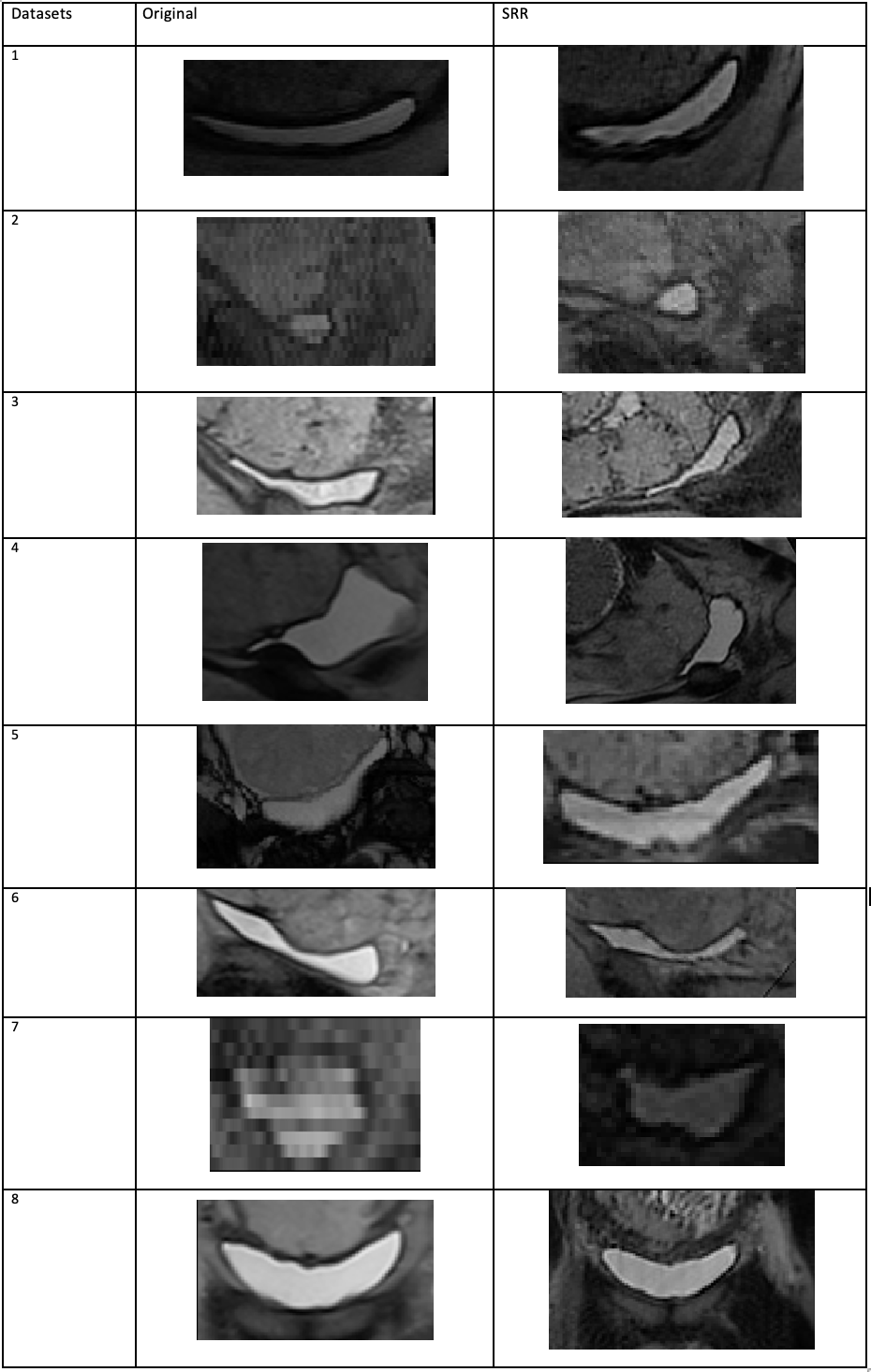
Figure 2 shows the direct comparison of the 2D original MRI images and the SRR reconstruction images for visual observation.For each of the images the bladder is the Region of Interest.
Figure 3 shows the statistical significance between the entropy values of the 2 sets of images, along with the means and standard deviations.
Discussion
In this study we found that the SRR images were statistically different from the original MRI images. SRR could be potentially useful clinically for diagnosis and treatment of PAS, if the anatomical features are more visibly defined.The results of our work correlate with previous findings from Gholipour et al6 applied to the fetal brain showing that the PSNR and the quality of the image have a negative correlation when comparing the results.However, there are limitations to the use of PSNR and SNR as both calculations make assumptions about the image intensity values, which can lead to degradations not being considered within the metric.The SSIM correlation also illustrates this same point, however for all of the datasets the value can be rounded to 1, this may be due to the SSIM being correlated with the HVS colour model and all the images being grey scale. Image quality metrics by themselves are not sufficient to assess the clinical utility of imaging, in order to address this limitation, qualitative radiological input is also required in order to rate the images and direct the clinical impact of work such as this.It can be seen that the entropy values for each of the cases shows statistical significance that the images are statistically different for all of the datasets between 2D and 3D reconstruction.Conclusion
In this study we applied Image Quality metrics to compare Super-Resolution Reconstruction images to 2D of pregnancies complicated by PAS in late gestation.We find that SRR enhances image quality which may increase the utility of MRI to manage PAS.Acknowledgements
This research was supported by the Wellcome Trust (210182/Z/18/Z, 101957/Z/13/Z, 203148/Z/16/Z) and the EPSRC (NS/A000027/1).References
1. Aughware, R. et al. Placental MRI and its application to fetal intervention. Prenatal Diagnosis, 2020. 40(1): p. 38-48
2. Melbourne, A. Aughwane, R. et al. Separating fetal and maternal placental circulations using multiparametric MRI. Magnetic Resonance in Medicine, 2019. 81(1): p.350-361
3. Torrents-Barrena, J et al. Fully Automatic 3D Reconstruction of the Placenta and its Peripheral Vasculature in Intrauterine Fetal MRI. Med Image Anal, 2019. 54(1): p. 263-279
4. Ebner, M et al. An automated framework for localization, segmentation and super-resolution reconstruction of fetal brain MRI. Neuroimage, 2020. 206(1)
5. Mufti N, Ebner M, Patel P, et al. Super-resolution Reconstruction MRI Application in Fetal Neck Masses and Congenital High Airway Obstruction Syndrome. OTO Open, 2021.
6. Tsai, D. Lee, Y and Matsuyama, E. Information Entropy Measure for Evaluation of Image Quality. Journal of Digital Imaging, 2008. 21 (2): p. 338-347.
7. Plenge, E. Poot, D. Niessen, W. Meijering, E. LNCS 8151 – Super-Resolution Reconstruction Using Cross-Scale Self-similarity in Multi-slice MRI. MICCAI, 2013: p. 123-130
8. Gholipour, A. et al. Super-resolution reconstruction in frequency, image and wavelet domains to reduce through-plane partial voluming in MRI. Medical Physics, 2015. 42(12): p. 6919-6932.
9. Obuchowicz, R. et al. Magnetic Resonance Image Quality Assessment by Using Non-Maximum Suppression and Entropy Analysis. Entropy, 2020. 22(2): p. 220
10. Ji, Q. Glass, J. Reddick, W. A novel, fast entropy-minimization algorithm for bias field correction in MR images. Magn Reson Imaging, 2007. 25(2): p. 259-264
Figures