0941
Preliminary study of amide proton transfer-weighted with intravoxel incoherent motion imaging in predicting bone metastasis of prostate cancer1Department of Radiology, the First Affiliated Hospital of Dalian Medical University, Dalian, China, 2Philips Healthcare, Beijing, China
Synopsis
Bone metastasis is an important issue in the management of prostate cancer, and can drastically alter the treatment strategy. PET/CT has been widely used in the diagnosis of bone metastasis; however, the high cost and radiation exposure limit its extensive clinical applications. APT-weighted imaging and IVIM, recent developed function-oriented MRI sequences, allow for a non-invasive visualization of tissue composition and microscopic information without the need for contrast agents. Results of this study indicate the APT value and IVIM parameters can effectively predict bone metastases in prostate cancer. A combination of APT and D* mono can further improve the prediction performance.
Introduction
Prostate cancer is the third most common malignancy after lung and breast malignancies1. Bone metastases (BM) are very common in prostate cancer. PET/CT is recognized as an effective method to detect BM, but a whole-body bone investigation will increase unnecessary radiation burden and diagnostic expenses for a high number of low risk prostate cancer patients. Therefore, looking for a reliable tool for predicting the probability of BM and determining the indication for bone nuclide scans is of great importance. Amide proton transfer-weighted (APTw) imaging generates image contrast using endogenous mobile proteins and peptides in tissue2. Intravoxel incoherent motion imaging (IVIM) can reflect both diffusion and micro-perfusion information of lesions3. The purpose of this study is to explore the value of APT and IVIM parameters in predicting BM of prostate cancer.Method
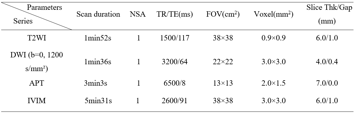
36 patients with pathologically confirmed prostate cancer were involved in this study. According to the state of the skeleton diagnosed by PET-CT, the patients were divided into two groups: group A (13 patients) with BM and group B (23patients) without BM. They underwent preoperative pelvic 3.0T MR scans (Ingenia CX, Philips Healthcare, Best, the Netherlands) with a 32-channel abdominal array coil. The scanned MR sequences included APT, IVIM, DWI, etc. Detailed parameters were listed in Table 1. With reference to T2WI and DWI images, two radiologists used a double-blind method to place a circular-like ROI on the largest layer of the lesion to measure APT value and parameters of IVIM. An unpaired t-test was used to analyze the differences of APT value and parameters of IVIM between the two groups, and the ROC curves were used to evaluate the diagnostic efficacy of these parameters in predicting BM of prostate cancer.Results
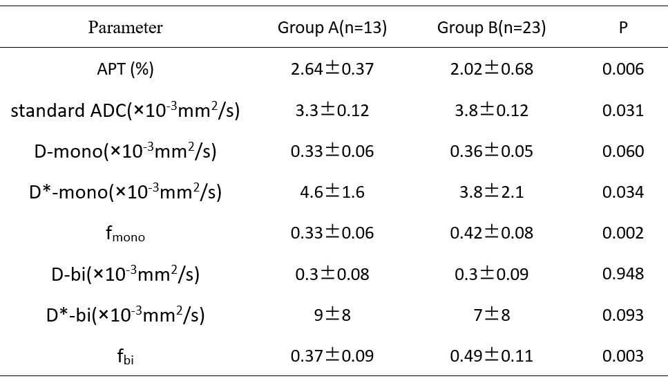
The consistency of the data obtained by the two observers was good (ICC value > 0. 75). The APT and D*-mono values of the PCa group A were significantly higher than those of group B (P<0.05). The standard ADC, fmono, and fbi values of group A were significantly lower than those of group B (P<0.05). No significant difference of D-mono, D-bi, D*-bi was observed between the two groups (P>0.05). The area under the ROC curve (AUC) of APT, standard ADC, D*-mono, fmono, fbi on predicting BM of prostate cancer were 0.774, 0.719, 0.694, 0.806 and 0.799, respectively. With combination of APT and fmono, the AUC of ROC analysis was improved to be 0.866.Discussion and Conclusion
The significant difference in APT value and IVIM parameters between the two groups may be due to the more production of mobile protein, higher cell density and greater capillary blood flow in PCa with BM than in non-metastases PCa. A combination of APT and IVIM parameters can greatly improve the differential diagnosis efficiency. In summary, APT and IVIM has great potential for clinical applications of individualized prediction of BM in PCa.Acknowledgements
NoReferences
1. Zhou J, Gou Z, Wu R, Yuan Y, Yu G, Zhao Y. Comparison of PSMA-PET/CT, choline-PET/CT, NaF-PET/CT, MRI, and bone scintigraphy in the diagnosis of bone metastases in patients with prostate cancer: a systematic review and meta-analysis. Skeletal Radiol. 2019;48(12):1915-1924
2. Ma B, Blakeley JO, Hong X, et al.Applying amide proton transfer-weighted MRI to distinguish pseudoprogression from true progression in malignant gliomas. J Magn Reson Imaging,2016, 44(2):456-462
3. Pesapane F, Patella F, Fumarola EM, et al. Intravoxel Incoherent Motion (IVIM) Diffusion Weighted Imaging (DWI) in the Periferic Prostate Cancer Detection and Stratification. Med Oncol. 2017;34(3):35.
Figures