0924
The value of APTw combined with DKI for prediction of pelvic lymph node metastasis of cervical cancer1the First Affiliated Hospital of Dalian Medical University, Dalian, China, 2Dalian Medical Imaging artificial intelligence engineering technology research center, Dalian, China, 3MSC Clinical & Technical Solutions, Philips Healthcare, Beijing, China
Synopsis
Cervical cancer is one of the most common malignant cancers in pelvic cavity, and trends in young adults. This study aims to investigate the value of amide proton transfer weighted (APTw) combined with diffusion kurtosis imaging (DKI) for prediction of pelvic lymph node metastasis of cervical cancer, the accurate diagnosis of which is significant for the treatment and prognosis of cervical cancer.
INTRODUCTION
Amide proton transfer weighted (APTw) can get the proton information of amino compounds of free proteins and amino acids without exogenous contrast agents [1]. Diffusion kurtosis imaging (DKI) reflects the non-Gaussian diffusion of water molecules, which is more sensitive to the heterogeneity of tissues [2,3]. APTw and DKI have been separately used in the diagnosis of pelvic diseases in women, such as cervix and endometrium. This work proposed a combination method of APTw and DKI to predict the pelvic lymph node metastasis in cervical cancer.METHODS
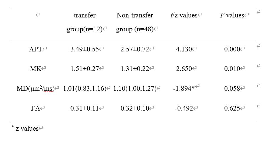
The data of 60 patients with cervical cancer confirmed by surgery and pathology were retrospectively analyzed, 12 patients with and 48 patients without pelvic lymph node metastasis included. All the patients underwent APTw and DKI scans at 3.0T MR (Ingenia CX, Philips healthcare, Best, the Netherlands) before surgery. The mean kurtosis (MK), fractional anisotropy (FA) and mean diffusivity (MD) in the parenchymal regions of DKI were measured by two observers. Consistency of the measurement results between the two observers were assessed using intra-class correlation coefficients (ICC) in SPSS (IBM). Kolmogorov-Smirnov test was adopted to test whether the data conformed to normal distribution. The data of normal distribution were expressed as x±s, and that of skewed distribution were expressed as median (P25, P75). Independent sample t test for normal distribution or Mann-Whitney U test for non-normal distribution was performed to compare the difference between the two groups with a significant threshold of P<0.05. The value of combination of APTw and DKI parameters was evaluated using logistic regression. The diagnostic performance of these parameters was evaluated by receiver operating characteristic (ROC) curves, and the difference was compared by Delong test.RESULTS
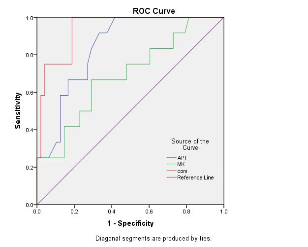
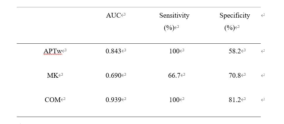
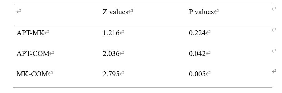
Measurements by the two observers were in good agreement (ICC>0.75). The APTw values from the lymph node metastasis group werr larger than those from the non-lymph node metastasis group, and the MK and MD values were smaller than those from the non-lymph node metastasis group, with statistically significant differences (P<0.05) (Table1, Figure1). The areas under ROC curves were observed as AUC(APT+MK) > AUC(APT) > AUC(MK), as shown in Table 2 and Figure 2. Delong test showed that APT and MK were significantly different from those of combination (p < 0.05). (Table 3).DISCUSSION AND CONCLUSIONS
Our results demonstrated the potential clinical application of the method of APTw combined with DKI to predict pelvic lymph node metastasis of cervical cancer. In the next step, further study should be carried out to investigate the clinical significance with a larger cohort.Acknowledgements
No acknowledgement.References
[1] He YL, Li Y, Lin CY, et al. Three-dimensional turbo-spin-echo amide proton transfer-weighted MRI for cervical cancer: a preliminary study. J MagnReson Imaging 2019; 50(4):1318-1325.
[2] Wu G, Li MM, Chen F, et al.Diffusion-kurtosis imaging predicts early radiotherapy response in nasopharyngeal carcinoma patients.Oncotarget, 2017, 8:66128-66136.
[3] He Yong-Lan, Li Yuan, Lin Cheng-Yu et al. Three-dimensional turbo-spin-echo amide proton transfer-weighted mri for cervical cancer: A preliminary study. [J] MagnReson Imaging, 2019, 50: 1318-1325.
Figures