0923
Application value of radiomics methods based on DTI sequence FA map for differentiating squamous cell carcinoma from cervix adenocarcinoma1Department of Radiology, the First Affiliated Hospital of Dalian Medical University, Dalian,China, China, 2Department of Radiology, the First Affiliated Hospital of Dalian Medical University, Dalian, China
Synopsis
Through high-throughput quantitative feature extraction, the imaging omics method achieves non-invasive analysis of tumor heterogeneity and improves the accuracy of imaging examination in assisting clinical decision making in tumor screening, diagnosis and prognosis prediction. This study intends to explore the value of imaging omics method based on DTI sequence FA map in differentiating CSCC and CA, and provide imaging basis for the formulation of individualized precision treatment plan for CC patients.
Introduction
To evaluate the application of diffusional tensor imaging (DTI) sequence anisotropy fraction (FA) map in the differential diagnosis of cervical squamous cell carcinoma (CSCC) and cervical adenocarcinoma (CA).Method
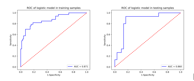
Retrospectively collected 74 patients with cervical cancer (CC) who underwent 1.5T MRI (included DTI sequence) in our hospital from January 2017 to September 2019 and confirmed by surgical pathology. And there were 50 cases of CSCC and 24 cases of CA. Patients were divided into training group (n=103) and test group (n=45). Using ITK-SNAP software, region of interest (ROI) was manually delineated on MK image layer by layer, and then volume of interest (VOI) of tumor was synthesized. Then it was imported into A.K. analysis software for feature extraction of high-dimensional imageology, spearman correlation test and gradient boosting decision tree (GBDT) were used for feature selection and dimension reduction. Then we built a multiple logical regression model and drawing ROC curves to evaluate the diagnostic effectiveness of the model and verify its effectiveness in the test group.Results
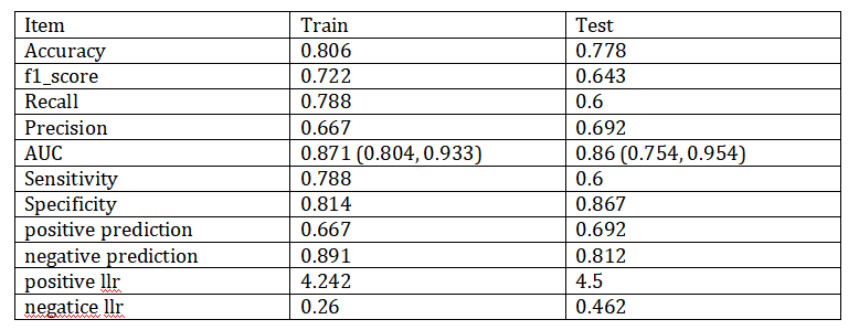
A total of 107 radiomics features were extracted, and 12 radiomics characteristics related to CC pathological classification were obtained by dimension reduction. The accuracy, AUC, sensitivity and specificity of the constructed model for differentiating different pathological types of CC were 80.6%,0.871,78.8% and 81.4% in the training group, and 77.8%,0.860,60.0% and 86.7% in the test group.Discussion and Conclusion
In this study, a total of 107 omics features of 7 categories were extracted from the global multi-category image of CC of different pathological types, and the omics information contained in MK map was deeply mined. Spearman correlation analysis and GBDT were used to reduce the dimensionality of the model to reasonably reduce the over-fitting effect of redundant features on the model. Finally, 12 omics prediction features with great discriminant value were selected and the prediction model was established. CA has a loose cellular structure, contains a large amount of glands and secretions, and has a high density of microvessels and strong generative capacity [1]. Compared with CA, CSCC has higher cell density, closer cell arrangement, smaller cell spacing and extracellular space, so the diffusion speed, direction and limitation degree of water molecules in CSCC and CA are theoretically different. Such differences in microenvironment level will be reflected in the uniformity, roughness, smoothness and other aspects of FA images, which cannot be detected by naked eyes and need to be extracted and quantified using omics methods.The 12 feature parameters finally used for modeling in this study include 4 categories, such as first-order statistics, GLRLM, GLSZM and NGTDM. These parameters comprehensively and effectively reflect the degree of gray scale distribution uniformity of MK maps between CSCC and CA, as well as the elemental direction, depth, thickness, non-uniformity and complexity of image texture. And the spatial arrangement relationship between image voxel gray levels and other diversified difference information [2]. The results show that the model has high diagnostic efficiency in both the training group and the control group.
Acknowledgements
No.References
[1] Saida T, Sakata A, Tanaka YO, et al. Clinical and MRI Characteristics of Uterine Cervical Adenocarcinoma: Its Variants and Mimics. Korean J Radiol, 2019, 20(3):364-377.
[2] Zhou J, Zhang Y, Chang KT, et al. Diagnosis of Benign and Malignant Breast Lesions on DCE-MRI by Using Radiomics and Deep Learning With Consideration of Peritumor Tissue. J Magn Reson Imaging, 2020, 51(3):798-809.
Figures