0922
Evaluation of microsatellite instability in endometrial carcinoma by amide proton transfer imaging and diffusion kurtosis imaging1the First Affiliated Hospital of Dalian Medical University, Dalian, China, 2Dalian Medical Imaging artificial intelligence engineering technology research center, Dalian, China, 3Philips Healthcare, Beijing, China
Synopsis
Microsatellite instability (MSI) is one of the four types of endometrial carcinoma (EC) molecular classification based on The Cancer Genome Atlas (TCGA) [1], which refers to the high mutation load caused by the failure of EC to correct certain errors in small sequence replication due to the abnormal function of DNA mismatch repair system [2]. The purpose of this study is to explore the value of multiple quantitative parameters of APT and DKI sequences in evaluating EC MSI status.
INTRODUCTION
At present, the commonly used method to determine the status of EC MSI is to detect the expression of four mismatch repair proteins in tumor tissues of EC patients by immunohistochemical method, such as MLH-1, MSH-2, MSH-6 and PMS-2[3], which were obtained by postoperative analysis. It is of great clinical significance to determine the MSI status of EC before surgery, including the evaluation of prognosis , the efficacy of PD-1&PD-L1 immunotherapy, and the screening of cases related to lynch syndrome (LS), etc. [4,5], which could provide a strong basis to implement precise personalized treatment for EC patients. Amide proton transfer (APT) imaging can indirectly reflect the levels of mobile protein and polypeptides in tissues and cells [6,7], which is quantified by APT value. Meawhile, Diffusion kurtosis imaging (DKI) quantifies water molecule deviation caused by non-Gaussian distribution diffusion with multi-parameters, and has the advantage of reflecting microenvironment change in tissues accurately [8,9].METHODS
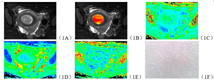
The data of 30 patients with EC confirmed by surgery and pathology were retrospectively analyzed, 12 cases of MSI and 18 cases of microsatellite stability (MSS) included. All patients underwent APT and DKI scans before surgery at 3.0T (Ingenia CX, Philips healthcare, Best, the Netherlands) with a body coil for RF transmitting and an anterior coil for signal receiving. The mean kurtosis (MK), mean diffusivity (MD) and fractional anisotropy (FA) maps of APT and DKI sequences were obtained by post-processing. The APT, MK, MD and FA values of both groups were measured by two observers. Consistency of the measurement results between the two observers were assessed using intra-class correlation coefficients (ICC) in SPSS (IBM). The independent-samples t-test or Mann-Whitney U test was performed to analyze the differences of each parameter values between the two groups. Receiver operator characteristic (ROC) curves were plotted and area under curve (AUC) was performed calculated to evaluate the differential diagnosis efficiency.RESULTS
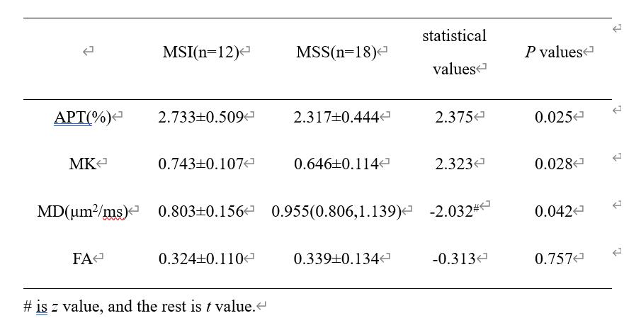
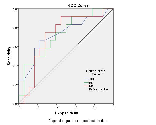
Measurements by the two observers were in good agreement (ICC>0.75). APT and MK values of MSI group were higher than those of MSS group, MD value was lower than that of MSS group (P<0.05), and FA value showed no significant difference between the two groups (P>0.05) (Table 1, Figure 1). The AUC of APT, MK and MD in distinguishing EC MSI from MSS were 0.729, 0.731, and 0.722, respectively. The corresponding sensitivity were 66.7%, 75.0%, 75.0%, and the specificity 77.8% , 61.1%, and 72.2%, respectively (Figure 2).DISCUSSION AND CONCLUSIONS
In this study, it shows that the APT and MK values of EC of MSI group are higher than those of MSS group, which could be caused by more lymphocytes infiltrating and undifferentiated complexes accumulating in the tumor tissues of MSI-related EC[10]. A large amount of protein, polypeptide, etc. in local areas may be the key cause of an increase in APT value. The complexity of water molecule diffusion microenvironment increases accordingly, which leads to an increase in MK value. In addition, the MD value of MSI group is smaller than that of MSS group due to the higher density of EC cells in MSI per unit volume, which leads to the decreased extracellular volume, and more restricted diffusion movement of water molecules.Acknowledgements
No acknowledgement.References
[1] Raffone A, Travaglino A, Cerbone M, et al. Diagnostic Accuracy of Immunohistochemistry for Mismatch Repair Proteins as Surrogate of Microsatellite Instability Molecular Testing in Endometrial Cancer. Pathol Oncol Res, 2020, 26(3):1417-1427.
[2] McEachron J, Zhou N, Spencer C, et al. Adjuvant chemoradiation associated with improved outcomes in patients with microsatellite instability-high advanced endometrial carcinoma. Int J Gynecol Cancer, 2021, 31(2):203-208.
[3] Gilson P, Levy J, Rouyer M, et al. Evaluation of 3 molecular-based assays for microsatellite instability detection in formalin-fixed tissues of patients with endometrial and colorectal cancers. Sci Rep, 2020, 10(1):16386.
[4] Kasherman L, Ahrari S, Lheureux S. Dostarlimab in the treatment of recurrent or primary advanced endometrial cancer. Future Oncol, 2021, 17(8):877-892.
[5] Deshpande M, Romanski PA, Rosenwaks Z, et al. Gynecological Cancers Caused by Deficient Mismatch Repair and Microsatellite Instability. Cancers (Basel), 2020, 12(11):3319.
[6] Takumi K, Nagano H, Kikuno H, et al. Differentiating malignant from benign salivary gland lesions: a multiparametric non-contrast MR imaging approach. Sci Rep, 2021, 11(1):2780.
[7] Wu B, Jia F, Li X, et al. Comparative Study of Amide Proton Transfer Imaging and Intravoxel Incoherent Motion Imaging for Predicting Histologic Grade of Hepatocellular Carcinoma. Front Oncol, 2020, 10:562049.
[8] Goryawala MZ, Heros DO, Komotar RJ, et al. Value of diffusion kurtosis imaging in assessing low-grade gliomas. J Magn Reson Imaging, 2018, 48(6):1551-1558.
[9] Li Z, Li X, Peng C, et al. The Diagnostic Performance of Diffusion Kurtosis Imaging in the Characterization of Breast Tumors: A Meta-Analysis. Front Oncol, 2020, 10:575272.
[10] Akhtar M, Al Hyassat S, Elaiwy O, et al. Classification of Endometrial Carcinoma: New Perspectives Beyond Morphology. Adv Anat Pathol, 2019, 26(6):421-427.
Figures