0921
A Pilot Evaluation of DKI in Characteristics and Diagnosis of Ovarian Cancer of p53 Status
Shiqi Chen1, Yulin Chen1, Ye Li1, and Ailian Liu1
1the First Affiliated Hospital of Dalian Medical University, Dalian, China
1the First Affiliated Hospital of Dalian Medical University, Dalian, China
Synopsis
The identification and treatment of ovarian cancer(OC) has always been a problem that plagues us[1]. Mutations in the p53 gene showed significantly up-regulation in human OC tissues and was closely associated with the development of human OC. This work aimed at exploring the value of Diffusion kurtosis imaging (DKI) for characteristics and diagnosis of ovarian cancer of p53 status. The results showed that FA provided a promising performance (AUC =0.742 , sensitivity = 100%, specificity = 43.7%) in evaluating p53 status and OC.
Purpose
Explore the value of Diffusion Kurtosis Imaging (DKI) for characteristics and diagnosis of ovarian cancer of p53 status.Introduction
The p53 gene on chromosome 17p is considered to be a tumor suppressor gene, and frequent mutations of the p53 gene have been found in a wide variety of human cancers[2]. Approximately one half of human epithelial ovarian cancers have mutations in the p53 gene and overexpress the mutant protein product[3]. A p53 immunohistochemistry can be used to assess the development of human OC. In clinic, it can be used for gene detection and targeted drug therapy. Diffusion kurtosis imaging (DKI) is a special DWI model which treats water diffusion as non-Gaussian behavior. Compared with traditional DWI, this model has been shown to provide greater sensitivity to tissue microstructural complexity with an extender b-value range[4]. Therefore, the purpose of this study is to evaluate the efficacy of DKI-derived parameters in the preoperative evaluation of p53 expression in OC patients.Methods
A total of 31 patients of ovarian cancer confirmed by surgery and pathology from 2015 to 2020 were retrospectively collected. including 16 p53 positive OC (age(45±21) years old; range:(24,66)) and 14 p53 negative OC (age, (61.5±14.5) years old; range:(47,76)). All patients underwent abdominal MR examinations (Signa HDxt, GE Medical Systems, USA)(Scan parameters show in table 1). The original axial digital images from the DKI sequence were transmitted to the GE SDC-ADW 4.6 workstation (Sun Microsystems, Santa Clara, Calif) and the post-processing was performed by Functool software. Referring to the anatomical location of lesion obtained on T2-weighted or DWI images, The FA, MD and MK maps were automatically constructed, and were reviewed by two observers who were blinded to clinical information and histopathologic results with 10 and 15 years of experience in pelvic imaging, respectively. The manual regions of interest (ROIs) were drawn along the edge of tumors on the slice with maximal solid area (we choose the solid in tumors)(Figure 1). The measurement was repeated for three times, and the averages of three measurements were calculated. The FA, MD and MK were recorded. Intra-group correlation coefficient (ICC) was used to test the measurement consistency between the two observers. Correlation between above values and the expression of p53 in OC was compared by Mann-Whitney U test. Receiver operating characteristic (ROC) analysis was performed to evaluate diagnostic performance.Results
The FA value of p53 positive group was higher than p53 negative group, and P value=0.022(table 2). The area under the curve (AUC) of MK for discriminating p53 positive group and p53 negative group was 0.742, when the FA value less than 0.17, the sensitivity was 73.3%, and the specificity was 62.5% (Figure 2).Discussion
A p53 immunohistochemistry can be used to assess the development of human OC[2]. Our findings suggest that FA is potentially the most specific parameter to reflect tumor biological behaviors. In the present study, we proposed that FA could be a promising imaging biomarker in evaluation of p53 expression.Conclusion
FA of DKI image could be a promising imaging biomarker in evaluation of p53 expression in OC.Acknowledgements
Thanks are due to the First Affiliated Hospital of Dalian Medical University for assistance with the experiments.References
[1]Al-Alem,CurryJr. Ovarian cancer: involvement of the martrixmetall oproteinases(J).Reproduction,2015,150(2):55-64[2]Okamoto, Sameshima, Yokoyama, Terashima, Sugimura, Terada, Yokota. Frequent allelic losses and mutations of the p53 gene in human ovarian cancer. Cancer research 51 (19), 5171-5176, 1991
[3]Kohler, Marks, Wiseman, Jacobs, Davidoff, Clarke-Pearson, Soper, Bast, Berchuck Jr. Spectrum of mutation and frequency of allelic deletion of the p53 gene in ovarian cancer. JNCI: Journal of the National Cancer Institute 85 (18), 1513-1519, 1993
[4] Likun Cao, Jie Chen, Ting Duan,et al. Diffusion kurtosis imaging (DKI) of hepatocellular carcinoma: correlation with microvascular invasion and histologic grade. Quant Imaging Med Surg. 2019 Apr; 9(4): 590–602.
Figures
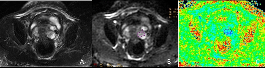
Figure 1: A 65-year-old woman with left side ovarian cancer. A is T2WI image. B is DKI image. C is FA map, it shows the FA value is 0.21.
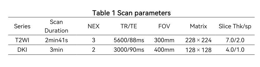
Table 1: Scan parameters
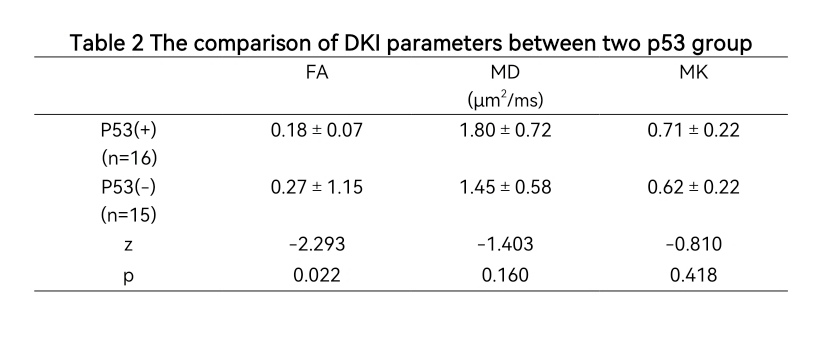
Table 2: The comparison of DKI parameters between two p53 group
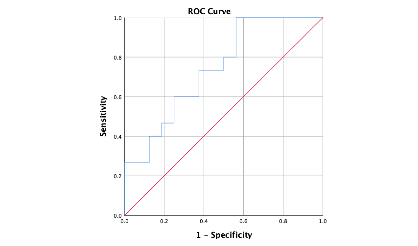
Figure 2: The ROC of FA value in differentiation of p53 positive OC.
DOI: https://doi.org/10.58530/2022/0921