0917
Performance Evaluation of high resolution T2-TSE sequence based on simultaneous multi-slice (SMS) in Female Pelvis MRI
Hongchao Wang1, Yueluan Jiang2, Zhuo Wang1, Lei Zhang1, Zhiqing Shao1, Yang Sun1, and Xiaoye Wang3
1The First Hospital of Jilin University, Changchun, China, 2MR Scientific Marketing, Siemens Healthineers, Beijing, China, 3MR Clinical Marketing, Siemens Healthineers, Beijing, China
1The First Hospital of Jilin University, Changchun, China, 2MR Scientific Marketing, Siemens Healthineers, Beijing, China, 3MR Clinical Marketing, Siemens Healthineers, Beijing, China
Synopsis
Simultaneous multi-slice (SMS) acquisition is a different option for accelerating 2D TSE MRI through the excitation of multiple slices simultaneously and is associated with much smaller SNR loss than parallel acceleration. In this study, we applied both parallel imaging and multiple slices simultaneous imaging technique to accelerate T2WI TSE of female pelvis MRI. Comparing to conventional TSE sequence, 25% of time could be reduced with more slices with thinner thickness using SMS acceleration with similar performances at 3T. SMS-PI-TSE is a feasible acceleration technique in Pelvis MRI.
Introduction
T2WI is essential for female pelvis MRI examinations, and turbo spin echo (TSE)-T2WI is the standard sequence owing to its high signal-to-noise ratio (SNR) and spatial resolution but suffers from motion artifacts caused by respiration and bowel movement during its long acquisition time [1]. Several approaches have been used to reduce acquisition time and motion artifacts, such as conventional in-plane parallel imaging (PI) method, administration of anticholinergic agents and the application of a saturation band to the subcutaneous fat of the anterior body wall [2]. However, in practice, the conventional in-plane techniques can usually perform with an acceleration factor of 2, because the more parallel acceleration reduces the SNR, and it reduces by the square root of the acceleration coefficient [3]. Simultaneous multi-slice (SMS) acquisition is a different option for accelerating 2D TSE MRI through the excitation of multiple slices simultaneously and is associated with much smaller SNR loss than PI. Parallel acceleration and simultaneous multi-slice acceleration can jointly accelerate TSE sequences. In this study, we compare the Image qualities and scanning time of normal PI-TSE and SMS-PI-TSE in female pelvis.Methods
Eight (age 53±22.4) healthy female volunteers were examined on a 3T system (MAGNETOM Vida, Siemens Healthcare, Erlangen, Germany) acquiring routine female pelvic MR examinations including T1WI, DWI,conventional PI-TSE (conv-PI-TSE) T2WI and SMS-PI-TSE T2WI in sagittal, coronal and transverse. The detailed imaging parameters are listed in Table 1. In SMS-PI-TSE T2WI sequence, keep the parameter of FOV, resolution, TE and parallel factor the same with PI-TSE T2WI sequence in each orientation. Adjusted TR, number of slices and slice thickness in SMS-PI-TSE T2WI sequence to reduce the scanning time and increase number of slices with thinner thickness. Two radiologists with 25 and 2 years of experience independently evaluated MR images in random order. Qualitative assessment of PI-TSE T2WI and SMS-PI-TSE T2WI was performed based on the following 4-point criteria: 0 = good, 1 = acceptable, 2 =poor but still interpretable, and 3 = non-diagnostic. Quantitative assessment was performed on a workstation (Syngo MR, Siemens Healthcare, Erlangen, Germany). Two radiologist independently placed regions of interest (ROI), which were as large as possible and devoid of severe artifacts, on the myometrium and junction zone muscle in all patients. Signal to noise ratio (SNR) and contrast-to-noise ratio (CNR) were calculated by the following equations: SNR=SI/SD and CNR =|SImyometrium−SIjunction |/|SImyometrium−SIjunction |, where SI is signal intensity and SD is standard deviation. Two radiologists performed the qualitative and quantitative analysis independently. The mean values of two readers were applied to further statistical analyses. Paired t-test was used for the comparison between groups of SNR, CNR, and Chi-square test was used for the comparison between groups of subject scores. All statistical analysis was performed with SPSS 22.0 (SPSS Inc., Chicago, USA). P values below 0.05 were considered statistically significant.Results and Discussion
Figure 1 shows PI-TSE T2WI and SMS-PI-TSE T2WI of a 36-year-old female pelvis MRI. The sagittal, coronal, and transverse MRI scans of the 8minute PI-TSE MRI protocol (upper row) and 6minute SMS-PI-TSE MRI protocol (lower row) were obtained. By jointly apply parallel acceleration and simultaneous multi-slice acceleration, T2WI scans of female pelvis acquisition time reduced from 8min11s to 6min04s. Besides, by combining SMS-PI-TSE, the number of slices is appropriately increased, and the layer thickness is appropriately decreased, which could reduce the volume effect to some extent. For qualitative assessment, there was no significant difference between PI-TSE T2WI and SMS-PI-TSE T2WI (table 1) in subjective score, p >0.05. For quantitative assessment, there was no significant difference in SNR and CNR in sag T2WI, cor T2WI, and tra fat saturate T2WI.Conclusion
Simultaneous multi-slice acceleration showed good performance for T2-weighted TSE MRI of female pelvis, as it allows both faster acquisition and more slicers with thinner thickness than conventional TSE with comparable image quality. Comparing to conventional TSE sequence, 25% of time could be reduced with more slices with thinner thickness using SMS acceleration with similar performances at 3T.Acknowledgements
N/AReferences
- Kubik-Huch RA, Weston M, Nougaret S, et al. European Society of Urogenital Radiology (ESUR) guidelines: MR imaging of leiomyomas. Eur Radiol.2018;28(1):3125–3137.
- Tsuboyama T, Takei O, Okada A, et al. Comparison of HASTE with multiple signal averaging versus conventional turbo spin echo sequence: a new option for T2-weighted MRI of the female pelvis. Eur Radiol. 2020 ;30(6):3245-3253.
- Fritz J, Guggenberger R, Del Grande F. Rapid Musculoskeletal
MRI in 2021: Clinical Application of Advanced Accelerated Techniques. AJR
AmJ Roentgenol 2021;216(3):718–733
Figures
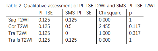
Table 2. Qualitative assessment of PI-TSE T2WI and SMS-PI-TSE T2WI
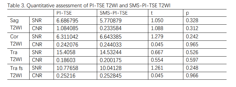
Table 3. Quantitative assessment of PI-TSE T2WI and SMS-PI-TSE T2WI
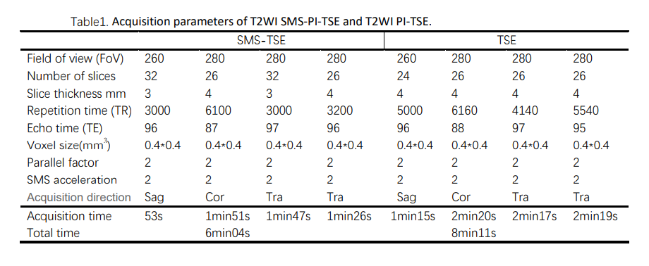
Table1. Acquisition parameters of T2WI SMS-PI-TSE and
T2WI PI-TSE.
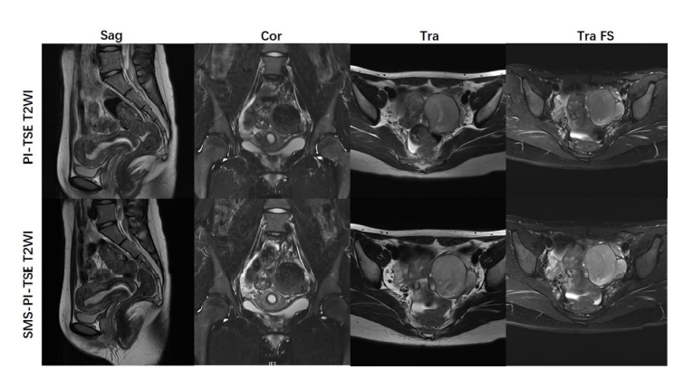
Fig. 1. T2-weighted images of pelvis in 36-year-old
female volunteers obtained with PI-TSE (top line) and SMS-PI-TSE (bottom line).
The uterine were equivalently depicted with good contrast by both sequences in sagittal,
coronal and transverse planes.
DOI: https://doi.org/10.58530/2022/0917