0915
Feasibility of high-quality uterus DWI with segmented phase-encoding and reduced FOV1MR Clinical Science, Philips Healthcare, Suzhou, China, 2Philips Healthcare (China), Shenzhen, China, 3Philips Healthcare (China), Beijing, China, 4MR Clinical Application, Philips Healthcare, Suzhou, China, 5MR R&D, Philips Healthcare, Suzhou, China
Synopsis
Diffusion-weighted imaging (DWI) of uterus has been crucial for providing useful information in differentiating pathologies such as uterine fibroids and adenomyosis. In this study, we aim to investigate the feasibility of high quality uterus DWI through a segmented phase-encoding acquisition with reduced FOV.
Introduction
Diffusion weighted imaging (DWI) of uterus is an important application in pelvis MR imaging. However, the conventional diffusion weighted imaging (DWI) by single shot EPI (ssEPI) has remained challenging due to the local B0 field variation and the result images often suffer from geometric distortion and signal inhomogeneity. A few techniques have been proposed to improve the image homogeneity and SNR. Zonally magnified oblique multislice (Zoom)1 allows to acquire image with reduced FOV with a single shot acquisition and has been proposed for spine imaging2. The Image Reconstruction with Image space Sampling (IRIS) scheme by 2D navigated multi-shot SENSE EPI3 can be combined with Zoom (Zoom-IRIS). An alternative way to achieve reduced FOV is by using outer volume suppression4 with segmented phase-encoding scheme (OVS-IRIS). In this work, we propose to test the feasibility of improved image quality in uterus imaging by using OVS-IRIS and Zoom-IRIS.Methods
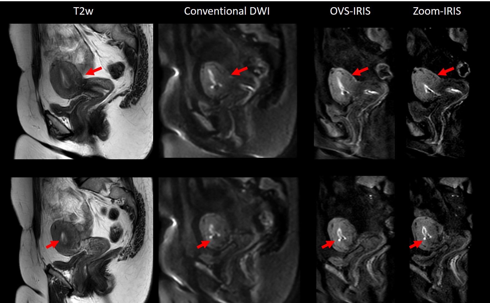
The imaging of uterus was performed on a 3.0 T Ingenia Elition system (Philips Healthcare, the Netherlands) with healthy volunteers with informed consent. The 16-channel torso array coil was used to acquire the anatomical T2w images with in-plane resolution of 1.1 x 1.1 mm2. The diffusion-weighted sagittal images using conventional ssEPI, OVS-IRIS and Zoom-IRIS schemes, respectively. The scanning parameters for ssEPI is as following: FOV = 260 x 200 mm2, voxel size = 2.2 x 2.2 mm2, 12 slices with thickness = 4 mm, TE/TR = 56/2200 ms, and b-value = 0, 1000 s/mm2, with SENSE factor set to 2.5. For OVS-IRIS and Zoom-IRIS imaging, FOV = 200 x 100 mm2, voxel size = 1.4 x 1.4 mm2, 12 slices with thickness = 4 mm, b-value = 0, 1000 s/ mm2, and TE/TR was 61/879 and 66/2618 ms, respectively. A 2D navigator was also acquired following each image acquisition train for motion correction for each k-space segment, and data was reconstructed as described in Jeong H et al3.Results
Figure 1 showed the images acquired using the proposed method with b = 1000 s/mm2. Signals in the uterus region (red arrowed) were more homogeneous in the images acquired from the proposed method of reduced FOV combined with segmented phase-encoding, when compared to those in the conventional images. A higher spatial resolution was achieved on reduced FOV imaging while preserving the image quality. Scan time for OVS-IRIS and Zoom-IRIS were 4min11s and 8min33s, respectively.Discussion and Conclusion
We demonstrate the feasibility of applying the segmented phase-encoding combined with reduce FOV to the imaging of uterus. Signal inhomogeneity and low image quality is a known problem for uterus DWI. The proposed method improves the image quality in two aspects: 1) a reduced FOV in phase encoding direction reduces the signal replacement distance; and 2) the segmented phase encoding provides increased sampling bandwidth. Zoom-IRIS adopts the benefit from multishot, because the reduced-FOV in combination with a multi-shot acquisition enables shorter EPI echo train lengths than the reduced FOV on its own.Acknowledgements
No.References
1. Wheeler-Kingshott, C.A.M., et al. Investigating Cervical Spinal Cord Structure Using Axial Diffusion Tensor Imaging. NeuroImage 2002, Vol16, Issue1: 93-102.
2. Furuya, S. et al. Highly Accurate Analysis of the Cervical Neural Tract of the Elderly Using ZOOM DTI. Neurospine 2018; 15(20): 169-174.
3. Jeong, H., et al. High resolution human diffusion tensor imaging using 2D navigated multi-shot SENSE EPI at 7 Tesla. Magn Reson Med. 2013, 69 (3): 793-802.
4. Wilm B.J., et al. Reduced Field-of-View MRI Using Outer Volume
Suppression for Spinal Cord Diffusion Imaging. Magn Reson Med 2007; 57: 625–630
Figures