0906
F0 determination at the labeling plane: the neglected factor for successful pCASL perfusion MRI1C.J. Gorter Center for High Field MRI, Department of Radiology, Leiden University Medical Center, Leiden, Netherlands, 2Philips Healthcare, Best, Netherlands
Synopsis
In perfusion imaging with arterial spin labeling (ASL), the labeling plane is located further away from the isocenter of the scanner, than the imaging volume. This can result in offsets in resonance frequencies which can lead to imperfect inversion and hence lower labeling efficiency. In this study, a perfusion phantom is used to study the influence of off-resonance on two arterial spin labeling schemes, in order to determine whether an additional F0 determination at the labelling plane is the redeeming factor for successful ASL MRI independent of the labeling scheme used.
Introduction
Pseudo-continuous arterial spin labeling (PCASL) enables the non-invasive assessment of brain perfusion by utilizing the inflowing blood as an endogenous tracer. The inversion of flowing spins is performed at the level of the neck, typically 90 mm below the center of the brain (the isocenter of the scanner). While the magnetic field in the brain region is typically shimmed to provide optimal quality, the labeling plane is often the overlooked or at least unspoken about factor for implementing successful ASL measurements. More specifically, off-resonance (ΔF0) effects at the location of the labeling plane are well-known causes of labeling errors leading to underestimation or even negative cerebral blood flow measurements. Proposed solutions include multi-phase PCASL1, Optimized PCASL2, unbalanced PCASL3 and use of a separate B0-map4. The use of the much simpler approach of an additional f0-measurement at the labeling plane, has maybe been used by many, but remains rather unspoken of in literature and ASL recommendations. The purpose of this study was to investigate the sensitivity of balanced (bPCASL) and unbalanced (ubPCASL) implementations to off-resonance at the labeling plane, and whether this depends on an F0 determination at the labelling plane.Methods
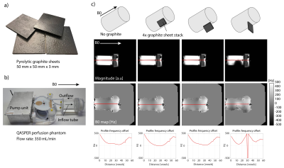
Images were acquired on a phantom as well as in a healthy female volunteer on a 3T clinical MR system (Achieva, Philips Healthcare, Best, NL) using a 32-channel head-coil and body-coil transmission. Two PCASL schemes were compared: 1) bPCASL, where both the control and label conditions have a net average gradient (Gmean) over the labeling duration, and 2) ubPCASL3, where the control condition has Gmean,control = 0. Gmean was 0.36 T/m, gradient strength was 5.0 mT/m and the RF interval of the Hanning-shaped RF pCASL pulses was 1.21 ms with a flip angle of 27.81°. The total label duration was 1800 ms, post-label delay was 2000 ms, and images were acquired with a 4-shot 3D GRaSE readout, with 3.75 x 3.75 x 6 mm3 voxels, TR/TE 4250/11 ms, flip angle 80°, 4 background suppression pulses, 5 inferior saturation pulses, and scan duration of 04:56 mins. B0 maps were acquired to estimate ΔF0 (Hz) using a delta TE of 1 ms, multi-slice FFE read-out with 7 slices, 3.75 x 3.75 mm2 voxels, 4 mm slice thickness, TR/TE 650/7 ms, flip angle 80° and scan duration of 02:20 mins. A perfusion phantom (QASPER, Gold Standard Phantoms, London, UK) was used to investigate the susceptibility-induced off-resonance effects of pyrolytic graphite sheets secured to the outer tube of the ‘neck’ region of the phantom as shown in Figure 1. The flow rate was set to 350 mL/min (default). The main metric for evaluation was the observed signal difference between control-label, and whether the difference resulted in positive ASL signalResults
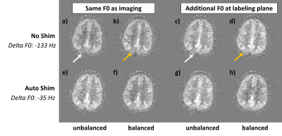
Figure 1 shows the effects of placing the pyrolytic graphite material on the neck of the phantom. We found that a stack of 4 sheets secured perpendicular to the B0 field had the strongest effect on the B0 maps. The red profile shown in the magnitude images (along the inflow tube) and B0 maps is plotted in the bottom row to illustrate the frequency offset induced by the diamagnetic graphite material for each orientation of the graphite stack of sheets with respect to the B0 field.The PCASL results for different conditions are shown in Figure 2 for a single slice/location of the perfusion phantom. First, Figure 2(a) shows that when no shimming was performed and the same F0 was assumed for labeling as for imaging, that ubPCASL performed well, despite the -246Hz off-resonance at the labeling plane, which was not optimal for bPCASL as seen by the negative ASL signal. 2(b) shows that the additional F0 determination in the labeling plane enabled positive ASL signal despite no shim. Similar observations were made in 2(c-d), where the additional F0 proved crucial for successful PCASL imaging. When graphite was introduced, the off-resonance changed, and without shim, only ubPCASL showed robust ASL signal, again, when the F0 determination was performed at the labeling plane.
Figure 3 shows the somewhat more subtle in vivo results with higher ASL signal when F0 was determined at the labeling plane compared to no additional F0 determination. This suggests a greater difference between control and label due to more optimal inversion conditions.
Discussion
Overall, unbalanced PCASL was more robust, but not immune, to off-resonance offsets at the labeling plane. The additional F0 determination greatly improved the ASL signal for both bPCASL and ubPCASL schemes. The lower sensitivity to off-resonance of ubPCASL is known from theory as well as experimental evidence5,6. However, applying balanced PCASL in combination with an F0 determination at the labeling plane also enabled robust perfusion imaging across the brain and in the phantom. The experiments carried out here were limited by the fact that the perfusion phantom only has a single inflow tube in the centre of the cylinder, which precludes left-right comparisons of off-resonance effects on ASL signal. Our findings could guide implementation of PCASL at higher field strengths, but future studies are also needed at clinical field-strengths to demonstrate the influence of more realistic off-resonance and non-uniform effects on different ASL implementations.Acknowledgements
No acknowledgement found.References
1. Jung, Wong, and Liu (2010), Multiphase pseudocontinuous arterial spin labeling (MP-PCASL) for robust quantification of cerebral blood flow. Magn. Reson. Med., 64: 799-810.
2. Shin, Liu, Wong, Shankaranarayanan, and Jung (2012), Pseudocontinuous arterial spin labeling with optimized tagging efficiency. Magnetic Resonance Medicine, 68: 1135-1144.
3. Dai, Garcia, de Bazelaire, Alsop. Continuous flow-driven inversion for arterial spin labeling using pulsed radio frequency and gradient fields. Magn Reson Med 2008;60:1488–1497.
4. Jahanian, Noll, and Hernandez-Garcia (2011), B0 field inhomogeneity considerations in pseudo-continuous arterial spin labeling (pCASL): effects on tagging efficiency and correction strategy. NMR Biomed., 24: 1202-1209.
5. Zhao, Vidorreta, Soman, Detre, and Alsop, "Improving the robustness of pseudo-continuous arterial spin labeling to off-resonance and pulsatile flow velocity," Magn Reson Med, vol. 78, no. 4, pp. 1342-1351, Oct 2017
6. Wu, Fernandez-Seara, Detre, Wehrli, and Wang, "A theoretical and experimental investigation of the tagging efficiency of pseudocontinuous arterial spin labeling," Magn Reson Med, vol. 58, no. 5, pp. 1020-7, Nov 2007.
Figures


