0897
Investigation of asymmetric undersampling scheme in accelerated flow imaging for improving velocity and turbulence kinetic energy estimation1Electrical & Electronic Engineering, Yonsei university, Seoul, Korea, Republic of, 2Radiology (Cardiovascular Imaging), Asan Medical Center, University of Ulsan College of Medicine, Seoul, Korea, Republic of, 3Department of Mechanical and Biomedical Engineering, Kangwon National University, GANGWON-DO, Korea, Republic of, 4Radiology, Stanford University, Stanford, CA, United States
Synopsis
Compressed sensing (CS) technique has recently been used to accelerate long acquisition time in 4D Flow. However, it aggravates underestimation in velocity calculation and overestimation in turbulent kinetic energy (TKE) estimation, when under-sampling is applied. An asymmetric undersampling scheme method of the reference image and velocity encoding images could alleviate problems. The purpose of this paper is investigation of improving velocity and turbulence kinetic energy estimation with an asymmetric reduction for two motion encoding schemes (i.e., conventional 4D Flow vs ICOSA6 Flow)
Introduction
A variety of techniques has been widely used to accelerate acquisition time of 4D Flow MRI1-4. However, reduction technique may lead to the result of velocity underestimation and turbulent kinetic energy (TKE) overestimation, since under-sampled data can cause partial volume effect in phase difference5 and decrease of signal to noise ratio (SNR). Until now, reduction factors of reference and VENC images have been the same. But VENC data might be more important than reference data in velocity mapping. In this study, we investigate an asymmetric reduction method for CS technique to alleviate the problems in 4D Flow and Icosahedral6 (ICOSA6) Flow has more VENC bipolar directions to estimate TKE precisely using a carefully designed flow phantom experiment, which can generate pulsatile flow.Method
[Pulsatile jet flow phantom & Data acquisition]Flow Phantom:
Working Fluid was generated 6:4 mixture of water and glycerol (Density= 1053.8 kg/m3, Viscosity= 3.72x10⁻³ kg·m-1s-1). Rectangular model which is narrowed in the middle as demonstrated in Fig 1. Moreover 30 mL of Contrast agent (0.5 mmol/kg, Meglumin gadoterate, Guerbet, Paris, France) was added to increase the signal intensity6.
Scan protocol for full acquisition:
Fully sampled data was acquired on a clinical 3 Tesla MRI scanner (Ingenia, Philips Medical Systems, Best, The Netherlands) using 32ch torso coil. 4D Flow & ICOSA6 Flow sequence was acquired as followed: FOV= 256x140x70mm3, spatial resolution= 2mm iso-voxel, flip angle= 10°, TR= 4.5ms, TE= 2.5ms, cardiac retrospective phase= 30, VENC= 300m/s for velocity, 150m/s for TKE.
[Retrospective symmetric and asymmetric under-sampling and Compressed sensing-based reconstruction]
Asymmetric acceleration factors were calculated as follows:
$$R_{effective} = \frac{N_{total}\times R_{reference}\times R_{VENC}}{N_{VENC}\times R_{reference}+R_{VENC}}$$
Where, Ntotal is the number of total encodings and NVENC$ is the number of velocity encodings. Full sampled k-space data from 4D Flow and ICOSA6 Flow were under-sampled using 2D variable density patterns with symmetric (R = 3, 1:1 proportion) and asymmetric (Proportion of reference and VENC = 3:1) acceleration factors as shown in Figure 2. CS reconstruction using total-variation regularization was used for the study.
[ICOSA6 encoding schemes & TKE estimation]
ICOSA6 velocity encoding schemes set 6 directions with golden angle7: ψ=(1+√5)/2
Turbulent Kinetic Energy was estimated from the reconstructed images. MRI signal S(kv) in 4D flow velocity can be expressed as follows8-9:
$$S(k_{v})=C\int_{-\infty}^{\infty} s(v) e^{-ik_{v}V}dv$$
where kv is level of flow sensitivity (kv=π/VENC) and C is a constant scaling factor. When turbulent flow is present in 4D flow, the intravoxel velocity variance (IVVV) of turbulent flow is expressed as follows [8,9]:
$$\sigma IVSD=sqrt[\frac{2}{k_v^2} ln(\frac{\mid S(0)\mid}{\mid S(k_{v})\mid})] = sqrt \overline{u_i'u_i'}$$
where S(0) is MRI signal of reference encoding (no bipolar encoding) and ui' velocity fluctuation portion. Using this equation TKE was estimated from IVVV with fluid density ρ as follows10:
$$TKE = \frac{1}{2}\rho(\overline{u_1'u_1'}+\overline{u_2'u_2'}+\overline{u_3'u_3'}) (J/m3)$$
[Evaluation]
We evaluated the peak velocity underestimation and total TKE overestimation between symmetric and asymmetric reduction methods for 4D Flow and ICOSA6 Flow respectively. Peak velocity and total TKE from fully sampled data were used as a reference for evaluation.
Results
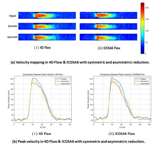
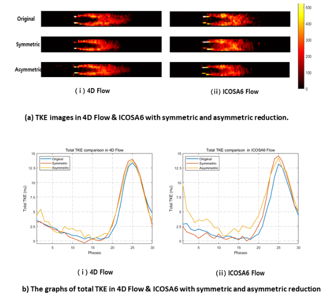
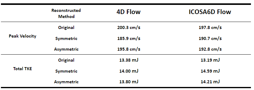
Reconstructed velocity mapping and graphs from symmetric and asymmetric are shown in Figure 3. Peak velocity was underestimated from 200.3 cm/s to 185.9 cm/s in 4D Flow, from 197.8 cm/s to 190.7 cm/s in ICOSA6 Flow when the symmetric method was applied. In contrast, peak velocity of the asymmetric reduction method was 195.8 cm/s in 4D Flow, 192.8 cm/s in ICOSA6 Flow. Underestimation of velocity was improved from 7.19% to 2.25% in 4D Flow and from 7.20% to 5.55% in ICOSA6 Flow by using asymmetric reduction.Images and graphs of TKE estimation were shown in Figure 4. Total TKE were 13.38 mJ and 13.19 mJ respectively. Overestimation of total TKE were 13.80 mJ, 14.21 mJ with asymmetric reduction slightly lower than 14.00 mJ, 14.59 mJ with symmetric method in 4D Flow and ICOSA6 Flow at a peak. However, the variance of total TKE with asymmetric reduction was higher than symmetric reduction method.
Peak velocity and total TKE in 4D Flow and ICOSA6 Flow with symmetric and asymmetric reduction are summarized in Figure 5
Discussion and Conclusion
In this study, we improved the underestimation of velocity in 4D Flow & ICOSA6 Flow by using asymmetric reduction method for CS technique. 4D Flow can give a more effectiveness since it has less under-sampled data with VENC images versus ICOSA6 Flow. On the other hand, the asymmetric reduction had no effect because TKE is estimated from magnitude and noise distribution. Further study for noise compensation method for 4D flow or ICOSA6 is needed.Acknowledgements
This work was supported by the National Research Foundation of Korea(NRF) grant funded by the Korea government(MSIT) (NRF-2019R1A2C1090635)References
1. Lustig M, Donoho D, Pauly JM. Sparse MRI: the application of compressed sensing for rapid MR imaging. Magn Reson Med. 2007; 58: 1182– 1195.
2. Ma LE, Markl M, Chow K, Huh H, Forman C, Vali A, Greiser A, Carr J, Schnell S, Barker AJ, Jin N. Aortic 4D flow MRI in 2 minutes using compressed sensing, respiratory controlled adaptive k-space reordering, and inline reconstruction. Magn Reson Med. 2019 Jun;81(6):3675-3690.
3. E.S., Gottwald, L.M., Zhang, Q. et al. Highly accelerated 4D flow cardiovascular magnetic resonance using a pseudo-spiral Cartesian acquisition and compressed sensing reconstruction for carotid flow and wall shear stress. J Cardiovasc Magn Reson 22, 7 (2020).
4. Tariq U, Hsiao A, Alley M, Zhang T, Lustig M, Vasanawala SS. Venous and arterial flow quantification are equally accurate and precise with parallel imaging compressed sensing 4D phase contrast MRI. J Magn Reson Imaging. 2013; 37: 1419– 1426.
5. Lew C, Alley MT, Spielman DM, Bammer R, Chan FP. Breathheld autocalibrated phase-contrast imaging. J Magn Reson Imaging. 2010;31(4):1004-1014. doi:10.1002/jmri.22127
6. Dyverfeldt, P., Bissell, M., Barker, A.J. et al. 4D flow cardiovascular magnetic resonance consensus statement. J Cardiovasc Magn Reson 17, 72 (2015). https://doi.org/10.1186/s12968-015-0174-5
7. Haraldsson H, Kefayati S, Ahn S, et al. Assessment of Reynolds stress components and turbulent pressure loss using 4D flow MRI with extended motion encoding. Magn Reson Med. 2018;79(4):1962-1971. doi:10.1002/mrm.26853
8. Dyverfeldt P, Sigfridsson A, Kvitting JPE, Ebbers T. Quantification of intravoxel velocity standard deviation and turbulence intensity by generalizing phase-contrast MRI. Magn Reson Med 2006;56(4):850–8.
9. Binter C, Gülan U, Holzner M, SJMrim Kozerke. On the accuracy of viscous and turbulent loss quantification in stenotic aortic flow using phase-contrast MRI. 76(1). 2016. p. 191–6.
10. Pope SB. Turbulent flows. IOP Publishing; 2001.
Figures