0851
A deep learning based direct mapping method for EPI image reconstruction1Gachon University, Incheon, Korea, Republic of, 2Daegu Gyeongbuk Medical Innovation Foundation, Daegu, Korea, Republic of
Synopsis
Echo planar imaging (EPI) is one of the fastest MRI pulse sequences that can acquire whole k-space data during a single excitation. In EPI, partial acquisition can be used for reducing the TR and the effective TE can be further reduced as the portion of the acquired k-space is reduced. However, the quality of the image decreases drastically as the acquired k-space portion is reduced. In this study, we proposed a solution for image reconstruction from partially acquired EPI based on a deep-learning based direct mapping method, where the images are directly reconstructed from input k-space data.
Introduction
As echo planar imaging (EPI) allows acquisition of the whole k-space data during a single excitation, EPI has shown strengths in many applications such as diffusion imaging, perfusion imaging, neurofunctional brain mapping, cardiac imaging, dynamic studies, and real-time imaging. Although it is already one of the fastest MRI pulse sequences, further acceleration will allow EPI to be applied to more challenging MR applications such as fMRI with higher temporal resolution. In EPI, partial acquisition of k-space can be considered for reducing TR. As the portion of the acquired k-space is reduced, the TR and the effective TE can be further reduced. On the other hand, as the acquired k-space portion is reduced, the quality of the image decreases drastically. To obtain only a predetermined part of the k-space and to reconstruct an image from it, the following two approaches are mainly used. First, EPI can be acquired by undersampling the k-space with regular intervals and the missing (or the un-acquired) lines can be generated by using the reconstruction methods such as GRAPPA. Alternatively, a partial k-space can be acquired and the missing region can be regenerated by utilizing the conjugate symmetry such as partial Fourier techniques. For image reconstruction using deep-learning based direct mapping, AUTOMAP[1] and ETER-net[2] were introduced. AUTOMAP performs domain transformation through fully connected layers. On the other hand, ETER-net uses bi-directional RNN for the same purpose[3,4]. Another approach is to reconstruct images using U-net in k-space domain[5]. In this study, we propose a solution for EPI image reconstruction using a deep-learning based direct mapping method called ‘ETER-net’[2], where images are directly reconstructed from input k-space data.Method
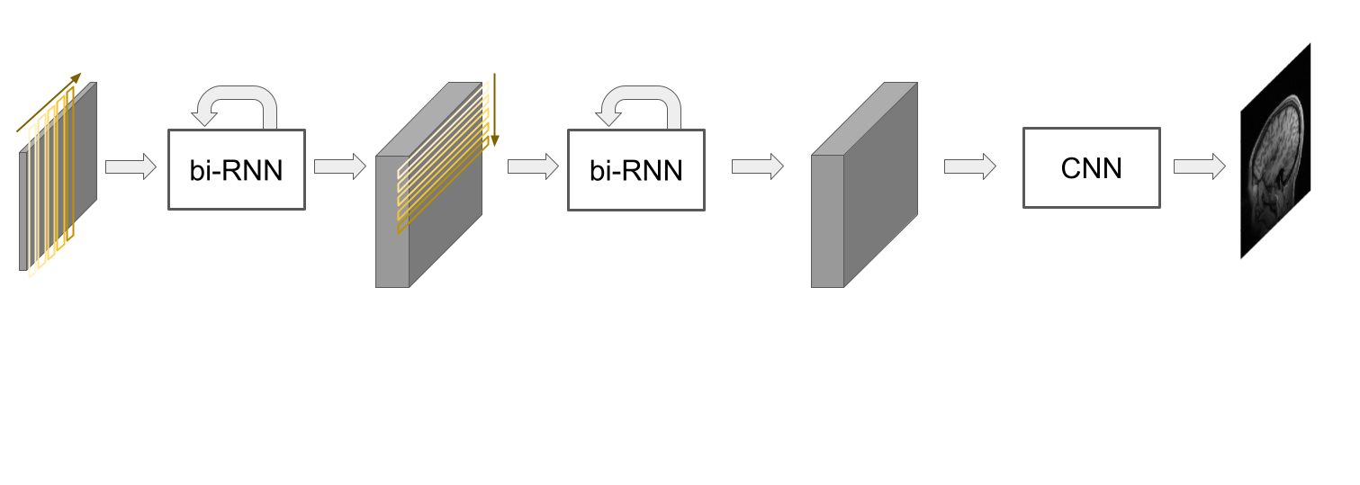
Figure 1 illustrates a diagram of the ETER-net architecture. It consists of two bi-directional recurrent layers and a CNN block. The first layer, horizontal bi-directional recurrent layer, takes the k-space data sequentially in the horizontal direction and produces the first latent matrix. In this procedure, recurrent units store features in a hidden state from the previous data. In addition, the hidden state is a bi-directionally computed. In this way, the entire k-space data can be integrated and processed in the recurrent units. The second layer, vertical bi-directional recurrent layer, takes the first latent matrix sequentially in the vertical direction and makes the second latent matrix. Finally, the CNN block reconstructs the final image from the input second latent matrix. To build the dataset for EPI image reconstruction, in-vivo human brain data were acquired with a 3T MRI scanner (Siemens, MAGNETOM Skyra) and a 20-channel head coil using the following imaging parameters: FOV=240 × 240 mm, matrix size=96 × 96, slice thickness=2.5 mm, flip angle=90 degree, number of slices=34, TR/TE=3000/35 ms, number of scans=104, bandwidth=1860Hz/pixel. To analyze different acceleration environments, we constructed three kinds of datasets: EPI without acceleration, EPI accelerated with regular subsampling (acceleration factor=2), and EPI accelerated with partial acquisitions (partial factor=6/8). We also generated datasets with partial factors of less than 6/8 by retrospective undersampling. For the reference images, the reconstructed images of EPI, which were acquired without acceleration and with average=3 were used. For the input k-space, EPI without acceleration was acquired with average=1. We trained the proposed deep learning models using the reference images as label, and k-space of each dataset as input. We compared the SNR of the reference image, the image reconstructed by the conventional approach and the image reconstructed by ETER-net. For quantitative evaluation, normalized mean square error (nMSE), structural similarity index (SSIM), and visual fidelity information (VIF) was used.Result and Discussion
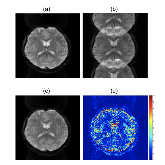
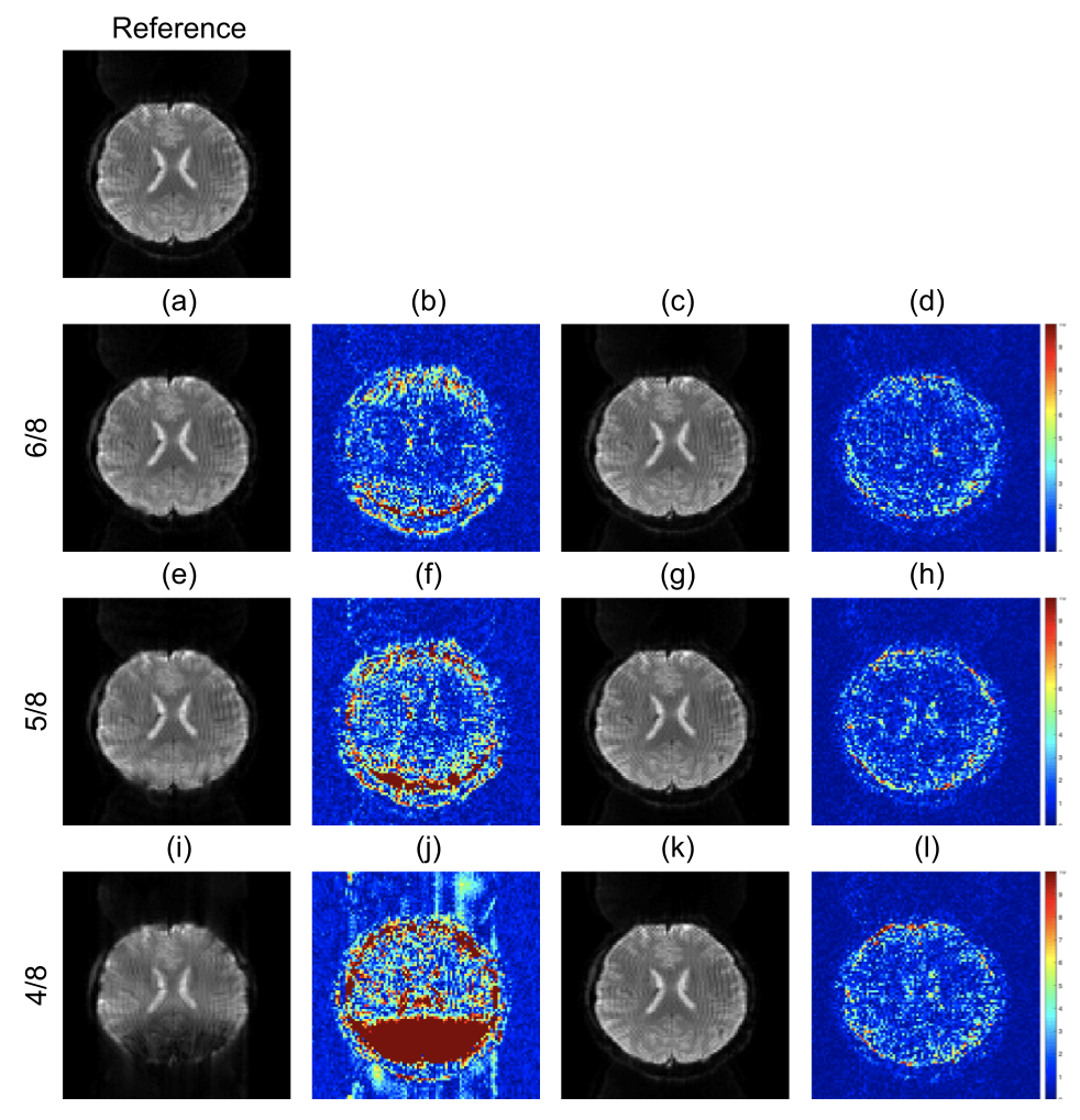
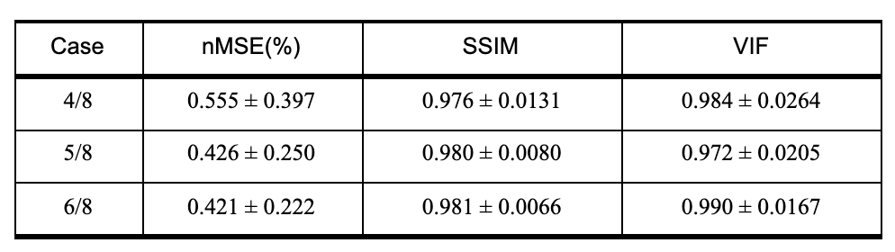
For quantitative analysis, we calculated the SNR at a sample slice of the images reconstructed from the full k-space; The SNR of the reference image, which was acquired with averages=1 and 3 was 134 and 263, respectively. When the full k-space data (average=1) was reconstructed using the ETER-net, the SNR was improved to 222. The images reconstructed by ETER-net from the regularly subsampled k-space are represented in figure 2. There is little difference between (a) the reference image and (c) the reconstructed image. In the ten times amplified error map (d), it is demonstrated that the ghost artifacts are almost eliminated. In figure 3, the images reconstructed from the k-space acquired with partial acquisitions are represented. The first column shows the images reconstructed after zero filling and the second column shows the corresponding error map with a 10x amplification. The third column shows the reconstructed images of the proposed ETER-net and the fourth column shows the corresponding error map with a 10x amplification. As the partial factor decreases, significant error and cancelation can be observed in the conventional method. However, the errors related to the reduced amount of k-space data cannot be observed in the images reconstructed with the ETER-net. The quantitative evaluation is presented in table 1. In this study, we have performed EPI reconstruction using ETER-net and the reconstructed image shows better SNR than conventional methods. As the proposed network can be used to reconstruct the k-space acquired with regular undersampling or partial acquisition, it can be utilized to further accelerate EPI acquisition. In further studies, we will compare the proposed direct reconstruction method with conventional parallel imaging and deep-learning based methods.Acknowledgements
This work was supported by the Brain Research Program through the National Research Foundation of Korea (NRF) funded by the Korea government, Ministry of Science and ICT (MSIT) (NRF-2017M3C7A1047228).References
[1] Zhu, B., Liu, J. Z., Cauley, S. F., Rosen, B. R., & Rosen, M. S. (2018). Image reconstruction by domain-transform manifold learning. Nature, 555(7697), 487.
[2] Oh, C., Kim, D., Chung, J. Y., Han, Y., & Park, H. (2018, September). ETER-net: End to end MR image reconstruction using recurrent neural network. In International Workshop on Machine Learning for Medical Image Reconstruction (pp. 12-20). Springer, Cham.
[3] Oh, C., et al. “A direct MR image reconstruction from k-space via End-To-End reconstruction network using recurrent neural network (ETER-net)” In International Society for Magnetic Resonance in Maedicine, ISMRM 2020.
[4] Oh, C., et al. “A k‐space‐to‐image reconstruction network for MRI using recurrent neural network.” Medical Physics 48.1 (2021): 193-203.
[5] Lee, Juyoung, et al. "k‐Space deep learning for reference‐free EPI ghost correction." Magnetic resonance in medicine 82.6 (2019): 2299-2313.
Figures