0836
IVIM MRI and PET in Locally Advanced Cervical Carcinoma undergoing Concurrent Chemoradiation Therapy1University of California, San Francisco, San Francisco, CA, United States, 2Stanford University, Stanford, CA, United States
Synopsis
IVIM MRI provides measurements of diffusion and perfusion, which has been shown to be predictive of prognosis, responsive to chemoradiotherapy, and correlated with DCE MRI in cervical cancer. We hypothesized that IVIM in cervical cancer patients would be sensitive to CCRT, and hence our objective was to first investigate changes in IVIM in cervical cancer patients undergoing adjuvant CCRT and evaluate the relationship between these metrics with changes in tumor volume and PET. Preliminary results show that IVIM measurements are related to both PET and changes in overall tumor volume and may reflect changes in the tumor microenvironment during treatment.
Introduction
Dynamic contrast enhanced (DCE) computed tomography (CT) and positron emission tomography (PET) have been utilized to non-invasively evaluate tumor hypoxia and metabolic activity in cervical carcinoma patients, respectively.1–3 While only weak correlations between PET and DCE CT were observed in a previous study4, more recently, combining these metrics of the tumor microenvironment via parametric response mapping (PRM) have shown to better identify radioresistant sub-volume, over PET and DCE CT alone.5 Although promising, a challenge with DCE CT is the need for contrast agents as well as additional imaging dose. Accordingly, this has motivated the use of intravoxel incoherent motion (IVIM)6 MR imaging, which provides both measurements of perfusion and diffusion, as an alternative to DCE CT for predicting response to treatment in combination with PET. Here, in this retrospective evaluation, our objective was to first investigate changes in IVIM in cervical cancer patients undergoing adjuvant concurrent chemoradiation therapy (CCRT) and evaluate the relationship between these metrics with changes in tumor volume and PET. We hypothesized that greater changes in IVIM MRI would be associated with larger changes in tumor volume and related to PET imaging for patients receiving CCRT.Methods
Subjects: In this retrospective evaluation, eight patients underwent MRI at both pre- and mid-treatment timepoints while they were treated with concurrent chemotherapy, external beam pelvic radiation, and brachytherapy as well as FDG PET/CT imaging at pre-treatment as part of their standard of care. Patients were 18 years or older, non-pregnant women with biopsy proven cervical cancer (squamous, adenocarcinoma or adenosquamous). All participants were classified according to the International Federation of Gynecology and Obstetrics (FIGO) 2009 staging system.7Image Acquisition and Analysis: Diffusion weighted images was acquired on a 3T MRI from various manufactures (GE, Siemens, Philips) using a free-breathing single shot spin echo planar imaging (EPI) sequence with 11 b-values of 0, 10, 20, 30, 50, 80, 100, 150, 300, 500, 800 s/mm2 were used (TR = 11000 ms, TE = 86 ms, flip-angle = 90 deg, slice thickness = 3.0 mm). Image analysis was performed using MeVisLab (www.mevislab.de/), Matlab R2021a (Mathworks, Natick, USA), and Python 3.6 (www.python.org/). Briefly, MRI, gross tumor volume (GTV), and PET images were exported from Varian’s Eclipse and PACS software and pre-processed as to register all images and contours into the same frame of reference. IVIM MRI maps were generated from the diffusion weighted MR images using both a Bayesian fitting of a simplified IVIM model (the “Matlab” IVIM method)8 and a Variable Projection model that uses the MIX approach (the “Python” IVIM method, https://dipy.org/documentation/1.1.1./examples_built/reconst_ivim/).9 Specifically, IVIM maps were generated based on the two-compartment model6:
$$$ S(b)=S_0 ((1-f) e^{(-bD)}+fe^{(-bD^* )}) $$$
where S(b) is the signal at a diffusion weighted image (based on b-value), S0 is the signal without diffusion weighting, D is the diffusion coefficient, f is the perfusion fraction, and D* is the pseudo-diffusion coefficient. The mean measurement for each parameter within the GTV as well as differences between pre- and mid-treatment timepoints were determined. Additionally, the mean and max standard uptake value (SUV) from PET imaging was determined.
Statistical Analysis: Wilcoxon matched pairs signed rank tests were performed to compare measurements acquired pre- and mid-treatment. Univariate relationships were evaluated using Spearman correlation coefficients. Statistics were performed using GraphPad Prism version 9 (La Jolla, USA).
Results
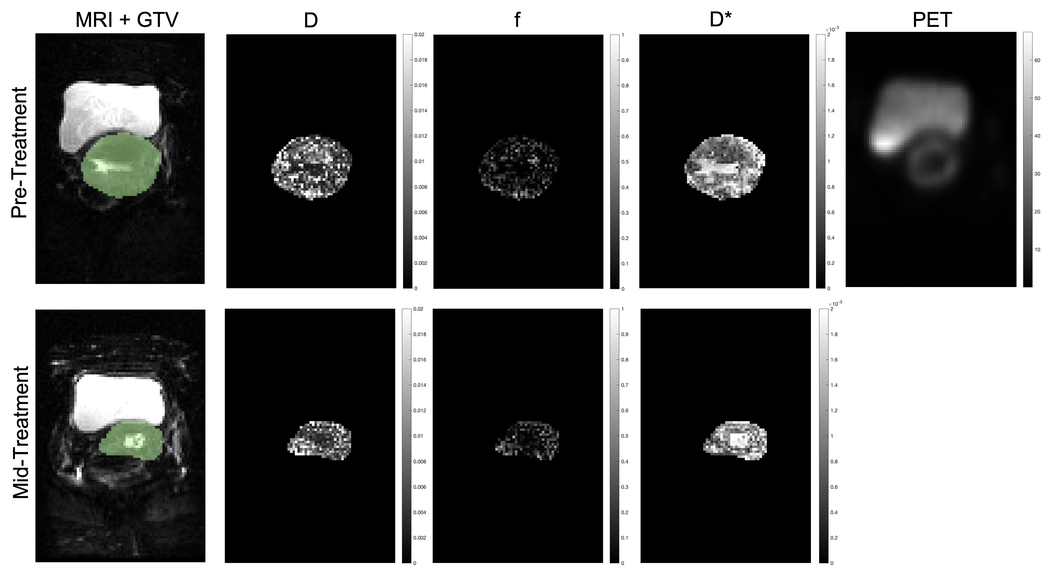
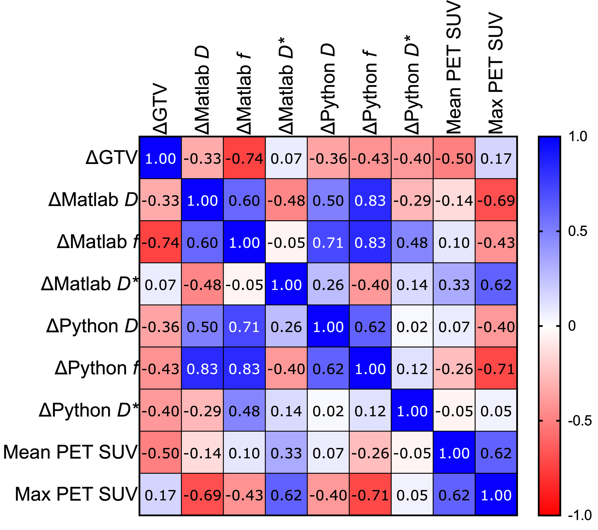
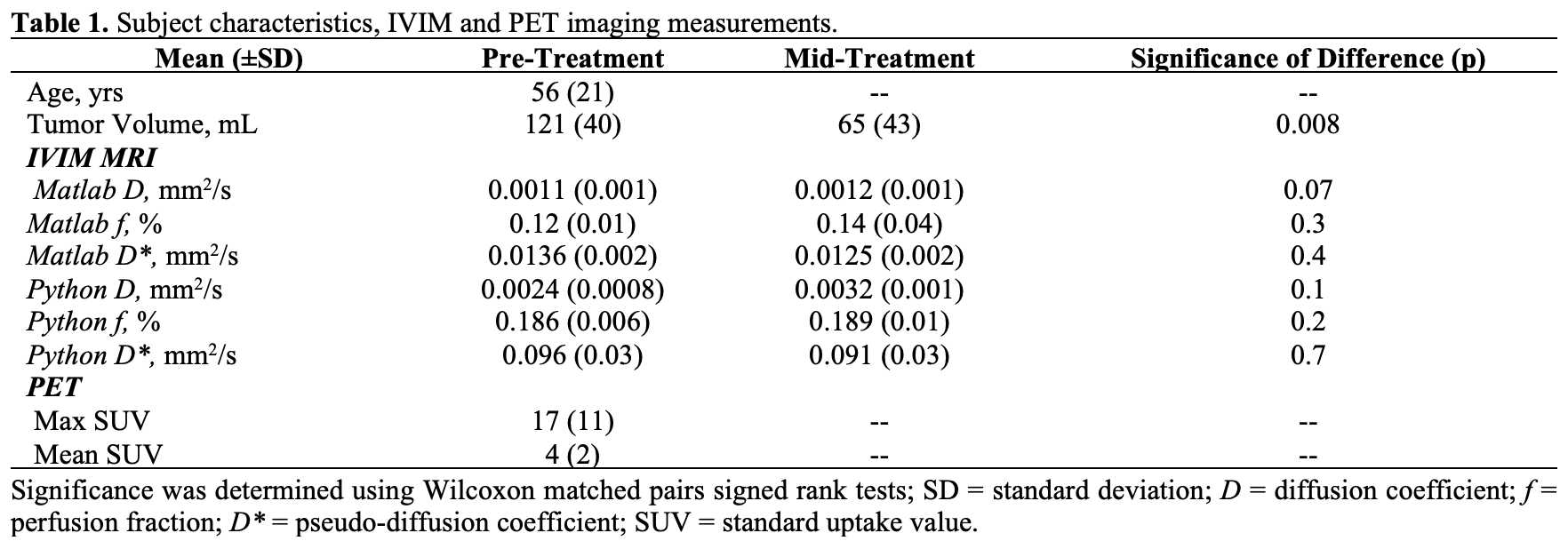
Table 1 summarizes patient characteristics as well as imaging measurements for the eight cervical cancer patients. As compared to pre-treatment, GTV and IVIM pseudo-diffusion decreased while IVIM measurements of diffusion and perfusion increased mid-treatment. Figure 1 shows pre- and mid-treatment images for a representative subject. Figure 2 shows a correlation matrix comparing changes in GTV with changes in IVIM measurements and initial PET measurements. Negative moderate relationships were observed between changes in IVIM and both PET measurements and changes in tumor volume.Discussion
In this preliminary study, IVIM and PET imaging measurements were evaluated in cervical cancer patients undergoing CCRT. We observed changes in IVIM measurements, specifically both perfusion fraction and diffusion coefficient increased during treatment, and that these changes were correlated to changes in tumor volume. Differences in IVIM during treatment have previously been observed and align with our current findings.10 Furthermore, differences were observed in IVIM measurements generated using the two different models (Matlab and Python), which may be due to the differences in how the fitting is performed.8,9 Changes in IVIM measurements were related to both changes in GTV and initial PET imaging, specifically max SUV. Although a previous study has looked at the combination of PET and IVIM in cervical cancer to evaluate lymphovascular space invasion11, our study was specifically designed to investigate changes that occur in IVIM during CCRT and how they relate to PET. Future work will relate to combining metabolic activity and oxygenation using PET and IVIM to better understand tumor microenvironment as it changes during treatment and potentially identify radio-resistant sub-volumes, as previously demonstrated with PET and DCE CT.5Conclusions
Both perfusion fraction and diffusion coefficient IVIM measurements increased in cervical cancer patients undergoing CCRT and these changes were related to both PET and changes in overall tumor volume – these changes in IVIM may reflect changes in the tumor microenvironment during treatment.Acknowledgements
No acknowledgement found.References
1. Haider, M. A. et al. Assessment of the tumor microenvironment in cervix cancer using dynamic contrast enhanced CT, interstitial fluid pressure and oxygen measurements. Int. J. Radiat. Oncol. Biol. Phys. 62, 1100–1107 (2005).
2. Mirpour, S. et al. The role of PET/CT in the management of cervical cancer. AJR Am. J. Roentgenol. 201, W192-205 (2013).
3. Son, H. et al. PET/CT evaluation of cervical cancer: spectrum of disease. Radiogr. Rev. Publ. Radiol. Soc. N. Am. Inc 30, 1251–1268 (2010).
4. Banks, T. I., von Eyben, R., Hristov, D. & Kidd, E. A. Pilot study of combined FDG-PET and dynamic contrast-enhanced CT of locally advanced cervical carcinoma before and during concurrent chemoradiotherapy suggests association between changes in tumor blood volume and treatment response. Cancer Med. 7, 3642–3651 (2018).
5. Capaldi, D. P. I., Hristov, D. H. & Kidd, E. A. Parametric Response Mapping of Coregistered Positron Emission Tomography and Dynamic Contrast Enhanced Computed Tomography to Identify Radioresistant Subvolumes in Locally Advanced Cervical Cancer. Int. J. Radiat. Oncol. Biol. Phys. 107, 756–765 (2020).
6. Le Bihan, D. et al. Separation of diffusion and perfusion in intravoxel incoherent motion MR imaging. Radiology 168, 497–505 (1988).
7. Pecorelli, S. Revised FIGO staging for carcinoma of the vulva, cervix, and endometrium. Int. J. Gynaecol. Obstet. Off. Organ Int. Fed. Gynaecol. Obstet. 105, 103–104 (2009).
8. Jalnefjord, O. et al. Comparison of methods for estimation of the intravoxel incoherent motion (IVIM) diffusion coefficient (D) and perfusion fraction (f). Magma N. Y. N 31, 715–723 (2018).
9. Farooq, H. et al. Microstructure Imaging of Crossing (MIX) White Matter Fibers from diffusion MRI. Sci. Rep. 6, 38927 (2016).
10. Kato, H. et al. Predicting Early Response to Chemoradiotherapy for Uterine Cervical Cancer Using Intravoxel Incoherent Motion MR Imaging. Magn. Reson. Med. Sci. MRMS Off. J. Jpn. Soc. Magn. Reson. Med. 18, 293–298 (2019).
11. Xu, C., Yu, Y., Li, X. & Sun, H. Value of integrated PET-IVIM MRI in predicting lymphovascular space invasion in cervical cancer without lymphatic metastasis. Eur. J. Nucl. Med. Mol. Imaging 48, 2990–3000 (2021).
Figures