0835
Usage of Dissolution DNP Magnetic Resonance Spectroscopy Depicts Efficacy of Chemotherapeutic and Radiotherapeutic Anticancer Interferences1Department of Radiology, Frontier Science for Imaging, Gifu University, Gifu, Japan, 2Department of Food Hygiene and Control, Faculty of Veterinary Medicine, Suez Canal University, Ismailia, Egypt, 3Department of Radiology, Gifu University, Gifu, Japan, 4Department of Tumor Pathology, Gifu University, Gifu, Japan, 5Innovation Center for Medical Redox Navigation, Kyushu University, Kyushu, Japan
Synopsis
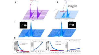
DNP highly improves the sensitivity of magnetic resonance spectroscopy allowing for greater insight into in vivo metabolic activity. We used our dissolution 13C DNP system to monitor the Warburg effect in MIA PaCa human pancreatic carcinoma model mice subjected to radiotherapy or chemotherapy. In vivo production of 13C lactate in tumors showed a significant reduction after radiation therapy. Mice subjected to anticancer drugs also showed a significant reduction in the relative production of 13C lactate at an early stage of the treatment course before any anatomical changes in the structure or size of the tumors were detectable by MRI images.
Introduction
DNP (dynamic nuclear polarization) is a powerful technique for highly improving the sensitivity of magnetic resonance spectroscopy (MRS). When compared to thermal equilibrium, it can increase the signal by more than 10000 times, allowing for greater insight into the in vivo metabolic activity. Hyperpolarized pyruvate MRS provides the capacity to evaluate metabolic state in cancer tissue rapidly, noninvasively, and directly. Additionally, it can determine the efficacy of therapeutic interference1.Generation of new therapeutic agents targeting tumor metabolism is becoming available for treating pancreatic cancer. DNP technique combined with MRS is ideally suited to measure the metabolic effects of these emerging treatments noninvasively. Moreover, it can determine the efficacy of an anti-cancer medical interference earlier than the usual method that depends on the follow-up of morphological condition which takes a relatively long time. Thus, it may help in saving the patients’ invaluable time to shift to another line of therapy if it was found non-effective. DNP-MRS of 13C metabolites measures the metabolic effects of anti-cancer treatments by monitoring the Warburg effect, a characteristic increase of lactate production by aerobic glycolysis in most tumors. The system provides rapid acquisition of in vivo 13C MR data of agents such as [1-13C]-pyruvate and its metabolic products lactate, alanine, in a matter of seconds.Purpose
This study aimed to compare the efficacy of the traditionally used radiation therapy and two other chemotherapeutic agents, Bevacizumab and Gemcitabine, in the treatment of pancreatic cancer using our dissolution DNP-MRS system after injection of hyperpolarized [1–13C]-pyruvate in MIA PaCa-2 pancreatic tumor mice models. Moreover, we aimed to investigate the feasibility of our system using hyperpolarized [1-13C]-pyruvate with a 1.5 T MRI scanner for early evaluation of the efficacy of anticancer interference by real-time probing of the metabolic state of cancer as a direct readout of treatment effect.Methods
Hyperpolarization of 13C Pyruvate was done by a 3.3T HyperSense DNP polarizer (Oxford Instruments, UK) at 1.4 K. in vitro 13C NMR acquisition was done by a 1.4 T Spinsolve 60 Carbon High-Performance benchtop NMR apparatus (Magritek, New Zealand). In vivo MRS experiment and T2-weighted anatomical images were acquired using an MRI scanner with a 1.5 T permanent magnet (Japan Redox Ltd. Japan).In vitro and ex vivo 13C lactate production from 13C Pyruvate by lactate dehydrogenase enzyme or tumor tissue homogenate were initially confirmed in an NMR test tube. Nude mice with leg xenografts of MIA PaCa-2 human pancreatic carcinoma were used for this study. In vivo DNP-MRS scanning of the tumor-bearing legs was performed before and after administration of radiation therapy or intraperitoneal injection of the anti-cancer mediations: Bevacizumab (10 mg/Kg every 2 days) or Gemcitabine (120 mg/Kg every 2 days). Real-time DNP-MRI was performed immediately after the intravenous injection of hyperpolarized 13C pyruvate solution. MRI acquisition of tumor tissue was also conducted before and after cancer treatment. Analysis of MRI images and the spectra of 13C Pyruvate and 13C Lactate were performed using Medalist (Japan Redox Ltd. Japan), Delta NMR (JEOL, Japan), and MestreNova (Mestrelab Research, Spain).
Results
In vitro and ex vivo data of lactate dehydrogenase enzyme and tumor tissue homogenate confirmed that 13C pyruvate and its metabolite 13C lactate were detected at measurable levels until these spectra decayed within a few minutes. In vivo production of 13C lactate in tumor tissue showed a significant difference before and after radiation therapy. Mice subjected to Bevacizumab or Gemcitabine intraperitoneal injection for 7 days also showed significant differences in the relative production of 13C lactate to 13C pyruvate. On the other hand, the T2-weighted anatomical images acquired by a 1.5 T MRI scanner on day 7 of treatment showed no significant differences in the anatomical structures or sizes of the tumor tissues after radiation therapy or treatment with the anticancer drugs Bevacizumab or Gemcitabine.Conclusion
Both radiation therapy and the chemotherapeutic agents, Bevacizumab and Gemcitabine proved effective in the treatment of cancer in MIA PaCa-2 pancreatic tumor mice models using our in vivo DNP-MRS system. Our system using dissolution DNP-MRS together with hyperpolarized 13C pyruvate has also proved its feasibility for real-time probing of the metabolic state of cancer indicated by the Warburg effect as a direct readout for the early evaluation of the efficacy of anticancer interference.Acknowledgements
No acknowledgement found.References
1. Ardenkjaer-Larsen JH, Fridlund B, Gram A, et al. Increase in signal-to-noise ratio of 10,000 times in liquid-state NMR. Proc Natl Acad Sci U S A 2003;100(18): 10158–10163.
Figures
