0804
High-resolution Spiral First-pass Myocardial Perfusion Imaging at 1.5 T with DEep learning-based rapid Spiral Image REconstruction (DESIRE)1Biomedical Engineering, University of Virginia, Charlottesville, VA, United States, 2Medicine, University of Virginia, Charlottesville, VA, United States, 3Radiology, University of Virginia, Charlottesville, VA, United States, 4Medicine, Stanford University, Stanford, CA, United States
Synopsis
First-pass contrast-enhanced myocardial perfusion imaging is useful for evaluating coronary artery disease (CAD). Spiral perfusion imaging techniques, using a motion-compensated (SMS-Slice-) L1-SPIRiT reconstruction, are capable of whole-heart high-resolution perfusion imaging. However, this reconstruction is performed off-line and takes ~30 minutes per slice. To address this, we developed a DEep learning-based rapid Spiral Image REconstruction technique (DESIRE) for spiral perfusion imaging at 1.5 T, for both single-slice (SS) and simultaneous multi-slice (SMS) MB=3 and MB=4 acquisitions, to provide rapid and high-quality reconstruction and make rapid online reconstruction feasible. High image quality was demonstrated using the proposed technique.
Introduction
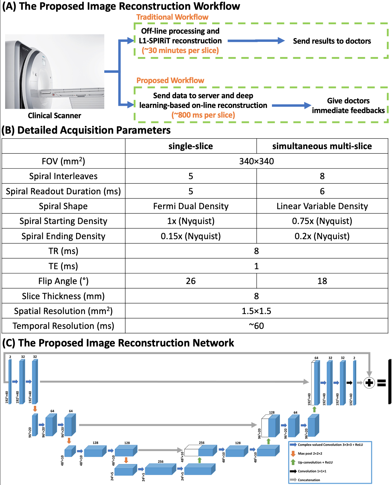
First-pass contrast-enhanced myocardial perfusion imaging is valuable for evaluating coronary artery disease (CAD)1–4. Recently, we have developed spiral perfusion imaging sequences for both single-slice (SS) and simultaneous multi-slice (SMS) imaging at 3 T and a corresponding motion-compensated (SMS-Slice-)L1-SPIRiT reconstruction capable of whole-heart high-resolution perfusion imaging5. However, this compressed-sensing (CS) based reconstruction technique is time-consuming and takes ~25 minutes per slice location. Hence, it can’t provide immediate feedback to doctors and impedes the clinical translation. Moreover, 1.5 T has been the dominant field strength for cardiac MR imaging as it has better and homogeneity than at 3 T. Here, we aim to develop high-resolution spiral perfusion imaging at 1.5 T for both SS and SMS imaging with high SMS acceleration factors (MB=3 and MB=4) with DEep learning-based rapid Spiral Image REconstruction technique (DESIRE), to provide fast and high-quality image reconstruction (Figure 1-A).Methods
Data Acquisition and Preprocessing: SS (N=5), SMS MB=3 (N=9), and MB=4 (N=3) golden-angle spiral perfusion data sets with 1.5×1.5 mm2 in-plane spatial resolution and whole-heart coverage (6-9 slices) were acquired from 17 patients undergoing clinical studies on a 1.5 T SIEMENS Aera scanner6,7. Detailed pulse sequence parameters are listed in Figure 1-B.Before inputting the data into the network, coil-selection8, motion-correction9, and adaptive phase combination10 were performed on the NUFFT-gridded11 multi-coil image series at each slice location. Specifically, for SMS data, perfusion image series that were input to the network were filtered by the through-plane kernels5,12 at each slice location to reduce the slice leakage.
Experimental Setup: Figure 1-C illustrates the proposed 3D U-Net13 based image denoising and enhancement network where the complex-valued convolution was enforced. The real and imaginary values of inputs and outputs were concatenated into two channels.
The training data were SS golden-angle spiral perfusion data sets with whole-heart coverage acquired from 18 healthy volunteers and 4 patients undergoing our prior clinical studies on 3 T SIEMENS Skyra/Prisma scanners5. The reference images were L1-SPIRiT reconstructions. 156 slices from 20 subjects were used for training. The training was conducted using PyTorch on four NVIDIA Tesla P100 GPUs with 150 epochs using an L1 loss.
To test the performance of the proposed technique, prospective SS and SMS images were also reconstructed using the GPU-accelerated (SMS-Slice-)L1-SPIRiT where the through-plane kernel was utilized for SMS imaging to reduce the slice leakage.
Image Analysis: Prospective SS and SMS images reconstructed by both (SMS-Slice-)L1-SPIRiT and the proposed DESIRE technique were blindly graded by an experienced cardiologist (5, excellent; 1, poor).
Results
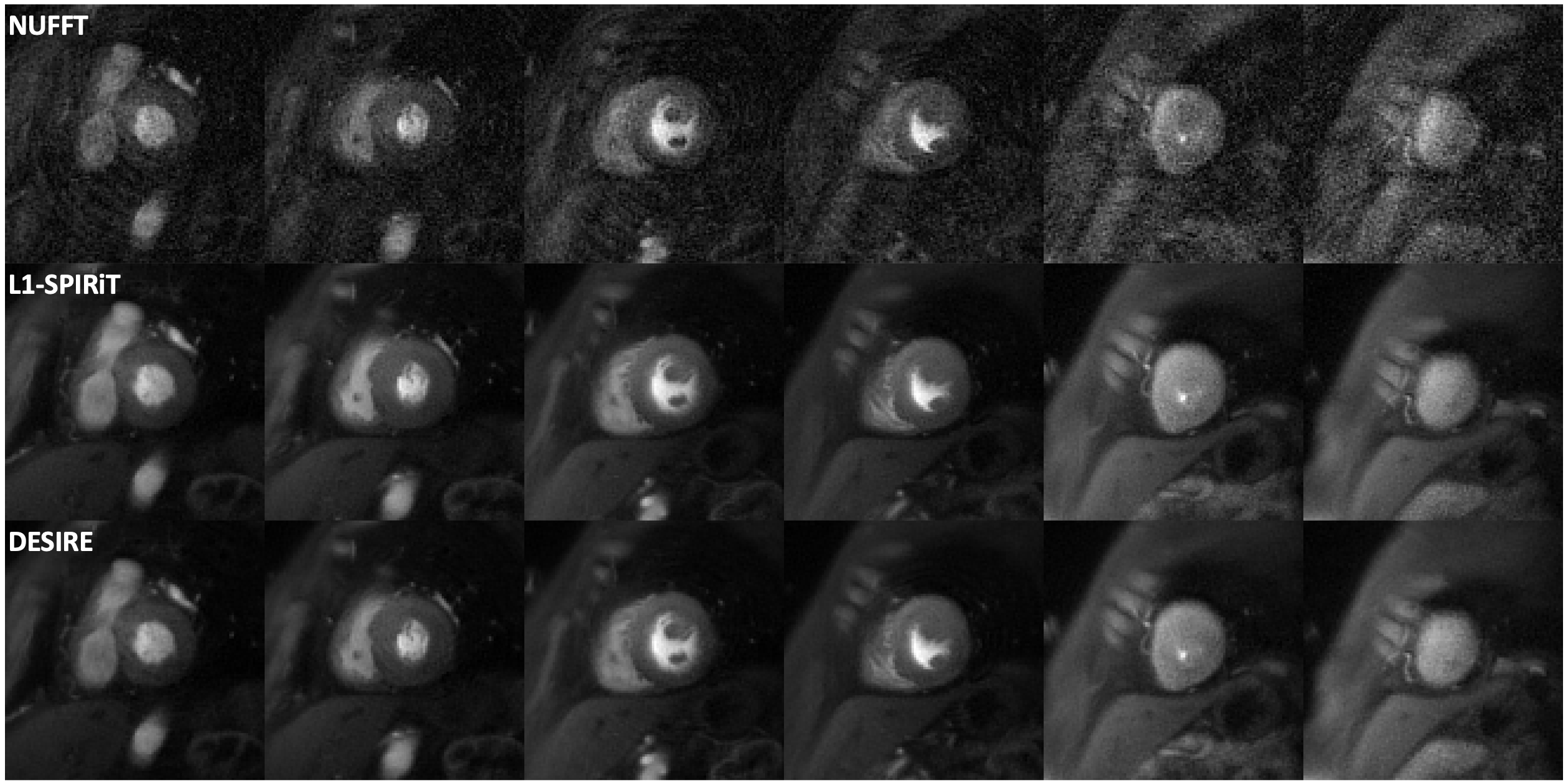
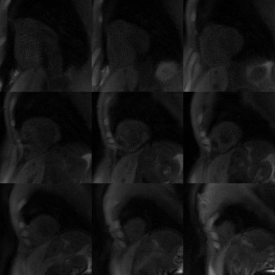
Good image quality was demonstrated with the proposed DESIRE technique. For (SMS-Slice-) L1-SPIRiT reconstruction, image quality scores were 4.5±0.7, 4.0±0.8 and 3.0±0.0 for SS, SMS MB=3 and MB=4 acquisitions. While for the proposed DESIRE technique, image quality scores were 4.4±0.8, 4.1±0.6 and 3.2±0.3 for SS, SMS MB=3 and MB=4 acquisitions (5 excellent, 1 poor).Figure 2 shows an example case from a patient undergoing clinical high-resolution spiral perfusion imaging with SS acquisitions. Excellent image quality was demonstrated with a score of 5 and 5 (5 excellent, 1 poor) for L1-SPIRiT and the proposed DESIRE technique, respectively. The first-pass perfusion video of this case reconstructed using the proposed DESIRE technique is shown in Video 1.
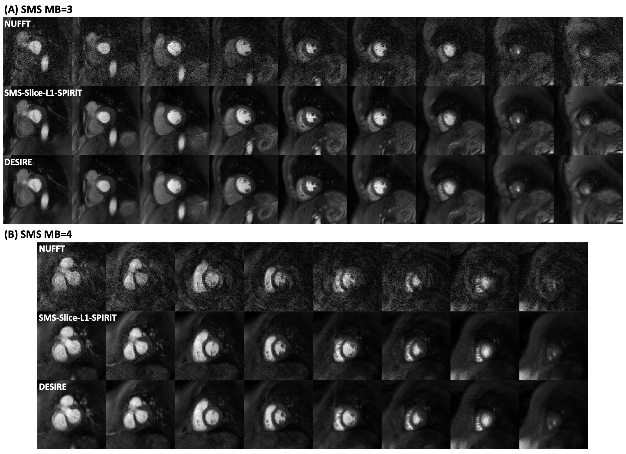
Figure 3 shows example cases from patient undergoing clinical high-resolution spiral perfusion imaging with SMS MB=3 (Figure 3-A) and MB=4 (Figure 3-B) acquisitions.
For SMS MB=3 (Figure 3-A), excellent image quality was demonstrated with a score of 5 and 5 (5 excellent, 1 poor) for SMS-Slice-L1-SPIRiT and the proposed DESIRE technique, respectively. The corresponding first-pass perfusion video reconstructed using the proposed DESIRE technique is shown in Video 2.
For SMS MB=4 (Figure 3-B), image quality scores were 3 and 3 (5 excellent, 1 poor) for SMS-Slice-L1-SPIRiT and the proposed DESIRE technique, respectively.
The reconstruction time was ~800 ms per slice on a NVIDIA Tesla P100 GPU, while the reconstruction time of using (SMS-Slice-)L1-SPIRiT with GPU-accelerated NUFFT on an Intel Xeon CPU (2.40 GHz) was ~30 minutes per image series.
Discussion and Conclusion
The proposed deep learning-based image reconstruction technique (DESIRE) enabled rapid and high-quality image reconstruction for whole-heart high-resolution first-pass spiral perfusion imaging at 1.5 T. Further validation will be required in patients undergoing adenosine stress studies.Acknowledgements
This work was supported by NIH R01 HL131919 and Wallace H. Coulter Foundation Grant.References
1. Greenwood JP, Maredia N, Younger JF, et al. Cardiovascular magnetic resonance and single-photon emission computed tomography for diagnosis of coronary heart disease (CE-MARC): a prospective trial. The Lancet 2012;379:453–460.
2. Salerno M, Beller GA. Noninvasive Assessment of Myocardial Perfusion. Circ Cardiovasc Imaging 2009;2:412–424.
3. Jaarsma C, Leiner T, Bekkers SC, et al. Diagnostic Performance of Noninvasive Myocardial Perfusion Imaging Using Single-Photon Emission Computed Tomography, Cardiac Magnetic Resonance, and Positron Emission Tomography Imaging for the Detection of Obstructive Coronary Artery Disease: A Meta-Analysis. Journal of the American College of Cardiology 2012;59:1719–1728.
4. Lipinski MJ, McVey CM, Berger JS, Kramer CM, Salerno M. Prognostic Value of Stress Cardiac Magnetic Resonance Imaging in Patients With Known or Suspected Coronary Artery Disease: A Systematic Review and Meta-Analysis. Journal of the American College of Cardiology 2013;62:826–838.
5. Wang J, Yang Y, Weller DS, et al. High spatial resolution spiral first-pass myocardial perfusion imaging with whole-heart coverage at 3 T. Magnetic Resonance in Medicine 2021;86:648–662.
6. Wang J, Yang Y, Zhou R, et al. High resolution spiral simultaneous multi‐slice first‐pass perfusion imaging with whole-heart coverage at 1.5 T and 3 T. In: Proceedings of the 28th Annual Meeting of ISMRM, 2020. p 1313.
7. Yang Y, Kramer C, Salerno M. Whole heart First-pass spiral perfusion imaging with 1.25mm resolution at 3T. In: Proceedings of the 26th Annual Meeting of ISMRM, Paris, France, 2018. p 3321.
8. Zhou R, Yang Y, Mathew RC, et al. Free-breathing cine imaging with motion-corrected reconstruction at 3T using SPiral Acquisition with Respiratory correction and Cardiac Self-gating (SPARCS). Magnetic Resonance in Medicine 2019;82:706–720.
9. Zhou R, Huang W, Yang Y, et al. Simple motion correction strategy reduces respiratory-induced motion artifacts for k-t accelerated and compressed-sensing cardiovascular magnetic resonance perfusion imaging. J Cardiovasc Magn Reson 2018;20:6.
10. Walsh DO, Gmitro AF, Marcellin MW. Adaptive reconstruction of phased array MR imagery. Magnetic Resonance in Medicine 2000;43:682–690.
11. Fessler JA, Sutton BP. Nonuniform fast Fourier transforms using min-max interpolation. IEEE Transactions on Signal Processing 2003;51:560–574.
12. Wang J, Yang Y, Zhou R, Jacob M, Weller DS, Salerno M. SMS Slice L1-SPIRiT: auto-calibrated image reconstruction for spiral simultaneous multi-slice first-pass perfusion imaging with 1.25 mm resolution and whole heart coverage at 3T. In: Proceedings of the SCMR 23rd Annual Scientific Sessions, Orlando, Florida, USA, 2020. Abstract 714001.
13. Ronneberger O, Fischer P, Brox T. U-Net: Convolutional Networks for Biomedical Image Segmentation. arXiv:1505.04597 [cs] 2015.
Figures