0790
Multimodal MRI Investigation of Basal Forebrain Damage in Patients with Mild Cognitive Impairment1Department of Radiology, Drum Tower Hospital, Clinical College of Nanjing Medical University, Nanjing, China, 2Philips Healthcare, Shanghai, China, 3Department of Radiology, Drum Tower Hospital, Medical School of Nanjing University, Nanjing, China
Synopsis
Cholinergic degeneration in the basal forebrain (BF) plays a crucial role in Alzheimer’s disease. In the present study, reduced BF volumes and damaged BF-associated tracts were observed in patients with mild cognitive impairment (MCI) compared to those with normal aging. Correlation and mediation analyses highlighted that volume reductions in the BF mainly accounted for deficits in global cognition, memory, and visuospatial ability, while disruption in BF-cortical tracts mainly contributed to executive and processing speed dysfunction. Collectively, this study may provide a better understanding of BF damage underlying cognitive impairment and provide guidance for intervention targets in MCI patients.
Introduction
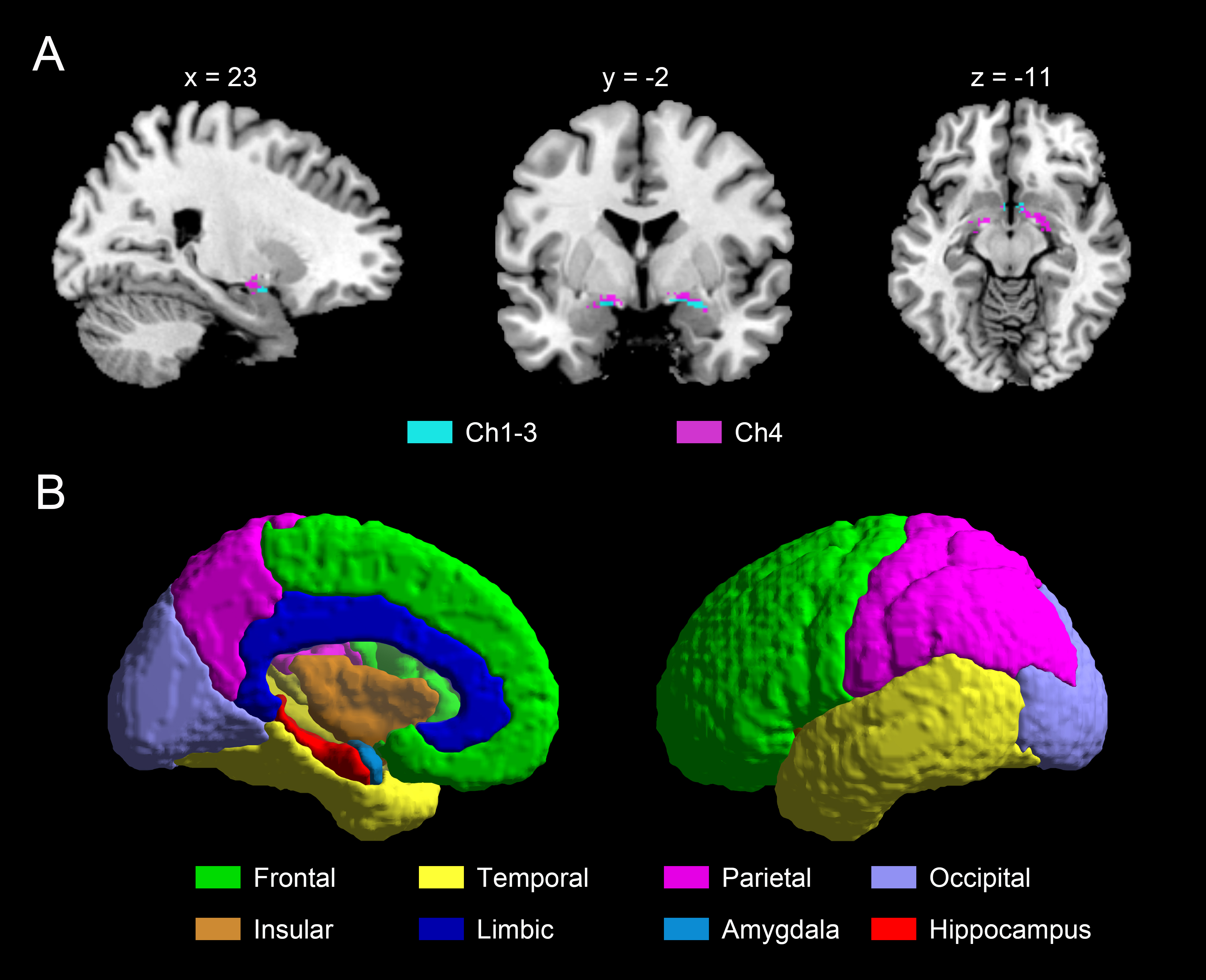
Alzheimer’s disease (AD) continues to be a global concern. Mild cognitive impairment (MCI), the prodromal stage of AD, is critical for the early diagnosis and targeted intervention of AD1. Cholinergic degeneration in the basal forebrain (BF) plays a crucial role during the progression of AD2. The BF consists of different nuclei, with Ch1 and Ch2 projecting to the hippocampus, Ch3 projecting to the olfactory bulb, and Ch4 projecting to the cerebral cortex and amygdala3. To date, the effects of BF damage, assessed with multimodal magnetic resonance imaging (MRI), on cognitive dysfunction have not been well characterized4. We aimed to investigate BF alterations in MCI patients using volumetry, probabilistic tractography, and functional connectivity (FC) analyses and to explore the relationships among groups, multimodal MRI measures, and cognition by correlation and mediation analyses.Methods
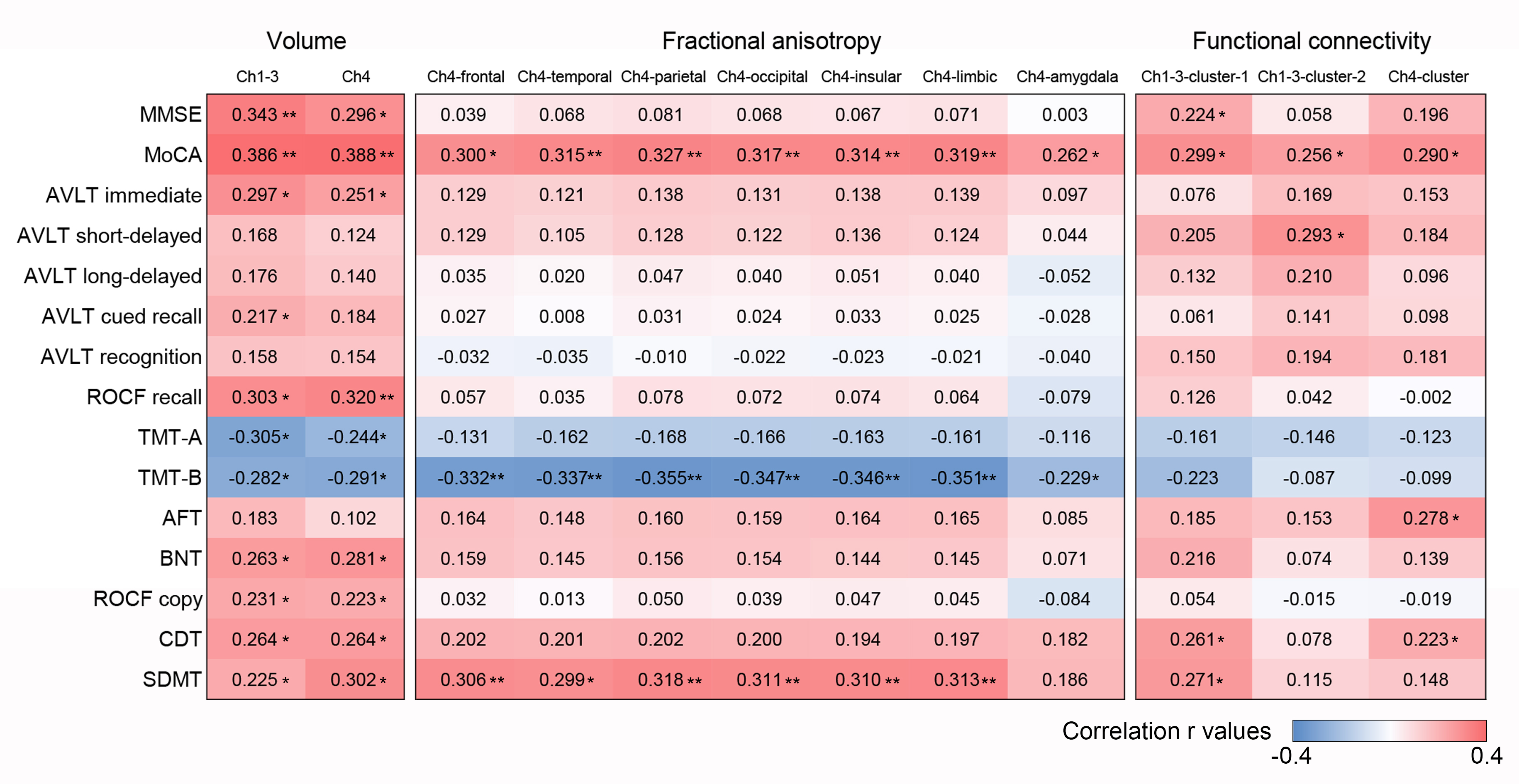
Fifty-two normal controls (NCs) and forty-two MCI patients were enrolled and performed the neuropsychological evaluation and brain MRI scanning in the Ingenia CX 3.0 T System (Philips Healthcare, Best, The Netherlands). A cytoarchitectonic BF mask derived from histological sections of a postmortem brain in Montreal Neurological Institute (MNI) space was applied5. Calculation of the individual Ch1-3 and Ch4 volumes was acquired by summing up the modulated grey matter (GM) voxel values within the respective masks. The spatially averaged fractional anisotropy (FA) and mean diffusivity (MD) values within the thresholded tracts from Ch1-3 to hippocampus and from Ch4 to the frontal, temporal, parietal, occipital, insular, limbic cortex, and amygdala were extracted from individuals’ native diffusion images. For the seed-based FC analyses, FC was calculated based on Pearson correlation coefficients between each masks’ time series (Ch1-3 and Ch4) and each other voxels’ time series. The MRI metrics that showed significant differences between the two groups were subsequently assessed associations with cognitive measures by correlation and mediation analyses.Results
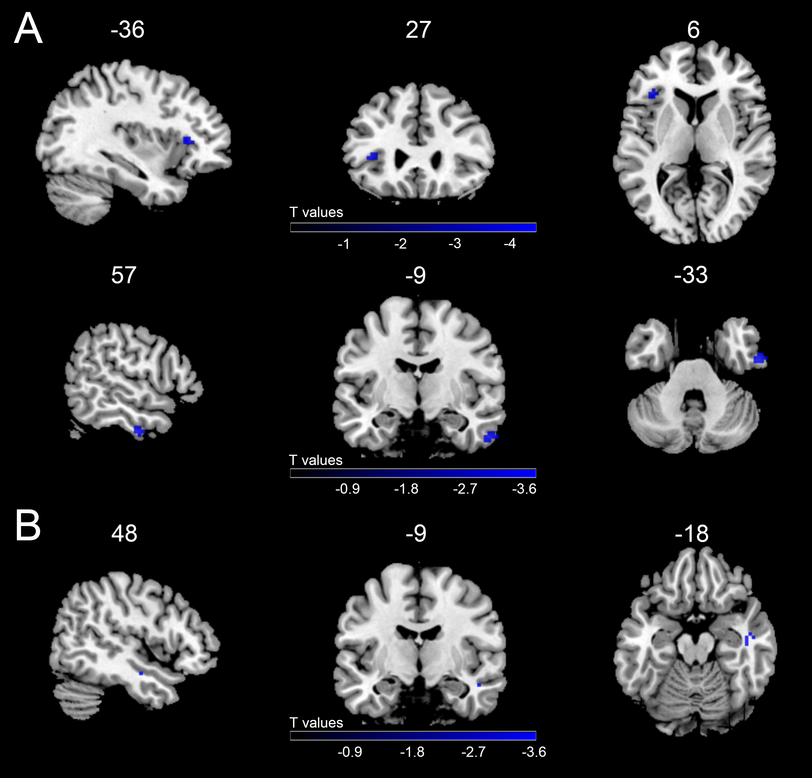
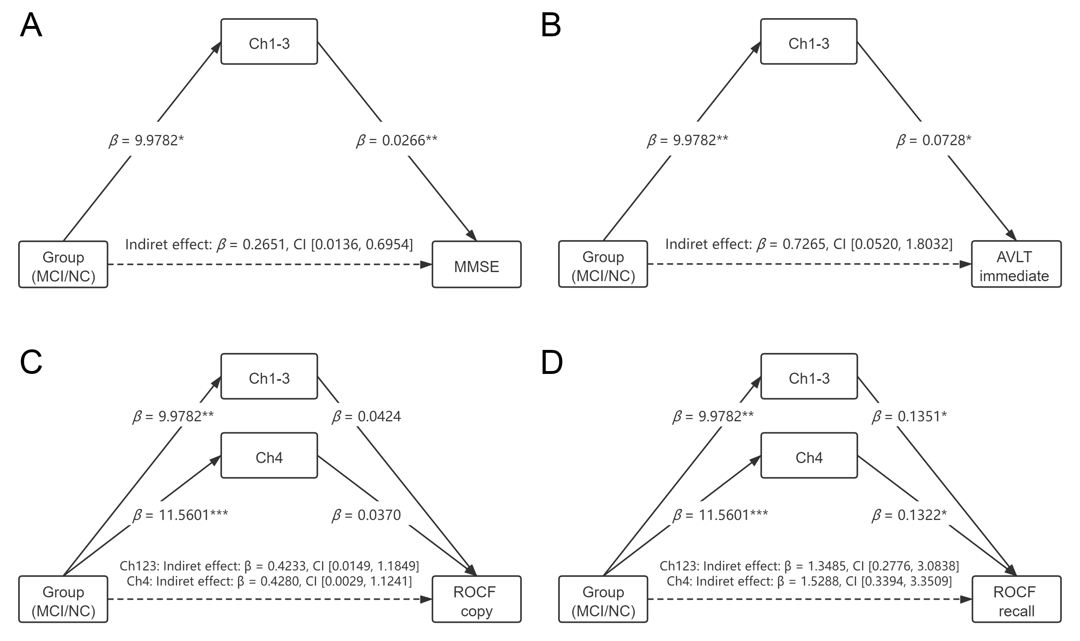
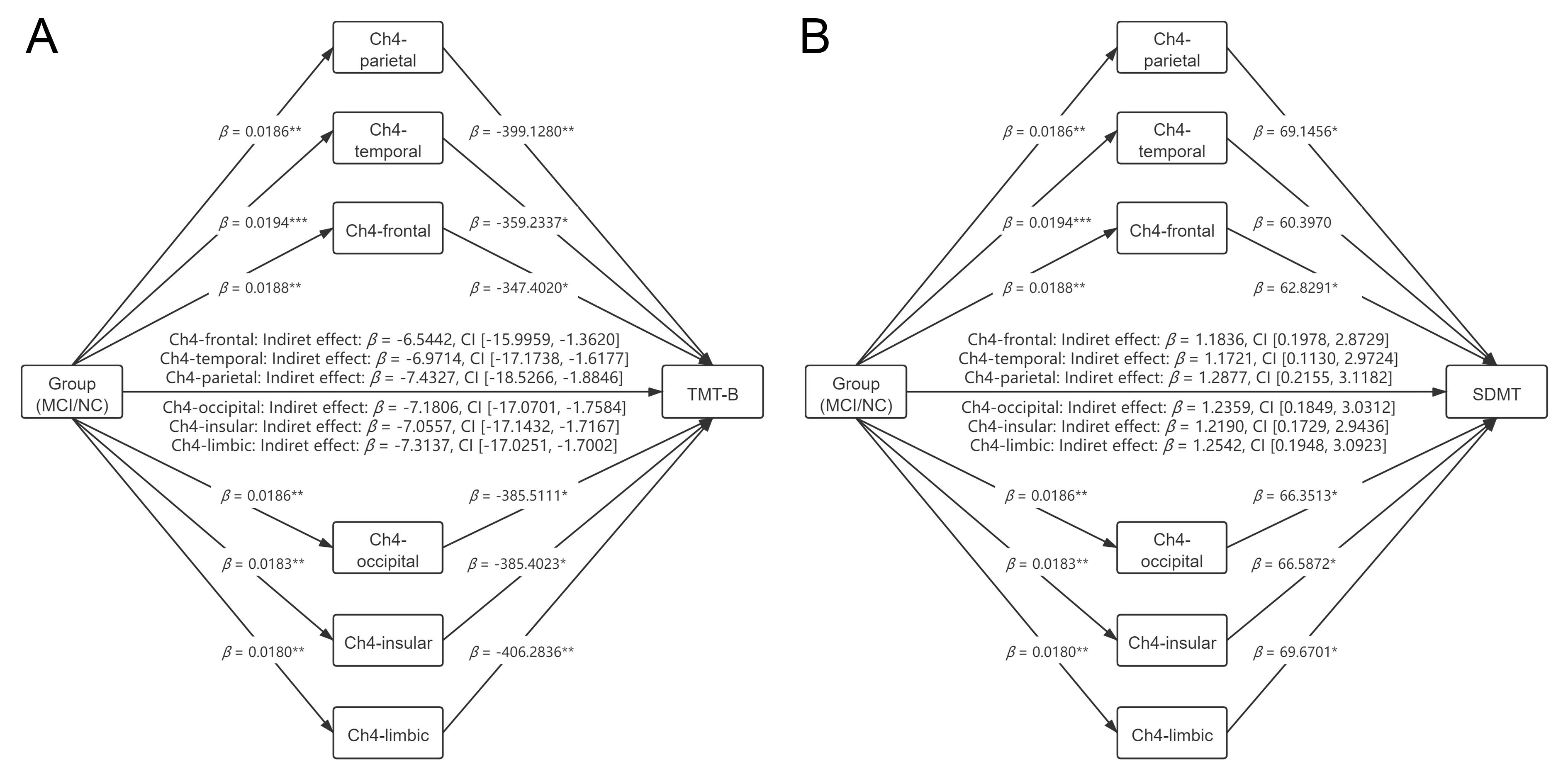
The MCI group showed smaller volumes than the NC group in Ch1-3 and Ch4. The MCI group also had significantly decreased FA values in the Ch4-frontal, Ch4-temporal, Ch4-parietal, Ch4-occipital, Ch4-insular, Ch4-limbic, and Ch4-amygdala tracts. The MCI patients presented evidence of reduced FC between Ch1-3 and two small clusters: one contained 11 voxels located in the left inferior frontal gyrus and insular cortex, and the other contained 15 voxels located in the right inferior temporal gyrus. Regarding Ch4, the MCI group showed a small cluster of 11 voxels with reduced connectivity in the right middle temporal gyrus. Mediation analyses showed that Ch1-3 volumes fully mediated the correlations between group and Mini-Mental State Examination (MMSE) scores and between group and auditory verbal learning test (AVLT) immediate memory scores. Ch1-3 and Ch4 volumes both fully mediated the associations between group and Rey-Osterrieth complex figure (ROCF) copy scores and between group and ROCF recall scores. FA values in the Ch4-cortical tracts showed partial mediating effects on the correlations between group and trail making test part B (TMT-B) performance, and between group and symbol digit modalities test (SDMT) performance. No significant mediating effects of FC on group-cognition associations were identified.Discussion
AD pathology gradually leads to the death of BF neurons, giving rise to atrophy that can be detected by MRI volumetry6. Evidence of associations between BF atrophy and cortical amyloid burden and hypometabolism has been identified7. Consistent with previous studies8, the MCI group showed significant volume reductions in Ch1-3 and Ch4 compared to the NC group. Cholinergic inputs to the cerebrum mainly originate from the BF, with Ch1-3 innervating the hippocampus and Ch4 innervating associative cortical regions and the amygdala9. The neurofibrillary tangles within cell bodies of the BF may eventually trigger a loss of cholinergic axons projecting to the cerebral cortex and of their presynaptic cholinergic terminals within the cerebral cortex6. By probabilistic tractography, we observed decreased FA in the Ch4-cortical and Ch4-amygdala tracts in MCI patients, suggesting disrupted microstructure of the Ch4-associated tracts. Under a relatively lenient statistical threshold of p < 0.001, uncorrected and k > 10 voxels, only three small clusters with reduced FC were identified in the MCI group, none of which would have survived the application of stricter levels of significance needed for multiple comparison corrections. Our results suggested that FC alteration in BF may not be a more sensitive biomarker compared to structural damage in the MCI stage. Mediation analyses help further illustrate whether the group-related cognitive differences were attributable to alterations in the BF assessed by multimodal MRI. The results demonstrated that GM atrophy in BF nuclei and white matter (WM) degeneration in Ch4-cortical tracts may underlie different patterns of cognitive deficits in MCI patients.Conclusion
MCI patients showed reduced volumes in Ch1-3 and Ch4 and damaged microstructure in Ch4-cortical and Ch4-amygdala tracts, while FC was relatively reserved compared to NCs. The volume reduction in Ch1-3 and Ch4 could fully explain deficits in global cognition, memory, and visuospatial ability, while WM disruption in Ch4-cortical tracts could partly account for executive and processing speed dysfunction. These findings may have further implications for BF structural damage as sensitive biomarkers for potential AD and provide novel insights into the specific role of GM atrophy in BF and WM disruption in BF-cortical tracts underlying cognitive impairment in prodromal AD patients.Acknowledgements
This work was supported by the National Natural Science Foundation of China (81720108022, B.Z.); the Fundamental Research Funds for the Central Universities, Nanjing University (2020-021414380462); and the key project of Jiangsu Commission of Health (K2019025).References
1. Petersen RC. Mild cognitive impairment as a diagnostic entity. J Intern Med. 2004;256(3):183-194.
2. Hampel H, Mesulam MM, Cuello AC, et al. The cholinergic system in the pathophysiology and treatment of Alzheimer's disease. Brain. 2018;141(7):1917-1933.
3. Gargouri F, Gallea C, Mongin M, et al. Multimodal magnetic resonance imaging investigation of basal forebrain damage and cognitive deficits in Parkinson's disease. Mov Disord. 2019;34(4):516-525.
4. Herdick M, Dyrba M, Fritz HJ, et al. Multimodal MRI analysis of basal forebrain structure and function across the Alzheimer's disease spectrum. Neuroimage Clin. 2020;28:102495.
5. Wolf D, Grothe M, Fischer FU, et al. Association of basal forebrain volumes and cognition in normal aging. Neuropsychologia. 2014;53:54-63.
6. Mesulam M. Cholinergic aspects of aging and Alzheimer's disease. Biol Psychiatry. 2012;71(9):760-761.
7. Grothe MJ, Ewers M, Krause B, et al. Basal forebrain atrophy and cortical amyloid deposition in nondemented elderly subjects. Alzheimers Dement. 2014;10(5 Suppl):S344-353.
8. Muth K, Schönmeyer R, Matura S, et al. Mild cognitive impairment in the elderly is associated with volume loss of the cholinergic basal forebrain region. Biol Psychiatry. 2010;67(6):588-591.
9. Mesulam MM. Cholinergic circuitry of the human nucleus basalis and its fate in Alzheimer's disease. J Comp Neurol. 2013;521(18):4124-4144.
Figures