0762
Microstructure-Informed Synthetic Axons (MISA): generating synthetic axons from 3D histology1* (Equal contributions), Lausanne, Switzerland and Copenhagen, Denmark, 2Copenhagen University Hospital - Amager and Hvidovre, Danish Research Centre for Magnetic Resonance, Copenhagen, Denmark, 3Department of Applied Mathematics and Computer Science, Technical University of Denmark, Kgs. Lyngby, Denmark, 4Signal Processing Laboratory (LTS5), École Polytechnique Fédérale de Lausanne (EPFL), Lausanne, Switzerland, 5Radiology Department, Centre Hospitalier Universitaire Vaudois and University of Lausanne, Lausanne, Switzerland, 6CIBM Center for Biomedical Imaging, Lausanne, Switzerland
Synopsis
Changes in diffusion properties of the intra-axonal space can be linked to brain health and function. Monte Carlo simulations of diffusion using realistic substrates are important for sequence design and for understanding diffusion MRI signals observed ex vivo and in vivo. However, it is difficult to construct tissue phantoms that accurately reflect compartment morphology. Here, we present the framework MISA for the generation of Microstructure-Informed Synthetic Axons. MISA uses statistical descriptions of axonal morphology from 3D histology to mimic real axons. We show here that MISA axons match axons from 3D histology in terms of their diffusion properties.
Introduction
Microstructural changes in the intra-axonal space (IAS) of brain white matter (WM) can occur due to healthy structural plasticity or disease. Sensitivity to such changes with diffusion magnetic resonance imaging (MRI) is key to understanding how diseases progress in the WM. Monte Carlo (MC) simulations of diffusion within realistic axonal phantoms, based on histological quantifications of axonal morphology, aid the design of diffusion MRI protocols that target IAS changes. Current frameworks1,2 aim to produce realistic phantoms by inputting a set of target parameters (e.g. axon diameter distribution and density) and packing axons. However, if the target density is high – as expected in WM – the packing algorithm may not succeed in maintaining the target parameters. This is problematic when trying to model the IAS with specific parameters.We present MISA, a simple framework to generate Microstructure Informed Synthetic Axons from descriptors of axon morphology. We show that this enables the generation of synthetic axons that match the properties of axons segmented from 3D histology in terms of morphology, and transverse and parallel apparent diffusion coefficients (ADC).
Methods
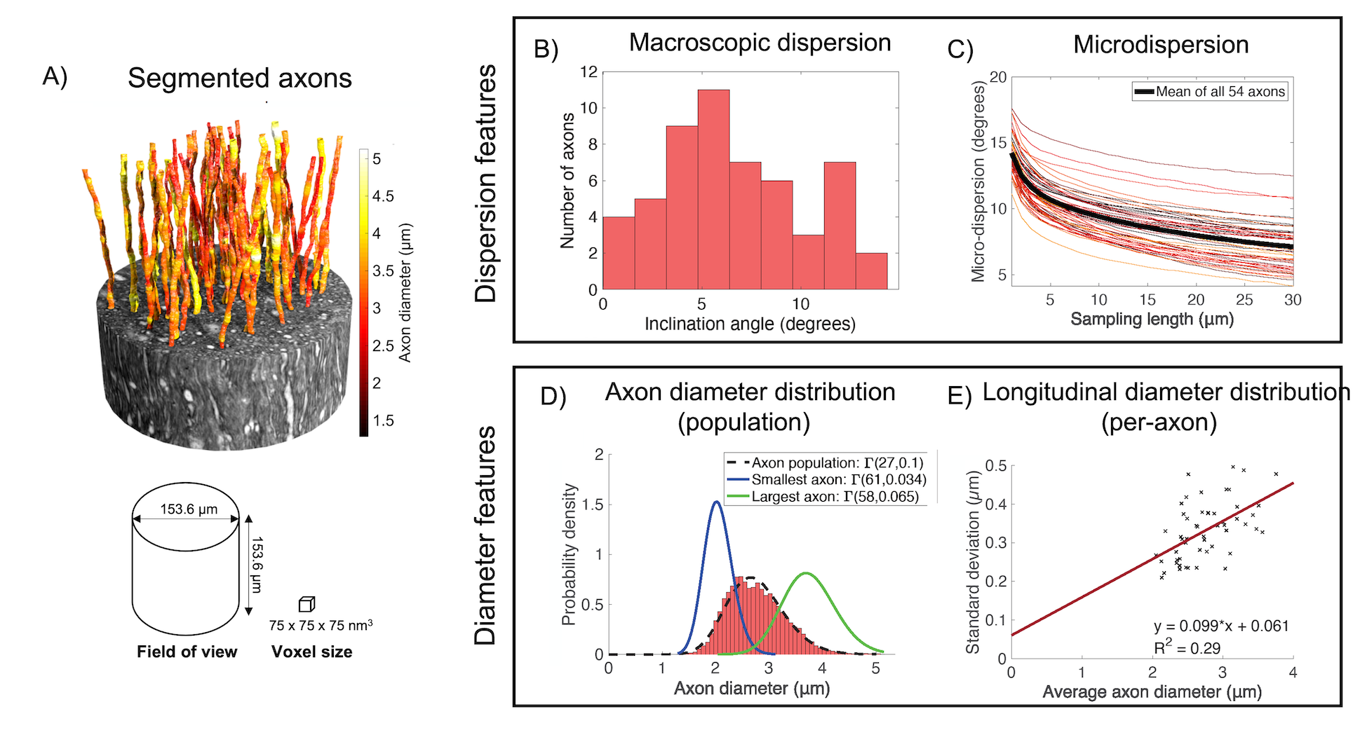
Morphological descriptors of axonsMISA axons were modelled on 54 segmented axons from a 3D x-ray nano-holotomography (XNH) volume of the vervet monkey splenium; the data and morphological analysis are described in Andersson et al.3 and are shown in Figure 1. In short, axonal “macrodispersion” was defined as the inclination angle between the average direction of the 54-axon bundle and the main direction of each individual axon. “Microdispersion” was defined as the mean inclination angle between the axon’s main direction and its trajectory divided into segments of length $$$L$$$. Diameters were evaluated at 150 nm intervals along the axonal trajectories. The pooled distribution of all diameters in the 54-axon population is shown in Figure 1D, and the relationship between mean and standard deviation of axon diameter variations along single axons is shown in panel E.
MISA algorithm
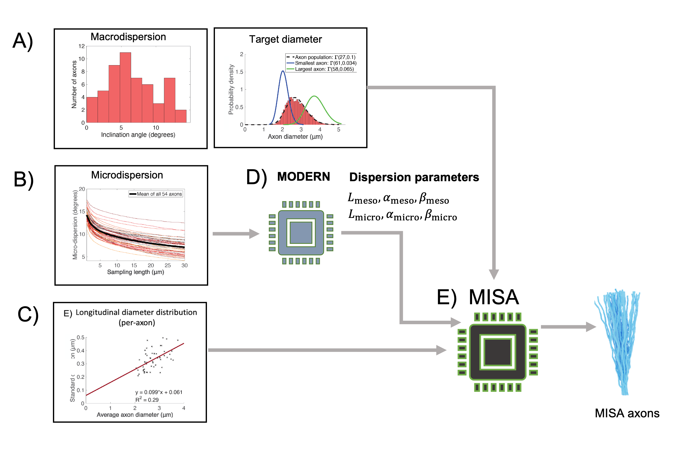
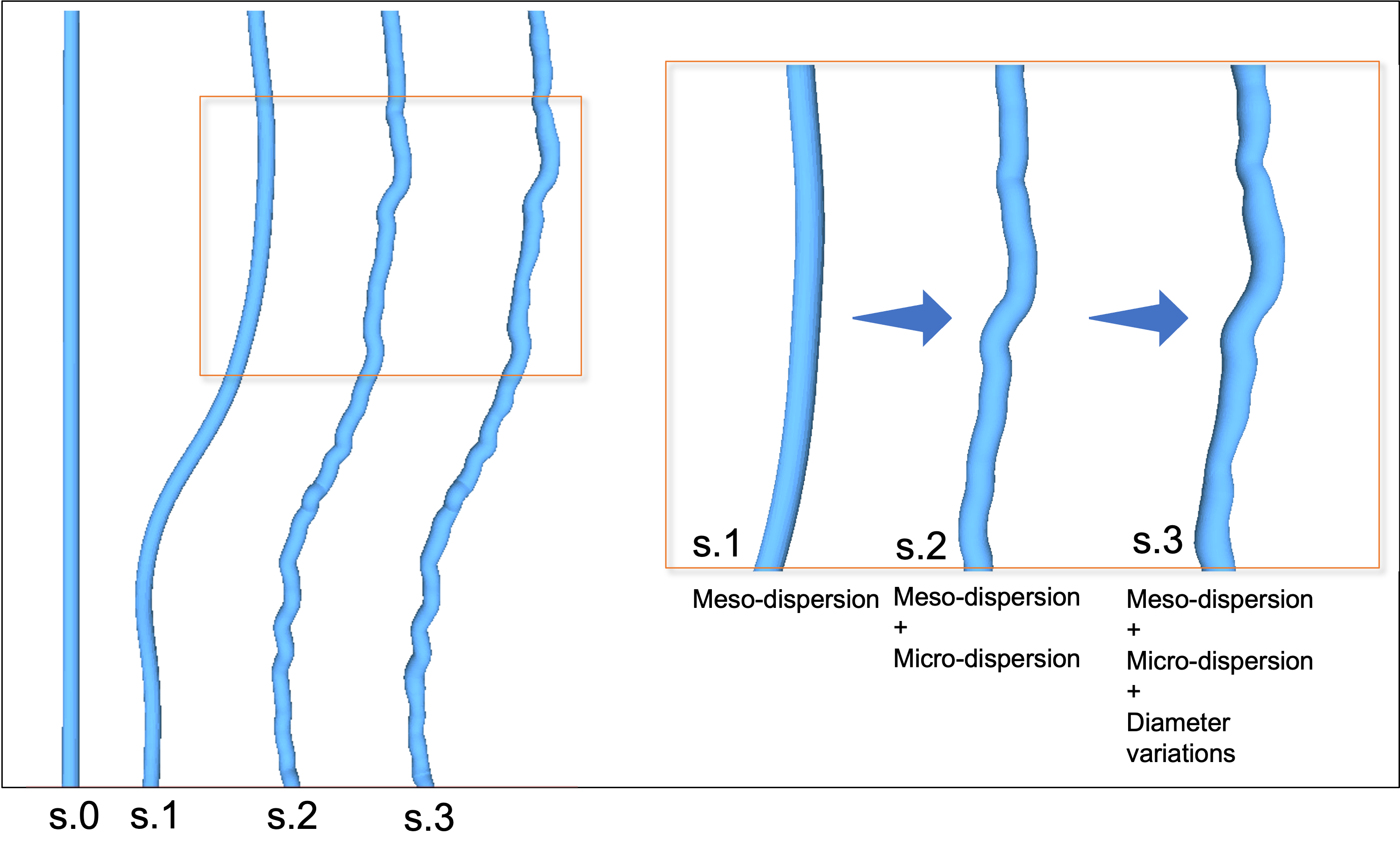
MISA (Figure 2) is a multistage algorithm that builds realistic axons by step-wise increasing the morphological complexity towards desired microstructural properties, as shown in Figure 3. In step 0, a cylinder with target diameter $$$d$$$ is given a random orientation from a macroscopic orientation distribution (e.g. a distribution fitted to Figure 1B). The target $$$d$$$ can be drawn from a distribution e.g. that in Figure 1D. In step 1, the cylinder midline is discretized into segments of length $$$L_{\text{meso}}$$$. Each segment is given an inclination angle sampled from a gamma distribution, $$$\Gamma_{\text{meso}}(\alpha_{\text{meso}},\beta_{\text{meso}})$$$. In step 2, the trajectory is further discretized into segments of length $$$L_{\text{micro}}$$$ where $$$L_{\text{micro}}< L_{\text{meso}}$$$. Again, the segments are assigned orientations that are picked from a second distribution, $$$\Gamma_{\text{micro}}(\alpha_{\text{micro}},\beta_{\text{micro}})$$$. We used the Multi-Objective Differential Evolution for Realistic Numerical Axons (MODERN) framework4 to learn the optimal discretization lengths ($$$L_{\text{micro}}$$$ and $$$L_{\text{meso}})$$$ and dispersion parameters ($$$\alpha_{\text{meso}}$$$, $$$\beta_{\text{meso}}$$$, $$$\alpha_{\text{micro}}$$$, $$$\beta_{\text{micro}}$$$) needed to replicate the mean microdispersion curve in Figure 1C, with 15 µm < $$$L_{\text{meso}}$$$ < 30 µm and 0.5 µm < $$$L_{\text{micro}}$$$ < 2 µm. Finally, in step 3, MISA applies diameter variations at intervals of $$$L_{\text{micro}}$$$, given a standard deviation of the diameter distribution along single axons – in this case provided by Figure 1E.
Monte-Carlo simulations
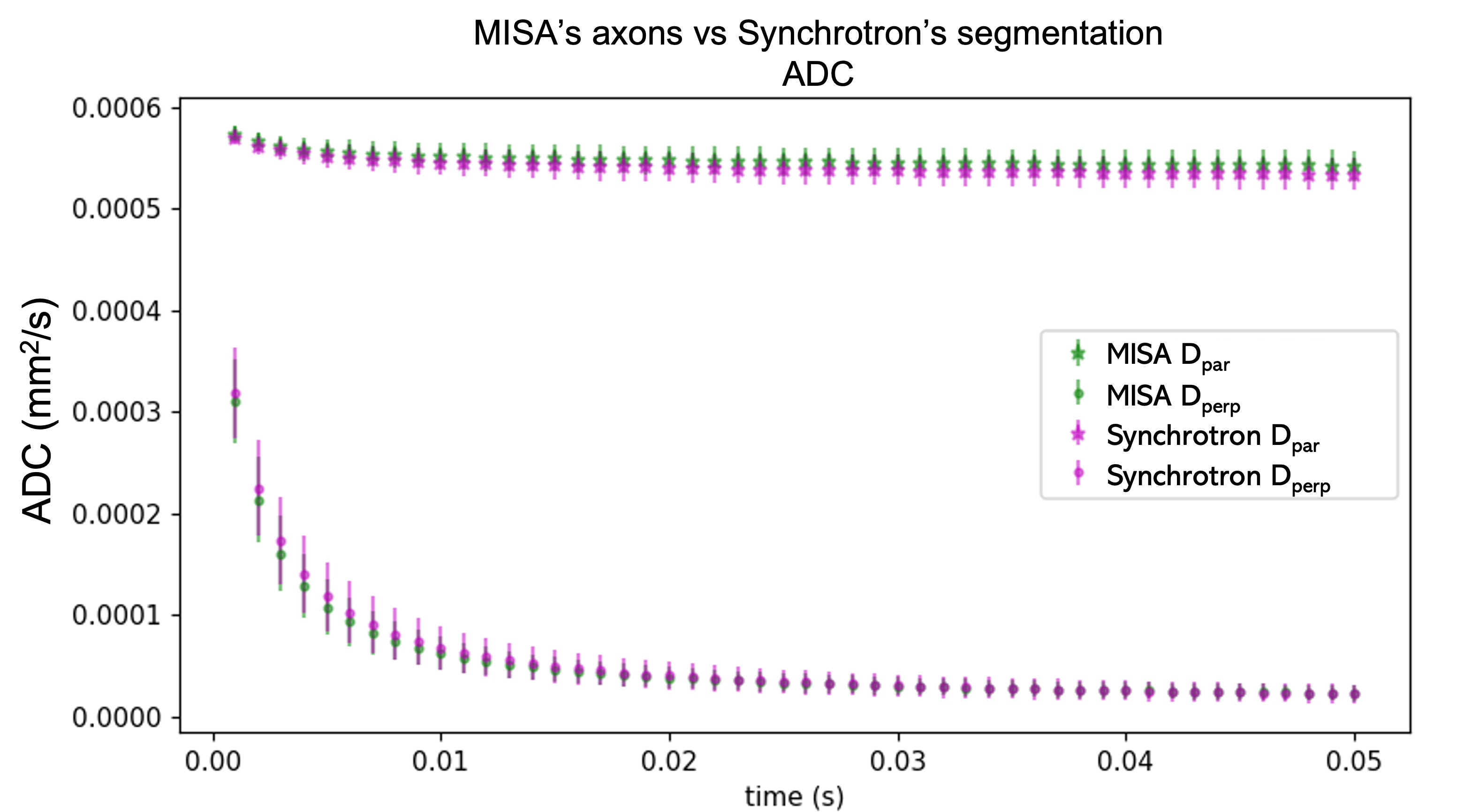
We generated 54 MISA axons using the inputs in Figure 1. Each was assigned the same target mean diameter as a corresponding XNH axon to focus only on the variability due to the randomly generated microdispersion and diameter variations. MC simulations5 of diffusion were performed for up to 50 ms in the XNH and MISA axons. Spins were uniformly initialised within each axon at a density of 1 µm$$$^{-3}$$$ and a minimum distance of 20 µm from the ends to prevent their escape. An ex vivo diffusivity of $$$D_0 = 0.6 \cdot 10^{-9} \text{m}^2\text{/s}$$$ was used as in Andersson et al.3. The ADCs parallel and transverse to the bundle main direction were computed using $$$ \left \langle (\Delta r)^2 \right \rangle = 2Dt $$$ where $$$(\Delta r)$$$ represents the mean-squared displacement in a given direction, $$$D$$$ is the ADC and $$$t$$$ is the diffusion time.
Results and Discussion
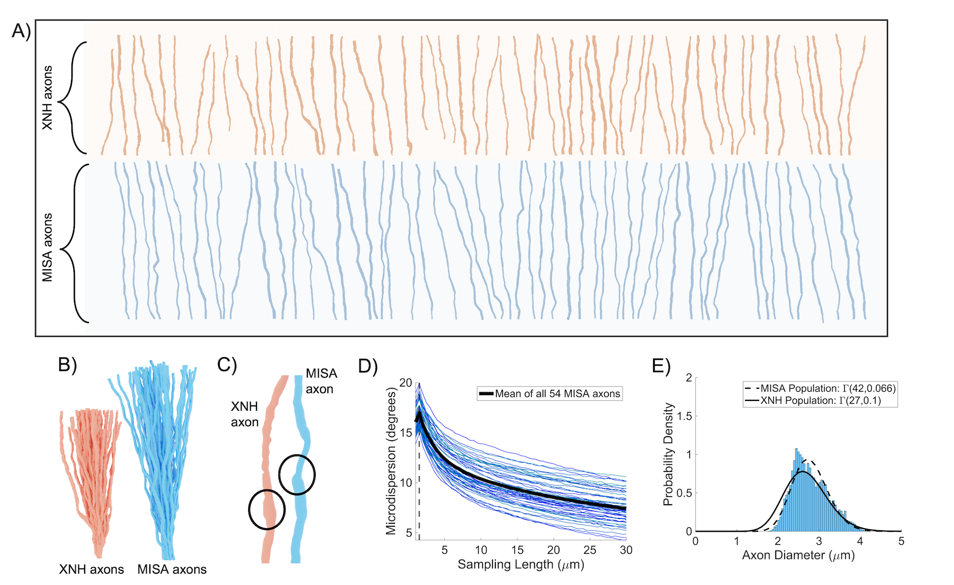
The MISA axons resemble those from XNH (Figures 4A-C) and capture their morphological variations. The mean trend in microdispersion across all MISA axons (black line, Figure 4D) is very similar to that in Figure 1C, including the spread around the mean. Small discrepancies between the pooled axon diameter distributions (<300 nm shift of the mean) in Figure 4E are expected due to limited sample size and the simple linear fit used to define the standard deviation of diameter variations in the generated axons (Figure 1E). Finally, Figure 5 shows how we obtain time-dependent ADCs similar to those of the XNH axons by mimicking the previously presented characteristics.Conclusion
The MISA framework allows for fast generation of realistic axons given statistical input parameters of morphology from 3D electron microscopy6–8 or XNH3. MISA can be used to generate anatomical region- or species-specific IAS substrates, as well as validate more complex frameworks that incorporate axonal packing. It outputs 3D meshes for MC simulations which are key to understanding the diffusion MRI signals from a given protocol. MISA will be freely available as a Blender add-on.Acknowledgements
M.A. and H.M.K. were supported by Capital Region Research Foundation Grant A5657 (principal investigator: T.B.D.) We acknowledge access to the facilities and expertise of the CIBM Center for Biomedical Imaging, a Swiss research center of excellence founded and supported by Lausanne University Hospital (CHUV), University of Lausanne (UNIL), Ecole Polytechnique Fédérale de Lausanne (EPFL), University of Geneva (UNIGE), and Geneva University Hospitals (HUG). This work was supported by EPFL through the use of the facilities of its Scientific IT and Application Support Center (SCITAS). This project has received funding from the European Union’s Horizon 2020 research and innovation programme under the Marie Skłodowska-Curie grant agreement No 754462 (MP).References
1. Callaghan R, Alexander DC, Palombo M, Zhang H. ConFiG: Contextual Fibre Growth to generate realistic axonal packing for diffusion MRI simulation. Neuroimage. 2020 Jul 2;220:117107.
2. Ginsburger K, Matuschke F, Poupon F, Mangin J-F, Axer M, Poupon C. MEDUSA: A GPU-based tool to create realistic phantoms of the brain microstructure using tiny spheres. Neuroimage. 2019 Jun;193:10–24.
3. Andersson M, Kjer HM, Rafael-Patino J, Pacureanu A, Pakkenberg B, Thiran J-P, Ptito M, Bech M, Bjorholm Dahl A, Andersen Dahl V, Dyrby TB. Axon morphology is modulated by the local environment and impacts the noninvasive investigation of its structure–function relationship. Proc Natl Acad Sci U S A. 2020 Dec 29;117(52):33649–59.
4. Rafael-Patino J, Yu T, Andersson M, Kjer HM, Andersen Dahl V, Pacureanu A, Bjorholm Dahl A, Dyrby TB, and Thiran JP. Phantoms for Diffusion Simulations: Multi-Objective Differential Evolution for Realistic Numerical (MODERN) Phantoms. In: Proceedings of the International Society for Magnetic Resonance in Medicine 27th Annual Meeting.
5. Rafael-Patino J, Romascano D, Ramirez-Manzanares A, Canales-Rodríguez EJ, Girard G, Thiran J-P. Robust Monte-Carlo Simulations in Diffusion-MRI: Effect of the Substrate Complexity and Parameter Choice on the Reproducibility of Results. Front Neuroinform. 2020;14:8.
6. Abdollahzadeh A, Belevich I, Jokitalo E, Sierra A, Tohka J. DeepACSON automated segmentation of white matter in 3D electron microscopy. Commun Biol. 2021 Feb 10;4(1):179.
7. Lee H-H, Yaros K, Veraart J, Pathan JL, Liang F-X, Kim SG, Novikov DS, Fieremans E. Along-axon diameter variation and axonal orientation dispersion revealed with 3D electron microscopy: implications for quantifying brain white matter microstructure with histology and diffusion MRI. Brain Struct Funct. 2019 May;224(4):1469–88.
8. Abdollahzadeh A, Belevich I, Jokitalo E, Tohka J, Sierra A. Automated 3D Axonal Morphometry of White Matter. Sci Rep. 2019 Apr 15;9(1):6084.
Figures