0757
Variable Flip, Blip-Up and -Down Undersampling (VUDU) Enables Motion-Robust, Distortion-Free Multi-Shot EPI1Athinoula A. Martinos Center for Biomedical Imaging, Charlestown, MA, United States, 2Department of Radiology, Harvard Medical School, Boston, MA, United States, 3Fetal-Neonatal Neuroimaging & Developmental Science Center, Boston Children’s Hospital, Boston, MA, United States, 4State Key Laboratory of Modern Optical Instrumentation, College of Optical Science and Engineering, Zhejiang University, Hangzhou, China, 5Radiological Sciences Laboratory, Stanford University, Palo Alto, CA, United States, 6Harvard/MIT Health Sciences and Technology, Cambridge, MA, United States
Synopsis
We demonstrate the motion-robustness of multi-shot diffusion imaging using VUDU (Variable flip, blip-Up, and -Down Undersampling) in the brain. VUDU employs FLEET-ordering to acquire all EPI shots of a given slice successively before proceeding to the next slice and maximizes the signal using variable flip angle (vfa) excitation. It also utilizes BUDA (Blip-Up and -Down Acquisition) to eliminate B0 distortion and employs the Hankel low-rank regularization to address shot-to-shot phase variations. VUDU was able to provide high-fidelity diffusion-weighted images from in vivo data acquired during intentional head motion.
Introduction
We introduce VUDU (Variable flip, blip-Up, and -Down Undersampling)1 for motion-robust and distortion-free multi-shot EPI (msEPI). msEPI can mitigate distortion and T2/T2*-related voxel blurring and voxel pile-ups by increasing acceleration and reducing echo-spacing time significantly. BUDA (Blip-Up/Down Acquisition)2 uses interleaved blip-up/blip-down phase encoding and incorporates B0 forward-modeling into structured low-rank reconstruction to enable distortion-free and navigator-free diffusion MRI. However, standard msEPI ordering requires a time gap of several seconds between multiple shots, which increases the vulnerability to motion. FLEET (Fast Low angle Excitation Echo-planar Technique)3 acquires all shots for a specific slice first before proceeding to the next slice. FLEET-ordering can bring motion-robustness in msEPI acquisition by reducing the acquisition timeframe for each slice, whereas variable flip angle (vfa)4 can maximize the signal, e.g. 45° and 90° RF pulses for 2-shot EPI. VUDU performs vfa-FLEET excitation, alternating blip-up/down phase encoding, and BUDA reconstruction to boost the motion-robustness and eliminate the B0 distortion. In this abstract, we further extend VUDU to diffusion-weighted imaging with rigid in-plane motion correction. Motion-robustness of VUDU was evaluated during in vivo diffusion imaging acquisition with intentional head motion.Data/Code: https://anonymous.4open.science/r/VUDU_ISMRM2022-8A07
Methods
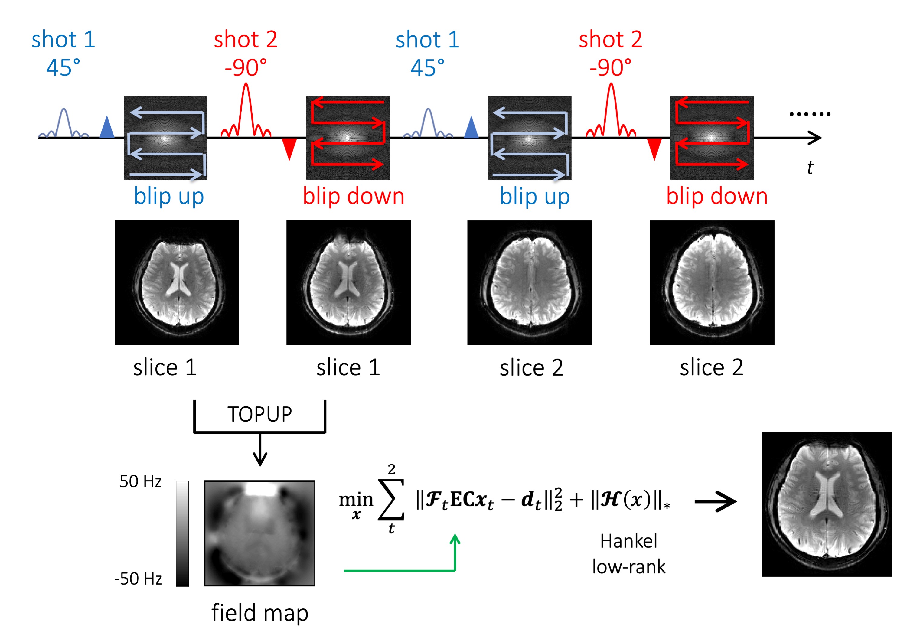
Figure 1 (top) shows the VUDU sequence diagram. VUDU excites a particular slice by a 45° pulse and encodes the signal by a blip-up readout. Immediately after, a second excitation made by a -90° pulse for the same slice is encoded with a blip-down readout. After the excited slice is fully encoded within a short timeframe (~280 msec), VUDU excites and encodes the next slice (FLEET-ordering). Figure 1 (bottom) shows the image reconstruction pipeline of VUDU. Each blip-up and blip-down image are separately reconstructed by SENSE5 and TOPUP6,7 estimates a field map $$$\mathbf{E}$$$. The distortion-free image can be calculated by the joint reconstruction of the two-shot, as follows.$$x=\underset{x}{\mathrm{argmin}}\sum_{t}^{2}{{\left\|\mathbf{\mathcal{F}}_t\mathbf{E}\mathbf{C} x_{t}-d_{t} \right\|}_2^2}+\lambda{\left\|\mathcal{H}(x)\right\|}_*$$
where $$$\mathbf{C}$$$ are sensitivities, t=1 stands for blip-up and t=2 for blip-down. $$$\mathbf{\mathcal{F}}_t$$$ is the undersampled Fourier transform in the tth shot, $$$x_t$$$ is the distortion-free image and $$$d_t$$$ are the k-space data for the tth shot. The constraint $$${\left\|\mathcal{H}(x)\right\|}_*$$$ is the nuclear norm to enhance Hankel low-rank prior between the two-shot in k-space.
Experiment
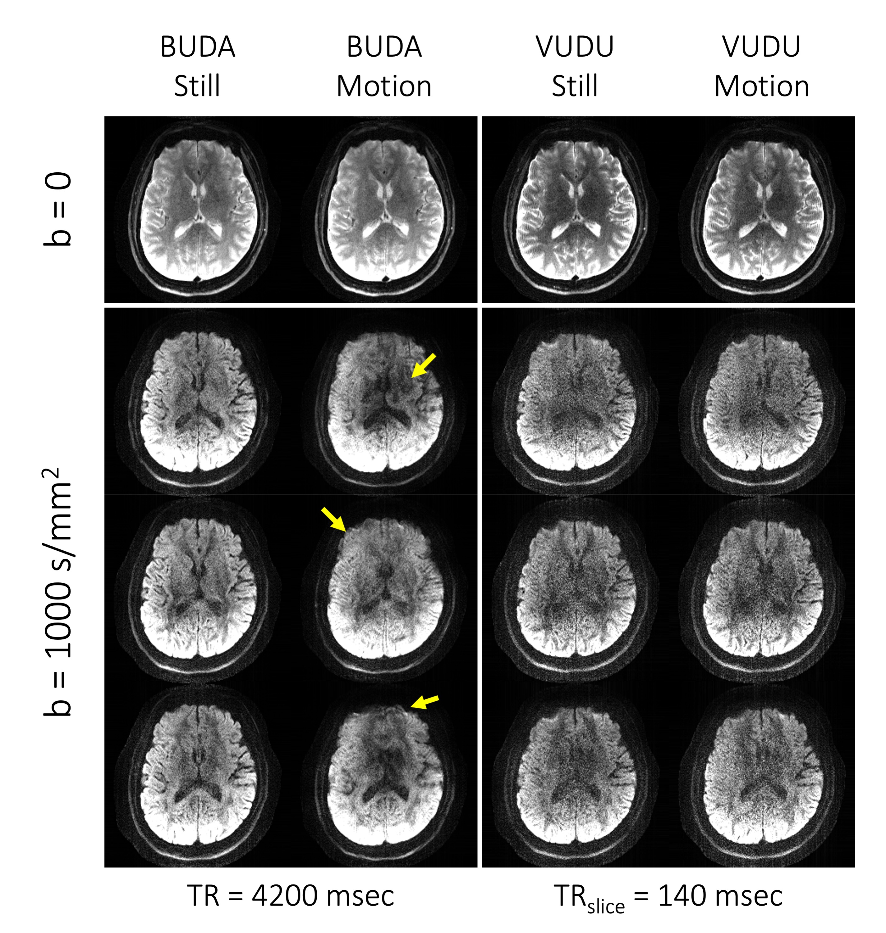
We conducted experiments on a Siemens 3T Prisma scanner equipped with a 32-channel head coil. A healthy adult volunteer was asked to move the head left-right intentionally throughout the scan. 2-shot spin-echo EPI acquisition was used at 4-fold acceleration per shot. The imaging parameters are voxel size=1x1x4mm3, b=1000 s/mm2 in 6-directions, TE=56msec, and TRslice=140msec (VUDU), and TR=4200msec (BUDA), respectively. We utilized FSL for image registration8,9 across the diffusion directions.Results
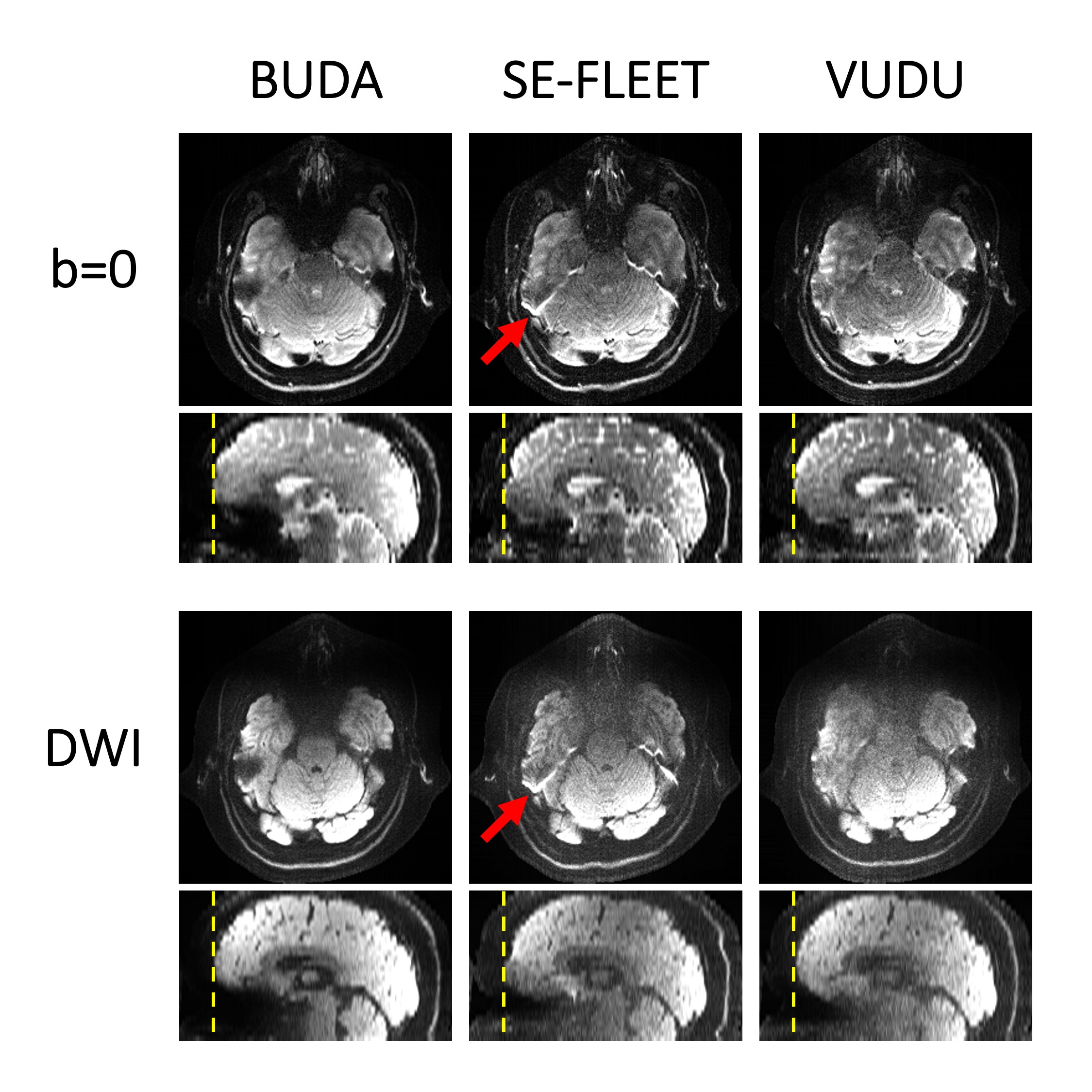
Figure 2 shows BUDA, vfa-FLEET, and VUDU images for b=0 and DWI at b=1000 s/mm2 in 6-directions. While vfa-FLEET encodes the signal by only blip-up readouts, BUDA and VUDU encode the signal using both blip-up and blip-down readouts. Therefore BUDA and VUDU were able to incorporate field map estimation and correct distortion by jointly reconstructing the two shots, as indicated by the dashed lines.Figure 3 shows BUDA and VUDU images acquired with and without motion at b=0 and b=1000 s/mm2. BUDA is vulnerable to motion as indicated by the arrows. VUDU took 280 msec to encode each slice, thereby showing the motion-robust images.
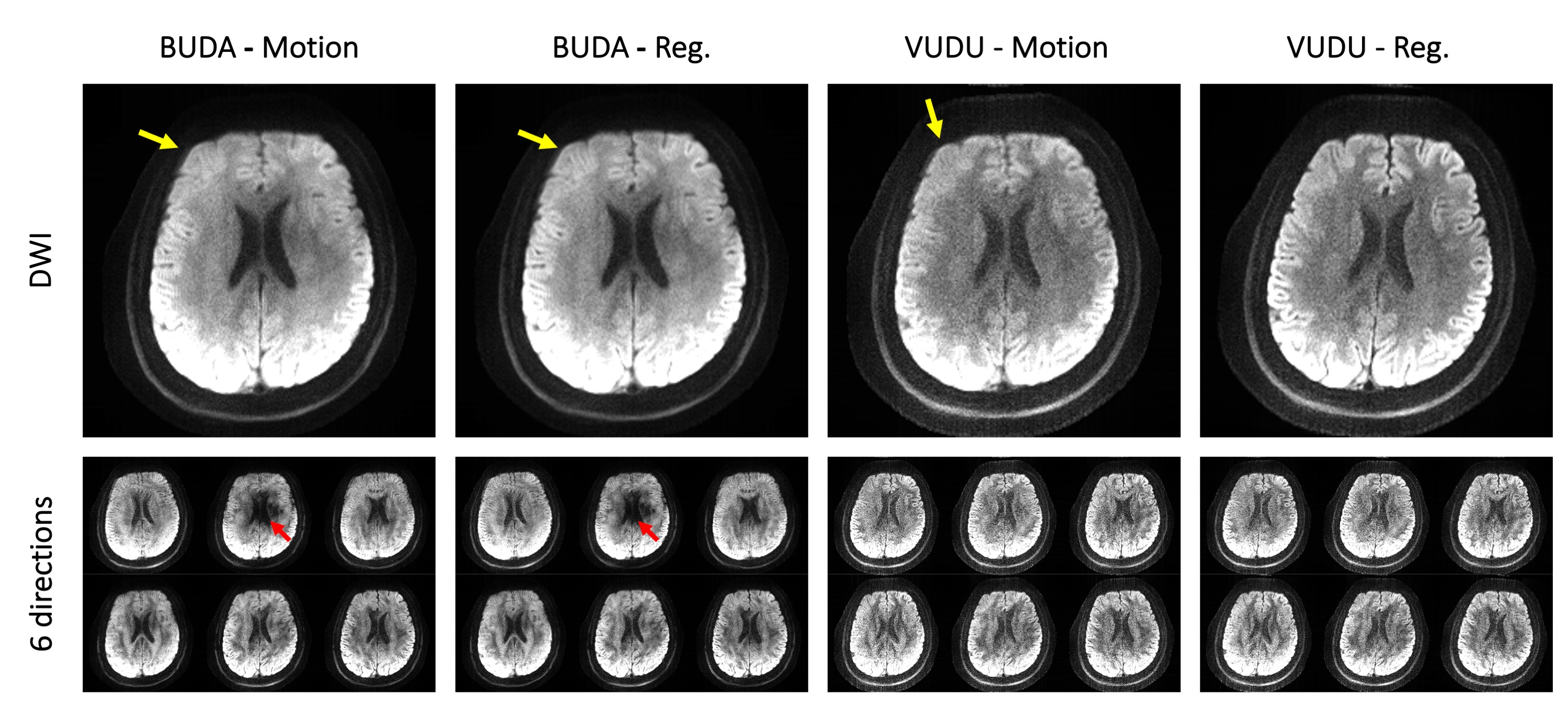
Figure 4 shows DWI images of BUDA and VUDU with and without the image registration across the diffusion direction. BUDA suffered from the motion-related artifact regardless of the image registration. VUDU can show high-fidelity images with the image registration across the diffusion directions.
Discussion & Conclusion
We presented VUDU, motion-robust, distortion-free msEPI. In-vivo brain experiment shows the motion robustness of VUDU in in-plane rigid motion. We think FLEET-ordered msEPI across the diffusion directions can further boost the motion-robustness. e.g) it would take 840 msec to encode each slice for the 3-direction diffusion with 6-shot EPI. By sacrificing the SNR, this approach should minimize the motion across the diffusion directions, thereby well-aligning images.The tradeoff made for the motion-robustness in the current VUDU acquisitions is ~30% of SNR reduction ($$$1-\sin45^{\circ}$$$) with respect to standard msEPI ordering. Taking account of T1 recovery during the inter-shot gap will improve the images4. We expect that VUDU can bring benefits of msEPI acquisition to fetal, cardiac, and abdominal imaging, where motion is unpredictable and non-rigid.
Acknowledgements
This work was supported by research grants NIH R01 EB028797, R03 EB031175, U01 EB025162, P41 EB030006, U01 EB026996, and the NVidia Corporation for computing support.References
1. Cho J, Berman AJ, Gagoski B, et al. VUDU: motion-robust, distortion-free multi-shot EPI. In: Proceedings of the 29th Scientific Meeting of ISMRM. Online Conference; p. 0002.
2. Liao C, Bilgic B, Tian Q, et al. Distortion-free, high-isotropic-resolution diffusion MRI with gSlider BUDA-EPI and multicoil dynamic B0 shimming. Magn. Reson. Med. 2021;86:791–803 doi: 10.1002/mrm.28748.
3. Polimeni JR, Bhat H, Witzel T, et al. Reducing sensitivity losses due to respiration and motion in accelerated echo planar imaging by reordering the autocalibration data acquisition: Reducing Losses in Accelerated EPI with FLEET-ACS. Magn. Reson. Med. 2016;75:665–679 doi: 10.1002/mrm.25628.
4. Berman AJ, Grissom WA, Witzel T, et al. Segmented spin-echo BOLD fMRI using a variable flip angle FLEET acquisition with recursive RF pulse design for high spatial resolution fMRI. In: Proceedings of 28th Annual Meeting of ISMRM. Paris, France. ; 2020. p. 5236.
5. Pruessmann KP, Weiger M, Scheidegger MB, Boesiger P. SENSE: sensitivity encoding for fast MRI. Magn. Reson. Med. Off. J. Int. Soc. Magn. Reson. Med. 1999;42:952–962.
6. Andersson JLR, Skare S, Ashburner J. How to correct susceptibility distortions in spin-echo echo-planar images: application to diffusion tensor imaging. NeuroImage 2003;20:870–888 doi: 10.1016/S1053-8119(03)00336-7.
7. Smith SM, Jenkinson M, Woolrich MW, et al. Advances in functional and structural MR image analysis and implementation as FSL. Neuroimage 2004;23:S208–S219.
8. Jenkinson M, Smith S. A global optimization method for robust affine registration of brain images. Med. Image Anal. 2001;5:143–156.
9. Jenkinson M, Bannister P, Brady M, Smith S. Improved optimization for the robust and accurate linear registration and motion correction of brain images. Neuroimage 2002;17:825–841.
Figures