0755
Real-time MRI of speech production at 0.55 Tesla1University of Southern California, Los Agneles, CA, United States
Synopsis
We demonstrate 2D mid-sagittal real-time MRI of speech production at 0.55T by employing a custom designed upper airway coil, spiral-based bSSFP and GRE sequences, and constrained image reconstruction. We found that bSSFP offered superior SNR and tissue contrast compared to GRE, and the quality of images at 0.55T was superior to that commonly reported at 1.5T and 3T field strengths. We attribute this improvement to the significantly reduced off-resonance from air-tissue boundaries at 0.55T, making it possible to leverage bSSFP without suffering from banding artifacts.
INTRODUCTION
Real-time MRI (RT-MRI) has deepened our understanding of the complex spatio-temporal coordination of vocal articulators during speech production1. Spiral acquisitions are widely used due to high scan efficiency and resilience to motion artifacts; they have produced high spatial (1~3mm) and temporal (12~40ms) resolution when combined with advanced image reconstruction2.Spirals are primarily limited by off-resonance adjacent to air-tissue interfaces (9.41 ppm, ∆f = ~600Hz at 1.5T)3. Blurring artifact and/or signal loss (banding artifact) due to off-resonance degrade image quality most significantly at air-tissue interfaces, which are the exact locations of interest for speech imaging, e.g., articulator boundaries4,5. For this reason, current speech RT-MRI studies are most often conducted using short readouts (2.5 ms at 1.5T) on low field strength (1.5T) commercial MRI scanners.
In this work, we develop and optimize speech production imaging at 0.55T. Lower-field imaging with high-performance gradients6 offers the advantage of reduced off-resonance, enabling longer spiral readouts7 with less blurring artifacts and making balanced steady-state free-precession (bSSFP) feasible again. The latter is especially important at a lower field because bSSFP provides superior SNR efficiency compared to traditional gradient recalled echo (GRE). While earlier work6,8 demonstrates the feasibility of longer readout GRE sequences for speech imaging at 0.55T, we explore the feasibility of bSSFP sequences and compare their performance against that of GRE.
METHODS
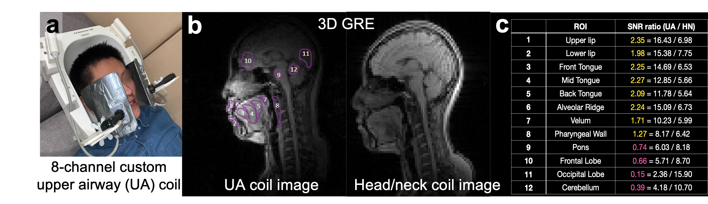
Experiments: Experiments were performed using a whole-body 0.55T system (prototype MAGNETOM Aera, Siemens Healthineers, Erlangen, Germany) equipped with high-performance shielded gradients (45 mT/m amplitude, 200 T/m/s slew rate). We used a custom 8-channel upper airway coil that has four elements on each side of the jaw (Figure 1). This provides SNR 1.71-2.35-fold superior to a 16-channel head/neck coil over the vocal tract articulators of interest including the lips, tongue, and velum. The imaging protocol was approved by our Institutional Review Board. Four healthy adult volunteers were scanned, after providing written informed consent.Image Contrast and Bloch Simulation: T1, T2, and T2* of the tongue (1 female and 1 male) were measured using protocols similar to those described by Campbell-Washburn et al6. Based on the estimated relaxation parameters (T1/T2/T2* = 662±69ms / 42±9ms / 31±3ms), the steady-state signal of bSSFP and GRE were simulated.
Acquisition: 2D spiral bSSFP and GRE pulse sequences were implemented within the RTHawk real‐time imaging platform (HeartVista, Inc., Los Altos, CA, USA). We tested sequences with 5, 8, 13-shot spiral trajectories, with corresponding imaging parameters listed in Table 1. We used temporal bit-reversed orders of spiral interleaves. For bSSFP, the through-slice equilibration compensation (dephasing of ± 18˚ along the slice direction) was used to annihilate eddy-current-induced signal oscillations9.
Reconstruction: A sparse SENSE reconstruction with temporal finite difference constraint10 was implemented in MATLAB (Mathworks, USA) and performed offline. Gradient impulse response function11,12 was estimated from a phantom-based measurement13 and used to correct B0 and linear eddy current artifacts for spiral trajectories prior to reconstruction.
RESULTS
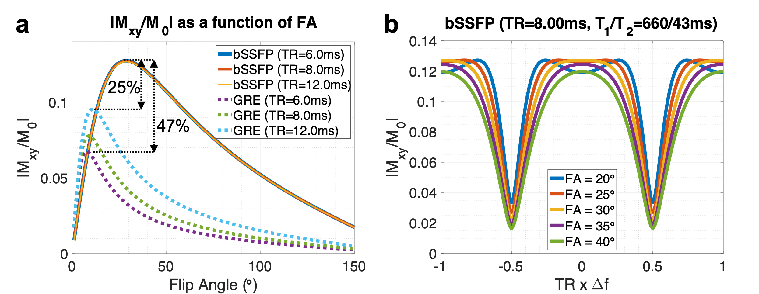
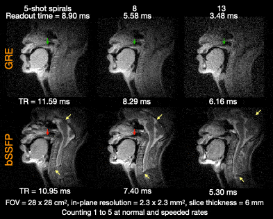
Figure 2a illustrates that bSSFP offers a relatively consistent signal profile (and an optimal FA of around 29˚) for TR values simulated (6~12ms) whereas GRE provides a higher signal intensity with longer TR. bSSFP offers a signal gain of 47% over GRE at TR=6ms and 25% at TR=12ms for the tongue muscle at 0.55T. From Figure 2b, bSSFP benefits from a short TR that provides a uniform signal over a broader range of ∆f. For example, for banding-free imaging of the air-tissue boundary (∆f =220Hz at 0.55T), the TR must be confined to approximately 4.54 ms at 0.55T, which would not be feasible at a higher field (e.g., ∆f =600Hz and TR=1.67 ms at 1.5T).Figure 3 shows reconstructed images acquired using bSSFP and GRE for varying readout durations. For GRE, a longer readout is more desirable in terms of SNR, but the tradeoff is that it suffers from increased blurring (green arrows). For bSSFP, a short readout may be desirable because the banding artifact due to off-resonance becomes dominant when using a longer TR (yellow arrows), especially around the articulator boundaries (red arrows).
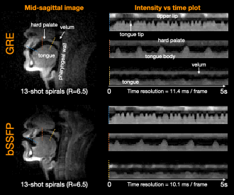
Figure 4 shows images from GRE and bSSFP reconstructed using 2 interleaves out of 13 (R=6.5) per frame at temporal resolutions of 11.4 ms and 10.1 ms, respectively. Both images are shown to be able to capture the rapid tongue motion, with the bSSFP image exhibiting a slightly clearer delineation of the tongue boundary than GRE. This is likely due to better SNR achieved by bSSFP.
DISCUSSION
Contrary to our initial expectation, short-readout spiral bSSFP outperformed long-readout spiral GRE at 0.55T by a large factor, providing 25% higher SNR and 60% less off-resonance blurring (based on the ratio of readout duration = 3.48/8.90ms).CONCLUSION
We demonstrate 2D mid-sagittal RT-MRI of speech production at 0.55T, with quality that is superior to that commonly reported at 1.5T and 3T field strengths. We attribute this improvement to the significantly reduced off-resonance from air-tissue boundaries, making it possible to leverage bSSFP without suffering from banding artifacts.Acknowledgements
We acknowledge grant support from the National Science Foundation (#1828736) and research support from Siemens Healthineers. We also acknowledge Helmut Stark for developing the 0.55T upper airway RF coil.References
1. Nayak KS, Lim Y, Campbell-Washburn AE, Steeden J. Real-time magnetic resonance imaging. J Magn Reson Imaging. 2020; Early view. doi:10.1002/jmri.27411
2. Lingala SG, Sutton BP, Miquel ME, Nayak KS. Recommendations for real-time speech MRI. J Magn Reson Imaging. 2016;43(1):28–44.
3. Schenck JF. The role of magnetic susceptibility in magnetic resonance imaging: MRI magnetic compatibility of the first and second kinds. Med Phys. 1996;23(6):815–850.
4. Lim Y, Lingala SG, Narayanan SS, Nayak KS. Dynamic off-resonance correction for spiral real-time MRI of speech. Magn Reson Med. 2019;81(1):234–246.
5. Lim Y, Bliesener Y, Narayanan SS, Nayak KS. Deblurring for spiral real-time MRI using convolutional neural network. Magn Reson Med. 2020;84(6):3438–3452.
6. Campbell-Washburn AE, Ramasawmy R, Restivo MC, Bhattacharya I, Basar B, Herzka DA, Hansen MS, Rogers T, Bandettini WP, McGuirt DR, et al. Opportunities in interventional and diagnostic imaging by using high-performance low-field-strength MRI. Radiology. 2019;293(2):384–393.
7. Restivo MC, Ramasawmy R, Bandettini WP, Herzka DA, Campbell-Washburn AE. Efficient spiral in-out and EPI balanced steady-state free precession cine imaging using a high-performance 0.55T MRI. Magn Reson Med. 2020;84(5):2364–2375.
8. Bhattacharya I, Ramasawmy R, Restivo MC, Campbell-Washburn AE. Dynamic speech imaging at 0.55T using single shot spirals for 11ms temporal resolution. ISMRM 27th Scientific Session, Montreal. 2019. p.0440.
9. Bieri O, Markl M, Scheffler K. Analysis and compensation of eddy currents in balanced SSFP. Magn Reson Med. 2005;54(1):129–37.
10. Lingala SG, Zhu Y, Kim Y-C, Toutios A, Narayanan S, Nayak KS. A fast and flexible MRI system for the study of dynamic vocal tract shaping. Magn Reson Med. 2016;77(1):112–125.
11. Robison RK, Li Z, Wang D, Ooi MB, Pipe JG. Correction of B0 eddy current effects in spiral MRI. Magn Reson Med. 2019;81(4):2501–2513.
12. Campbell-Washburn AE, Xue H, Lederman RJ, Faranesh AZ, Hansen MS. Real-time distortion correction of spiral and echo planar images using the gradient system impulse response function. Magn Reson Med. 2016;75(6):2278–2285.
13. Bruijnen T, Stemkens B, van den Berg CAT, Tijssen RHN. Prospective GIRF-based RF phase cycling to reduce eddy current-induced steady-state disruption in bSSFP imaging. Magn Reson Med. 2020;84(1):115–127.
Figures