0746
Virtual Reality Patient Training for Magnetic Resonance Imaging1Mathematics, Manhattan College, Riverdale, NY, United States, 2Radiology, Stanford University, Stanford, CA, United States
Synopsis
For many patients, particularly pediatric patients, MRI exams can be intimidating primarily due to the confined space and loud noises. This can result in failed, non-diagnostic, or degraded exams at cost and even risk to the patient. We present a virtual reality application for mobile devices, such as mobile phones, of a Magnetic Resonance Imaging simulation and training software to serve as a tool for medical professionals to prepare children for MRI scans. Initial evaluations show promising results for reducing movement in test subjects and potential for reducing worry or anxiety among individuals preparing for MRI.
Introduction
MRI is one of the most often and widely used imaging systems in medicine among both adult and pediatric patients. However, due to the nature of MRI procedure, this process can be especially frightening for pediatric patients. Specifically, the loud noise emitted by the machine, the requirement to remain still throughout the scan, the confined space inside the machine, and the use of IV needles are the main stressors that contribute to patient anxiety during MRI scans of children¹. This anxiety has costs both to the patient and the medical system. This is due to the need for repeated scans and more sedation among pediatric patients due to the effects of anxiety1,2. Here we describe the development of a virtual reality application that simulates MRI procedure to target the causes of patient anxiety, and help to prepare patients for MRI exams, both to help them remain still and to decrease the need for repeated MRI scans.Methods
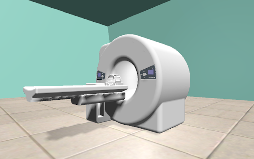
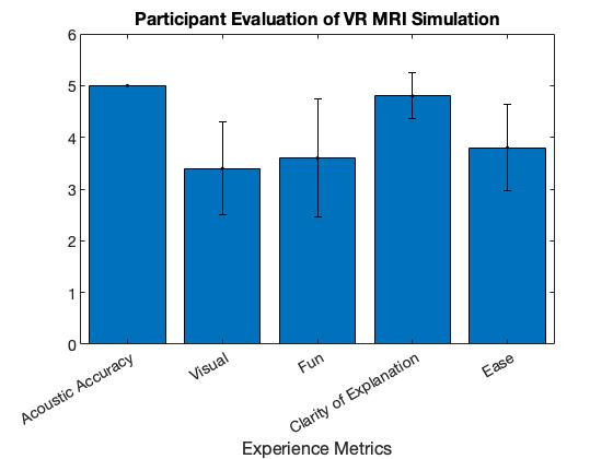
To simulate MRI, we have created a virtual reality environment in Unity (Unity3D, USA) that is aimed to acclimate patients to MRI procedure. Using simulated scanner sounds and a 3D model of an MRI machine and head coil, we consulted with MRI technologists to create a physical environment that emulates MRI procedure. This aspect of the application exposes the user to the loud noise and confined space that will be experienced when inside an actual MRI scanner. Additionally, a training program was created and integrated to teach patients the importance of laying still during scanning and provide practice for doing so. The challenge of staying still has been identified as especially difficult for pediatric patients, which can lead to motion artifacts and the need for additional scanning, sometimes using anesthesia. In this program, we create a VR head tracker to track the user’s head movement during simulated scanning. This tracker also provides feedback about how still the user was during the scanning procedure. We tested this application on five adult participants who had undergone an MRI scan before and evaluated five parameters: acoustic realism, visual realism, fun, accuracy in explanation of MRI, difficulty in staying still.Results
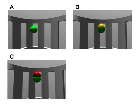
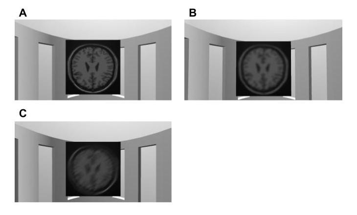
An application for iOS and Android was created to allow for wide distribution of this simulation. Once launched, the program runs for approximately six minutes. The resulting experience includes a brief background on MRI scanning with visuals of the entire scanner (Figure 1), a view from inside the scanner (Figure 2), a view of moving into the scanner, and three separate scans ascending in length with sound simulation. Head position is initialized at the beginning of each scan and movement is tracked with spheres in the center of the user's view (Figure 2). The spheres change colors based on the amount of user movement detected. This amount of movement is then used to give feedback after the scan is complete. A sample MRI image with noise levels is shown after scanning, with the amount of blur based on user movement during the scan (Figure 3). Additionally, there is an audio explanation of how the user performed and encouragement for improvement. Repetition of the procedure is encouraged for further training.The results from preliminary testing on the adult participants are depicted in Figure 4. All users felt that the experience replicated an actual MRI scan acoustically and that the explanation of the expectations of an MRI scan was clear. Furthermore, most users indicated that the program was easy to use and laying still to perform well was not challenging. Results indicate that improvements can be made to increase the enjoyment from the application and the visual accuracy of the MRI simulation.
Discussion
We have presented a virtual reality application for simulating MRI to introduce pediatric patients to MRI procedure and train patients for scanning. We provide a way for patients to comfortably and remotely become familiar with MRI. Further, this application monitors and provides feedback on user head movement. This method could have potential use in training patients prior to MRI and reducing movement during scanning. The main limitation of this study is the need for further testing in subjects, particularly in pediatric patients where such a training application might be most useful. The current VR application may not be appropriate for all age groups and would need modification for both very young and older children. For future work, the use of augmented reality where the user can see both the real and virtual world could be useful in pediatric patients. In such an environment, users could be interactive with their own environments, along with having parents, nurses or physicians in view to help them adapt in a more comfortable setting. To accurately train patients, simulations should be made for multiple age groups to ensure that explanations and training procedures are appropriate. Moreover, efforts to make the program entertaining for children would be beneficial. A proposed improvement is to show a video during scanning and pause the program if the child has significant head movement.Acknowledgements
No acknowledgement found.References
1. Carter, A. J., Greer, M.-L. C., Gray, S. E., & Ware, R. S. (2010). Mock MRI: reducing the need for anaesthesia in children. Pediatric Radiology, 40(8), 1368–1374.
2. Liszio, S., & Masuch, M. (2017). Virtual Reality MRI: Playful Reduction of Children's Anxiety in MRI Exams. Proceedings of the 2017 Conference on Interaction Design and Children.
Figures