0744
Improved geometric fidelity in brain DWI of pediatric patients using Multiplexed Sensitivity Encoding (MUSE)1Department of Radiology, Institute of Clinical Sciences, Sahlgrenska Academy, University of Gothenburg, Gothenburg, Sweden, 2Medical radiation sciences, Institute of Clinical Sciences, Sahlgrenska Academy, University of Gothenburg, Gothenburg, Sweden, 3Department of Medical Physics and Biomedical Engineering, Sahlgrenska University Hospital, Gothenburg, Sweden., Gothenburg, Sweden, 4Department of Radiology, Sahlgrenska University Hospital, Gothenburg, Sweden., Gothneburg, Sweden, 5Department of Radiology,, Brigham and Women's Hospital, Boston, MA, United States, Boston, MA, United States
Synopsis
This study demonstrates that DWI with MUSE and RPG correction significantly reduces the geometric distortions and results in increased overall image quality without altering ADC in a pediatric patient population, compared to single-shot EPI. This improvement was confirmed both through quantitative and qualitative analysis.
Introduction
Diffusion-weighted imaging (DWI) is a valuable tool for routine imaging of the pediatric brain1. However, the commonly used single-shot (ss) echo-planar imaging (EPI) DWI sequence is prone to geometric distortions and T2*-blurring2. One way to combat such artifacts is to lower spatial resolution. In a pediatric population, however, sacrificing resolution is undesirable, given that the structures of interest in very young patients are smaller. Multiplexed sensitivity-encoding (MUSE) is a new promising multi-shot technique for DWI with improved geometric fidelity, which uses self-navigation to correct for motion-induced shot-to-shot phase errors3. Another independent approach for distortion reduction is the reversed polarity gradient (RPG) method4. The RPG method with MUSE (RPG-MUSE) have been shown to effectively reduce distortions5, compared to MUSE alone6 in adults.This study aims to investigate the improvements of using MUSE and RPG-MUSE instead of ssEPI in terms of image quality, geometric fidelity, and ADC values in a pediatric population.
Method
This retrospective study compared image quality, geometric distortions, and ADC between three different approaches for DWI (ssEPI, MUSE, and RPG-MUSE) in 14 patients (median age = 4 (0.6-15) years, 11 males). DWI scan parameters identical for all patients were: 3T scanner (Signa Architect, GE Healthcare, Milwaukee WI, USA); 3 diffusion directions; 3.6/0.4 mm slice thickness/gap; and b-values of 0 and 1000 s/mm2. Other scan parameters are summarized in Table 1.Data analysis
Distortion levels were quantified and compared in two brain regions, i.e., the brain stem and the temporal lobes, using the Dice Coefficient and the Hausdorff Distance7, with T2-weighted images as reference (Fig. 1). Expected geometrical distortion was further evaluated by comparing the effective echo spacing between the DWI sequences. A lower effective echo spacing in echo-planar imaging corresponds to a higher phase encoding bandwidth and hence lower distortion artifacts in the final images. Apparent diffusion coefficient (ADC) values were determined in the genu of the corpus callosum and the optic nerves. Image quality was assessed by grading the images for overall image quality and image distortions on a Likert scale at two slice locations by two senior pediatric neuroradiologists. A one-way ANOVA and a Kruskal-Wallis test followed by a Tukey’s range post-hoc test was used to test for significant between DWI methods for the quantitative and qualitative assessment, respectively.
Results
Distortion levels assessed with Dice coefficient and Hausdorff distance were significantly lower for MUSE (p<0.05) and RPG-MUSE (p<0.01) compared to ssEPI (Table 2). No significant difference in ADC values was observed between methods. The RPG-MUSE method resulted in significantly higher overall image quality than ssEPI (p<0.05) and significantly lower levels of image distortions than both MUSE (p< 0.05) and ssEPI (p<0.05) (Table 3). These results agreed with the reduced effective echo spacing that was attained with MUSE and RPG-MUSE (Fig. 2).Discussion and Conclusion
This study showed that MUSE and even more so RPG-MUSE offer both improved geometric fidelity and image quality compared to ssEPI for DWI brain imaging in children. As Carney et al. have documented, diffusion-weighted imaging is a valuable tool in the diagnosis and evaluation of various pathologies in the brain of pediatric patients, such as intracranial or intra-orbital infection, brain tumors, demyelinating diseases, metabolic disorders, grey matter heterotopia, bone marrow infiltration, and dermoid cysts1. Particularly for neoplasms near bone structures of the skull base, e.g., in the brain stem, cerebellum, and along the optic pathways, for intra-orbital pathologies, and for dermoid cysts, distortions present in DWI can pose challenges in definite lesion characterization. These findings show that MUSE or RPG-MUSE, instead of ssEPI, are likely to aid in the diagnosis of pediatric brain pathologies due to the significant improvement in geometric fidelity and overall image quality.Moreover, in line with earlier publications, ADC was shown to be similar for ssEPI, MUSE and RPG-MUSE5,6,8. This is important as the ADC has been shown to be a potential biomarker for tumor grading or monitoring treatment response in glioma9,10. Another characteristic for MUSE was the lower effective echo spacing as a result of the faster k-space traversal. As it is favorable to traverse k-space as fast as possible for reduced T2*-blurring and lower levels of geometric distortions2, the lower echo spacing time implies that these artifacts must be lower for MUSE when compared to ssEPI.
In conclusion, significantly reduction of geometric distortions along with increased overall image quality was attained in pediatric brain DWI by using MUSE and RPG correction, compared to ssEPI, without altering the ADC.
Acknowledgements
This work was supported by the Swedish Childhood Cancer Fund [PR2016-0150] and by the Swedish state under an agreement between the Swedish government and the country councils: the ALF-agreement.References
1. Carney O, Falzon A, Mackinnon AD. Diffusion-weighted MRI in paediatric neuroimaging. Clin Radiol. 2018;79(12):999-1013. doi:10.1016/j.crad.2018.07.101Figures
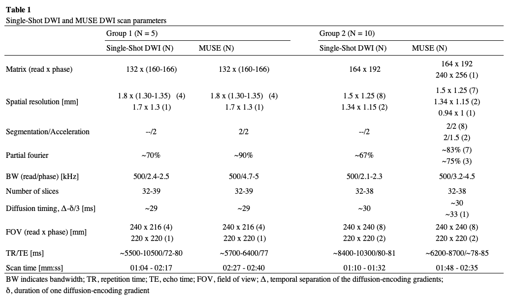
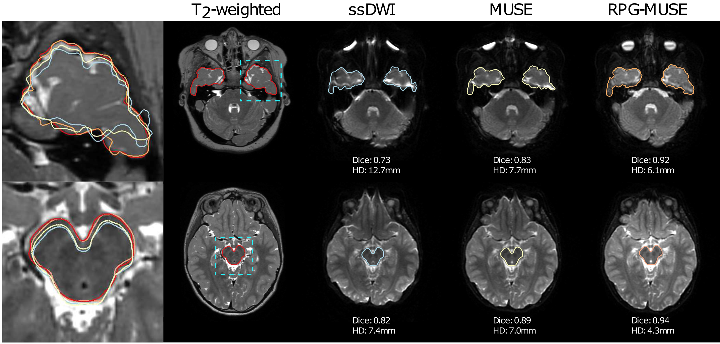
Figure 1: Delineations of the temporal lobes (top row) and brain stem (bottom row) for T2-weighted (red), ssEPI (blue), MUSE (yellow), and RPG-MUSE (orange). When comparing the outlines with the T2w-image as reference (sub-image on the left) the RPG-MUSE (orange) follows the T2w (red) better than both MUSE (yellow) and ssEPI (blue). Improvements are also evident in both the DICE and HD score for MUSE and RPG-MUSE, compared to ssEPI, using the T2-weighted image as reference.
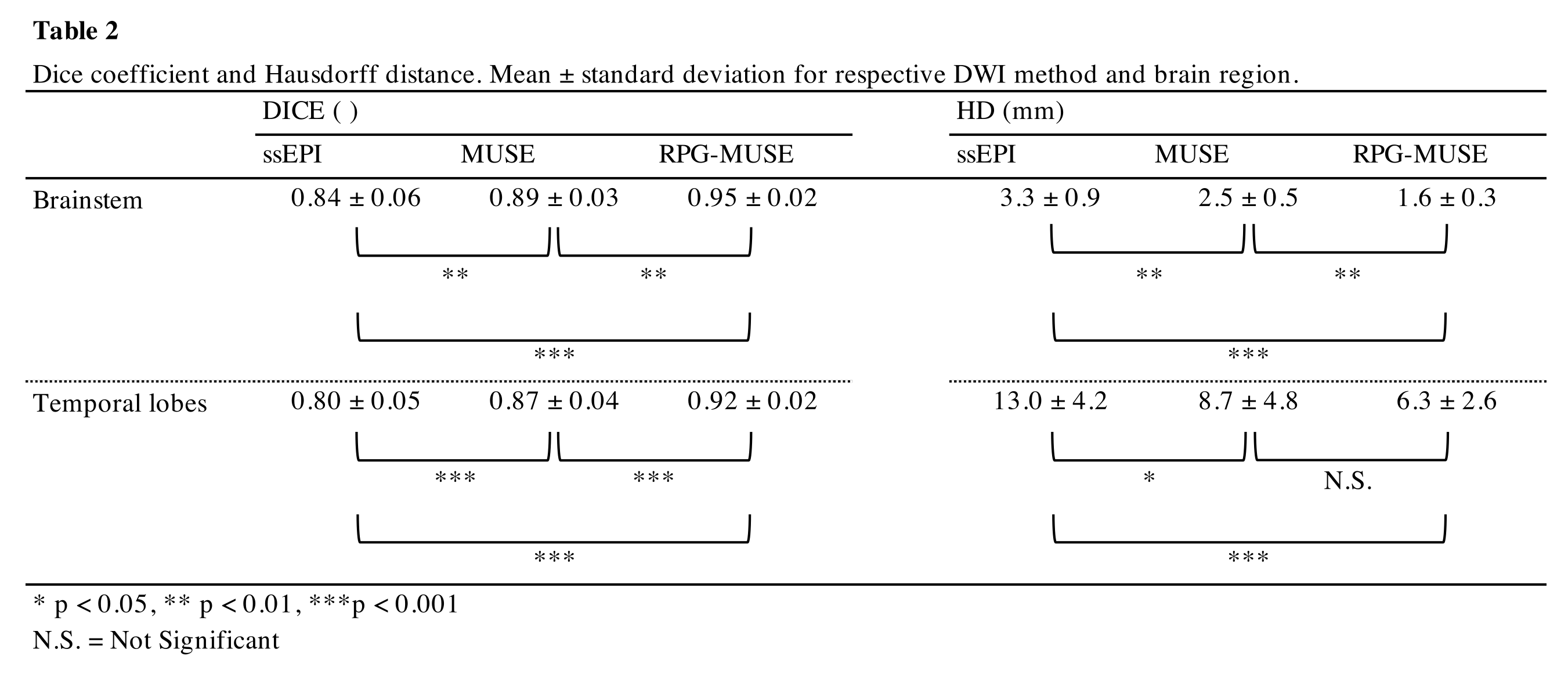
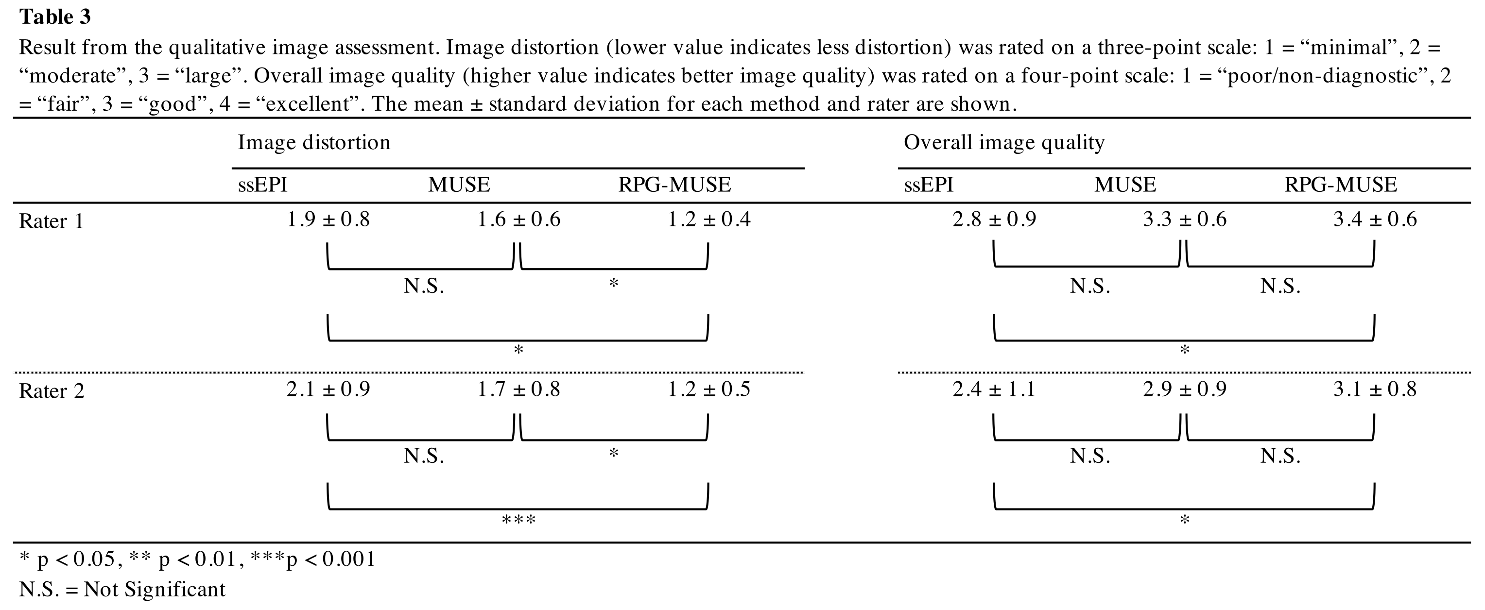
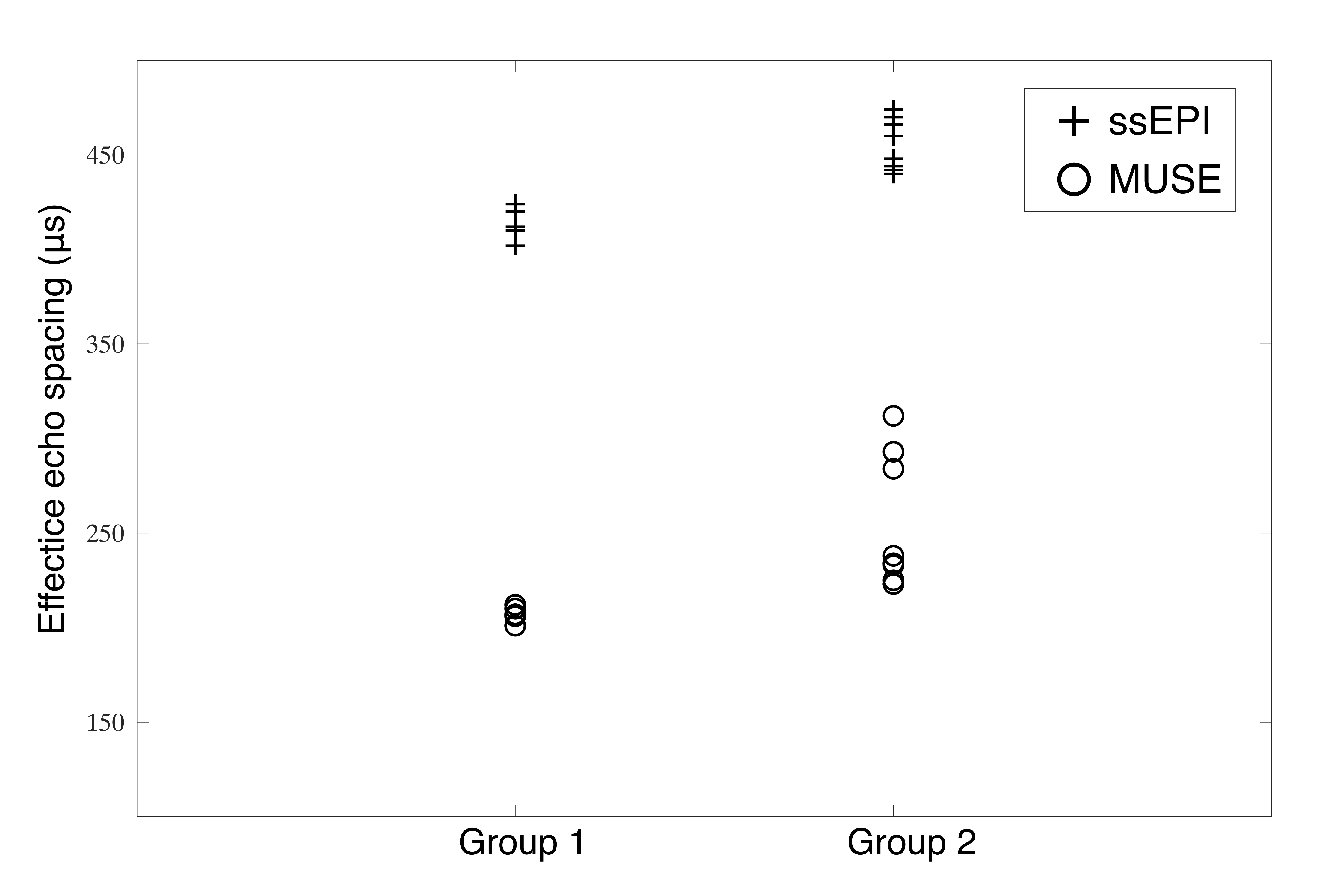
Figure 2: Plot showing effective echo spacing for all examinations included in this study for group 1 (acq matrix: 132x164) and group 2 (acq matrix 164x192). The MUSE sequence resulted in a lower effective echo spacing compared to ssEPI in all cases. This is consistent with the lower geometric distortions observed in the MUSE images.