0684
BladeNet: Rapid PROPELLER Acquisition and Reconstruction for High spatio-temporal Resolution Abdominal MRI1UC Berkeley, Berkeley, CA, United States, 2Stanford, Stanford, CA, United States
Synopsis
To improve bowel wall imaging in abdominal pediatric MRI scans, we propose a multi-phase single-shot fast spin echo (SSFSE) PROPELLER acquisition with novel deep learning reconstruction. This acquisition offers shorter scan time and thus higher temporal resolution, PROPELLER built-in motion correction, and alias-free images; these however exhibit spatial blurring. Our approach leverages the blurring-axis temporal rotation and data redundancy; we train a network to recover high-frequency spatial details from consecutive frames. Retrospective simulations with data from balanced SSFP scans show that this approach yields reconstructions with high spatio-temporal resolution and motion-correction, which are essential for pediatric abdominal imaging.
Introduction
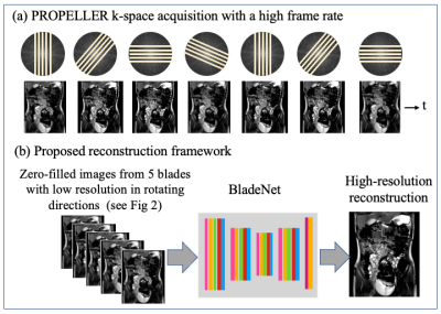
Detection of dynamic motility (digestive muscle contraction ability) is essential for diagnosis of pathology such as inflammatory bowel disease and short gut syndrome. While pediatric abdominal MRI exams are essential for this aim, their acquisition rate is typically not rapid enough for capturing the fast peristalsis dynamics. Moreover, patient motion and respiration induce motion artifacts. Methods for accelerating such scans while enabling high spatial resolution and motion robustness are therefore highly needed. Here we propose a framework that addresses these needs, which includes both acquisition and reconstruction strategies.Acquisition. We propose a multi-phase Single-Shot Fast Spin Echo (SSFSE) sequence with few (~100) echoes and PROPELLER rotation of the acquisition direction in every time frame. Propeller1 is a well-established technique in which k-space data are acquired using “blades” that rotate with time (Figure 1a); each blade consists of several parallel k-space lines. The proposed acquisition advantages are: (i) shorter scan time and thus higher temporal resolution; (ii) built-in motion correction ability due to the PROPELLER repetitive k-space center sampling; (iii) most importantly, alias-free images obtained from the PROPELLER blades. A disadvantage of PROPELLER is the low resolution along the Phase Encoding (PE) dimension. However, this is a local effect that occurs only in the image domain and can be corrected. This inverse-problem is highly suitable for Convolutional Neural Networks.
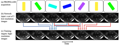
Reconstruction. We propose a Deep Learning (DL) reconstruction framework that leverages the PROPELLER advantages. In PROPELLER, the low-resolution blurring axis rotates with time; this reveals high-frequency details in different image parts at different times (Figures 1, 2). Our network receives a series of five consecutive single-blade images and a single target image corresponding to the fully-sampled data of the middle time frame (#3 of 5) (Figure 1b). This network is trained to optimally combine high-frequency information from the input images. Moreover, since respiration induces image-domain translation while PROPELLER provides data redundancy, the network learns to correct the respiratory motion. This results in high-resolution sharp images, with a high frame suitable for imaging rapid peristalsis motion.
Methods
Data acquisition. With IRB approval, data from four pediatric patients was acquired at a Hospital on 3T GE scanners using a custom balanced SSPF (bSSFP) sequence and single-slice acquisition; bSSFP was chosen because it is routinely used for clinical pediatric abdominal scans. The datasets were acquired in completely free-breathing scans, with 1-2ms TE, 3.1-5ms TR, and flip angle of 45 degrees. The frame rate was 2 Hz, and 500-700 frames were acquired from each patient. Images were reconstructed on the scanner and saved in DICOM format.Data preparation. Our study was based on simulated PROPELLER acquisition from fully-sampled k-space data generated from the DICOM images; the fully-sampled reference was needed for supervised training. To avoid subtle inverse crimes we follow the guidelines2: (1) we crop the zero-padded $$$512\times512$$$ k-space matrices to $$$320\times320$$$, and (2) we include here a full description of data preparation pipelines.
PROPELLER was simulated with a 36-degree rotation by interpolating k-space data to a non-Cartesian grid using a Non-Uniform Fast Fourier Transform (NUFFT) toolbox3. Blades sampling with 3x acceleration included 100 lines along the PE dimension and full readout sampling. Single-blade images were reconstructed from the zero-filled version of each k-space blade using the adjoint NUFFT operator. To mimic a real acquisition, only a single blade was sampled from the data of each time frame. Our training data included 1989 images from three pediatric patients, and the test data included 50 frames from a different patient.
Training. The proposed BladeNet architecture contains a Unet with ResNet blocks4,5. Training was done using Pytorch6, Adam optimizer7, L1-loss, and Nvidia Titan GPUs.
Results
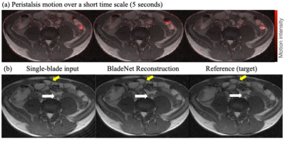
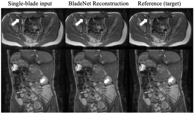
Figure 2 displays the single-blade images and targets. Notably, the single-blade images exhibit ringing (“Sinc”) due to the finite k-space sampling. More importantly, their spatial-blurring axis rotates with time, revealing fine details in different image locations over time.Figure 3a displays motion over a short time scale of 5 seconds (in red), which corresponds to 5 time frames. As can be seen, some areas remain still, while others exhibit significant motion, mainly due to the rapid peristalsis dynamics; this leads to temporal blurring in the single-blade images (e.g. Figure 3b (left)). Our framework exploits the rich dynamic data features and the PROPELLER redundancy for recovering high-frequency details. As can be seen, the reconstruction (Figure 3b, middle) exhibits sharp edges (yellow arrow) and fine details (white arrow), with a high fidelity to the reference image (Figure 3b, right). Figure 4 shows similar high-quality reconstruction results, from different patients.
Conclusion
Our innovative BladeNet framework offers acquisition and reconstruction strategies for rapid abdominal pediatric MRI. It combines the benefits of PROPELLER acquisition, with its built-in motion robustness, and powerful DL reconstruction. By leveraging the PROPELLER alias-free images and the rotation of its low-resolution blurring axis, BladeNet is able to restore fine details. The network is trained to optimally combine images into high-resolution reconstructions; advantageously, it also learns to corrects respiratory motion. Results from PROPELLER simulations performed with clinical bSSFP data indicate that this framework yields reconstructions with both high spatial and temporal resolutions, and is thus suitable for rapid peristalsis imaging.Acknowledgements
The authors acknowledge funding from grants U24 EB029240-01, R01EB009690, and R01HL136965.References
1. Pipe, James G. "Motion correction with PROPELLER MRI: application to head motion and free‐breathing cardiac imaging." Magnetic Resonance in Medicine: An Official Journal of the International Society for Magnetic Resonance in Medicine 42.5 (1999): 963-969.
2. Shimron, Efrat, et al. "Subtle Inverse Crimes: Na\" ively training machine learning algorithms could lead to overly-optimistic results." arXiv preprint arXiv:2109.08237 (2021)
3. Fessler, Jeffrey. "Michigan Image Reconstruction Toolbox," available at https://web.eecs.umich.edu/~fessler/code/
4. He, Kaiming, et al. "Deep residual learning for image recognition." Proceedings of the IEEE conference on computer vision and pattern recognition. 2016.
5. Ronneberger, Olaf, Philipp Fischer, and Thomas Brox. "U-net: Convolutional networks for biomedical image segmentation." International Conference on Medical image computing and computer-assisted intervention. Springer, Cham, 2015
6. Paszke, Adam, et al. "Pytorch: An imperative style, high-performance deep learning library." Advances in neural information processing systems 32 (2019): 8026-8037
7. Kingma, Diederik P., and Jimmy Ba. "Adam: A method for stochastic optimization." arXiv preprint arXiv:1412.6980 (2014).
Figures


