0656
Fixel-based analysis of aging white matter reveals selective fiber-specific degeneration1Rotman Research Institute, Baycrest Health Sciences, Toronto, ON, Canada, 2Department of Medical Biophysics, University of Toronto, Toronto, ON, Canada, 3Developmental Imaging, Murdoch Children's Research Institute, Melbourne, Australia
Synopsis
White matter (WM) microstructural aging is often studied with diffusion MRI via voxelwise metrics such as fractional anisotropy (FA). Decreasing FA with advancing age is frequently interpreted as WM microstructural degeneration, but relying on FA for this purpose only makes sense if a voxel contains a single fiber tract. In this work we use fixel-based analysis (FBA) to investigate the association of fiber-specific measures with age among each individual "fixel" within a voxel. We find intra-voxel differences in age associations of crossing fixels, demonstrating that traditional voxel-based analyses are inappropriate for probing age-related fiber degeneration in scenarios of multi-fiber crossings.
Introduction
White matter (WM) microstructural degeneration in aging is often studied with diffusion MRI via voxelwise metrics such as fractional anisotropy (FA). Decreasing FA with advancing age is frequently interpreted as WM microstructural degeneration.[1] However, relying on FA for this purpose only makes sense if a voxel contains a single fiber tract. Per contra, most voxels contain multiple crossing fiber tracts.[2] This is particularly problematic in the case of selective degeneration of secondary fibers that cross the primary diffusion orientation in the voxel, which can result in counterintuitive increases in FA with age.[3-5] Fixel-based analysis (FBA) was developed to study white matter microstructure in the presence of multiple fiber populations in voxels, by assigning measures specific to each individual crossing fiber (referred to as “fixels”) within the voxel.[5] In this work we conduct an FBA of normal white matter aging, with the goals of (i) identifying whether FBA-based fiber-specific measures suggest degeneration across the WM, despite the voxelwise FA metric being known to increase with age; and (ii) investigating the differential association of fiber-specific measures with age among each individual fixel within a voxel.Methods
This study consisted of 541 healthy participants aged 36-100 from the Human Connectome Project in Aging (HCP-A).[6] Diffusion MRI data were acquired on a Siemens 3T Prisma at TR=3.23s, TE=89.2ms, (1.5mm)3 resolution, MB=4. The selected data included 14 b=0 volumes and 92 gradient directions acquired at b=3000s/mm2 (a high b-value, for specific sensitisation to intra-axonal signal[7]). Data underwent denoising, motion correction, distortion correction, global intensity normalisation, and bias-field correction using MRtrix 3.0.1.Fiber orientation distributions (FODs) were estimated using single-shell, 3-tissue constrained spherical deconvolution (SS3T-CSD).[8] Data from a random sample of 50 participants were used to create a population template to which all FODs were registered. Fibre density (FD), fibre-bundle cross-section (FC) and the combined fibre density and cross-section (FDC) metrics were obtained. Correlations with age were assessed for each measure using a linear model via the tool fixelcfestats, with a significant association defined as p<0.05 after correction for multiple comparisons.
Results
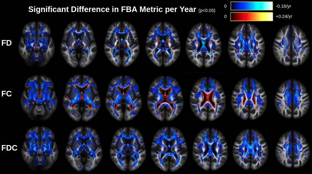
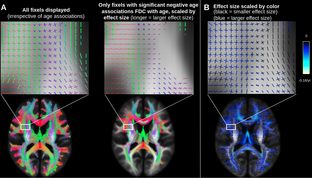
Figure 1 displays significant age associations of FD, FC and FDC across the WM. All three measures are negatively associated with age through most of the WM. FD exhibits some positive age associations in regions where FA is known to increase with age;[3-4] FC exhibits positive age associations throughout the periventricular WM; FDC, combining the effect of FD and FC,[5] does not exhibit positive age associations. Whereas Figure 1 displays the spatial layout of these age associations, Figure 2 focuses on the orientation of the fixels with that exhibit significant negative age associations of FDC.Results of the analysis of intra-voxel selective fibre-specific degeneration are visualised in Figures 3 and 4. Figure 3 reveals that fixels oriented medial-lateral (red) exhibit significant negative age associations of FDC, whereas fixels oriented anterior-posterior (green) and superior-inferior (blue) either exhibit no or small significant negative age associations of FDC. A similar trend is observed in Figure 4.
Discussion and Conclusion
In our study, the overwhelming trend of FDC decreases with advancing age is consistent with neurodegeneration. Our fixel-based analysis was additionally able to show intra-voxel differences in age associations of crossing fixels, demonstrating that traditional voxel-based analyses which analyze a single non-specific metric (e.g., FA) across the entire voxel are inappropriate for probing age-related fiber degeneration in scenarios of multi-fibre crossings. Notably, while FA is known to increase with age in crossing-fibre regions, we did not find such positive age associations of FDC; rather, we found negative age associations of FDC to often be specific to medial-lateral fixels. Therefore, this study provides direct evidence that increased FA with age can be seen as an artifactual result of selective degeneration of minor crossing fibers within a voxel. Fixel-based analysis will enable our future comparisons of the different rates of FDC decline between tracts in aging. Overall, this work suggests that a fixel-based approach is more appropriate to study age-related white matter microstructural decline than traditional voxel-based analyses.Acknowledgements
This research was funded by CIHR Project Grant #169688, in collaboration with the Human Connectome Project in Aging.References
1. Madden DJ et al (2012) Diffusion tensor imaging of cerebral white matter integrity in cognitive aging. Biochim Biophys Acta 1822(3):386-400.
2. Jbabdi S, Behrens TEJ, Smith SM (2010) Crossing fibres in tract-based spatial statistics. Neuroimage 49:249–256.
3. Miller KL et al (2016) Multimodal population brain imaging in the UK Biobank prospective epidemiological study. Nat Neurosci 19:1523-1536.
4. de Groot M et al (2016) White Matter Degeneration with Aging: Longitudinal Diffusion MR Imaging Analysis. Radiology 279:532-541.
5. Dhollander T, Clemente A, Singh M, et al (2021) Fixel-based Analysis of Diffusion MRI: Methods, Applications, Challenges and Opportunities. Neuroimage 241:118417.
6. Bookheimer SY, Salat DH, Terpstra M, et al (2019) The Lifespan Human Connectome Project in Aging: An overview. Neuroimage 185:335-348.
7. Genc S, Tax CMW, Raven EP, et al (2020) Impact of b-value on estimates of apparent fibre density. Hum Brain Map 41:2583-2595.
8. Dhollander T, Connelly A (2016) A novel iterative approach to reap the benefits of multi-tissue CSD from just single-shell (+b=0) diffusion MRI data. ISMRM 24:3010.
Figures