0637
A dedicated MRI/PET system for radiotherapy treatment simulation: pathophysiological guidance for small moving target volumes
Casper Beijst1, Woutjan Branderhorst1, Jan Lagendijk1, Bjoern Weissler2,3, David Schug2,3, Florian Mueller2, Harald Radermacher2, Martino Borgo4, Wout Schuth4, Jurgen Mollink5, Govaert Borst5, Marc Verheyen5, Frank van Duin4, Dennis Klomp1, Hugo de Jong1, and Volkmar Schulz2,3
1UMC Utrecht, Utrecht, Netherlands, 2Rheinisch-Westfaelische Technische Hochschule, Aachen, Germany, 3Hyperion Hybrid Imaging Systems, Aachen, Germany, 4Futura Composites, Heerhugowaard, Netherlands, 5Philips Medical Systems, Best, Netherlands
1UMC Utrecht, Utrecht, Netherlands, 2Rheinisch-Westfaelische Technische Hochschule, Aachen, Germany, 3Hyperion Hybrid Imaging Systems, Aachen, Germany, 4Futura Composites, Heerhugowaard, Netherlands, 5Philips Medical Systems, Best, Netherlands
Synopsis
The Unity 1.5T MR-linac has clinically been introduced to provide stereotactic precision for moving targets. We are now developing an integrated 1.5T MRI/PET system with identical MRI performance for planning of MR-linac treatments. The system offers 70 cm bore size and includes PET detectors mounted in the gap of a split gradient coil. Recently, the first ring of PET detectors was installed. We performed extensive interference tests and acquired the first simultaneous images using a brain phantom. Our vision is that the combination of MR-linac with MRI/PET will allow the use of PET information for stereotactic radiation therapy guidance.
Introduction
The Unity 1.5T MR-linac is designed to provide stereotactic precision for moving targets. Accurate multi-functional imaging for target definition is crucial for successful treatment with the MR-linac. PET provides highly sensitive pathophysiological information about the tumor, offering complementary information to MRI. Therefore, we are currently developing a dedicated integrated 1.5T MRI/PET system, which is a twin system of the MR-Linac in terms of MRI subsystems. This system is designed to transfer high-resolution, motion corrected, pathophysiological information of the PET signal to the workflow and treatment pipeline of the MR-linac, by intrinsic registration to the MRI. For the first time, sequences optimized for MR-linac therapy treatment can also be used and explored on our dedicated MRI/PET system, adding a high level of confidence to the overall therapy planning and workflow. The combination of MRI/PET and MR-Linac enables motion-corrected detection and treatment of small tumors and multiple poly/oligometastases in the thorax and abdomen.Methods
We designed and installed a dedicated integrated MRI/PET system for radiotherapy planning at the University Medical Center Utrecht. Our system is based on a 1.5T Philips Ingenia MRI. The PET detectors are integrated in the gap of a split gradient coil originally developed for the MR-Linac. In this way, we are able - for the first time - to ensure an identical bore size of MRI/PET and MR-Linac of 70cm. The RF shielding of PET is provided by specially designed carbon fiber housings (Figure 1). The PET detector electronics consist of non-magnetic detector stacks and a composite water cooling system of the detectors, which causes low eddy currents. Initial PET performance was evaluated with partial population of the first PET ring (Figure 2), which consisted of 24 LYSO PET detector stacks, each with an area of 48x48 mm2. Finally, the MRI/PET was equipped with the same integrated RF body coil as the MR-Linac, which was designed to be transmissive to high-energy gamma photons. We studied the interference between MRI and PET by performing constancy measurements of PET energy resolution, timing resolution and count rate during RF and gradient switching. In addition, we performed measurements of spikes and spurious during PET operation. Finally, simultaneous PET and MRI acquisitions of a multi-modal brain phantom were performed.Results
The first (partial) ring of PET detectors was installed in a 1.5T Philips Ingenia with a split gradient coil. Our extensive interference studies have shown that MRI and PET have no adverse effects on image quality of PET or MRI. PET energy resolution, timing resolution and count rate were not affected by RF or gradient operation as shown in Figure 3. Spikes and spurious signal were within the given tolerances (Figure 3). Simultaneous PET and MRI images of a human brain phantom were acquired successfully, as shown in Figure 4.Conclusion
A dedicated integrated MRI/PET system for radiotherapy planning was designed and installed. Experiments showed that there were no adverse effects of interference between the MRI and the first ring of PET detectors on image quality. Simultaneous PET and MRI images were acquired successfully. The combination of the MRI/PET and the MR-linac allows for treatment of small tumors and multiple poly/oligometastases in thorax and abdomen.Acknowledgements
No acknowledgement found.References
No reference found.Figures
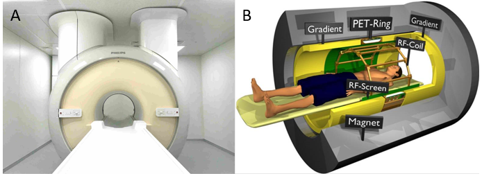
Figure 1: a)
Picture of the integrated MRI/PET system with covers shows the 70cm wide bore
character of our MRI/PET system. b) Schematic drawing of the MRI/PET system with
the PET detectors located in the gap of the split gradient coil originally
designed for the MRI-linac.
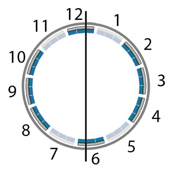
Figure 2:
Schematic drawing of the PET detector ring. 12 modules were distributed within
the gap of the split-gradient coil, with a small offset angle. Populated stacks
are shown in dark blue, unpopulated detector stack in light blue.
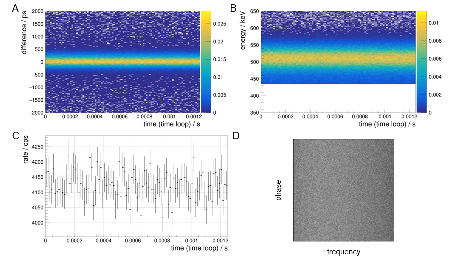
Figure 3: a)
PET coincidence timing difference, b) energy resolution and c) count rate as a
function of looped time during gradient operation (10 mT/m gradient strength, 65
mT/m/ms slew rate, 1.25ms repetition time, 49% duty cycle). d) Spurious noise
measurement (170kHz span and 63.9MHz central frequency) of MRI and PET. Absence of stripes/spots shows effective RF
shielding.Other
spurious noise measurements with central frequencies of 63.5, 63.7, 64.0 and
64.2 MHz show similar results (not shown).
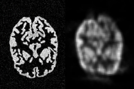
Figure 4: Simultaneously
acquired MRI (left) and PET (right) images of a human brain phantom filled with
25 MBq Ga-68.
DOI: https://doi.org/10.58530/2022/0637