0582
A web-accessible tool for Temperature Estimation from SAR Simulations (TESS)
Eros Montin1, Giuseppe Carluccio1, Christopher Collins1, and Riccardo Lattanzi1,2
1Center for Advanced Imaging Innovation and Research (CAI2R) Department of Radiology, New York University Grossman School of Medicine, New York, NY, United States, 2Vilcek Institute of Graduate Biomedical Sciences, New York University Grossman School of Medicine,, New York, NY, United States
1Center for Advanced Imaging Innovation and Research (CAI2R) Department of Radiology, New York University Grossman School of Medicine, New York, NY, United States, 2Vilcek Institute of Graduate Biomedical Sciences, New York University Grossman School of Medicine,, New York, NY, United States
Synopsis
TESS, Temperature Estimation from Sar Simulation, is a web-based application that performs safety assessments of MRI exams. It numerically solves modified versions of the bioheat equation that include parameters such as temporal variation of core body temperature and various physiological parameters. A web-based graphical user interface, accessible via the Cloud MR framework, enables users to set up simulations and display results.
Background and Purpose
Safety is a key component of MRI research. The interaction between the radiofrequency (RF) electric fields and conductive tissues generates heat which can be a source of discomfort or even tissue damage in the patient. Therefore, the whole-body and local Specific energy Absorption Rate (SAR) and the temperature should be monitored throughout the whole exam. Local SAR and temperature are mainly estimated through numerical simulations. Several methods to estimate the average SAR on a small volume and to compute temperature were recently proposed, with different levels of accuracy and computation speed 1-7.Most of the temperature computation methods rely on the bioheat equation 3:
$$ RC \frac{\partial T}{\partial t} = \nabla R_{bl}C_{bl}(T-T_{bl}) +Q +SAR$$
Where W represents the blood perfusion, R the material density, C the heat capacity, k the thermal conductivity, and Q the heat generated by metabolism (Figure 1). The subscripts bl denote a blood property.
Recently, there has been a growing interest in MRI safety because of the more common use of high-field scanners. Therefore, we have developed “TESS”, Temperature Estimation from SAR Simulation, a web-accessible numerical simulator that allows running thermal simulations of MRI scans without using local resources, and without the need to install additional software on local machines. TESS enables calculations of SAR and temperature for non-uniform spatial grids, and for temporal variation of the power levels (for example associated with different sequences). Furthermore, TESS allows the user to select which computation method to use, with different levels of accuracy and computation speed.
Calculations
In order to estimate the temperature with TESS, one must know the numerical model of the anatomy with the tissue properties (Figure 1), the SAR distribution, and the power levels of the sequences of the MRI scan. All this information is passed from the Graphical User Interface (GUI) to the simulator as a parameter file composed of two sections: the first one reports information about the image such as the dimensions of the field-of-view and the matrix size, while the second section contains all the links to the necessary files.Specifically, these files include the spatial distribution of the patient’s thermal properties that are in the bioheat equation, the spatial distribution of the SAR, the information on the spatial distribution of the mesh points (the mesh can be non-uniform), the temporal variation of the core body temperature and of the power levels of the sequences.
Graphical User Interface
TESS has been developed within the Cloud MR (http://cloudmrhub.com) framework, which is currently in beta testing. This framework allows developers and users to access software applications by means of a standardized web-based GUI 8-10, from which computations are run via Docker containers deployed locally or on the cloud.As for other applications in Cloud MR 8-10, TESS has a “Home” tab, where users can manage their data and results files. In the “Set Up” tab (Figure 2), users can customize simulations by choosing the spatial and temporal resolution of the model and the duration of the simulated exam. Furthermore, the users can import the geometry of the simulation with the corresponding thermal properties (W, R, C, K, and Q) needed for the bioheat equation.
Since the software allows simulations with non-uniform spatial grids, the user can also upload a file with the spatial variation of the resolution. The GUI can read the bounding box of the point cloud and the user is asked to input the working resolution, i.e., the final resolution of the thermal map.
In addition, the user can either upload as input the variation of SAR levels during an MRI scan in order to emulate the change in power levels among different sequences or let the software estimate that variation by providing information of the involved sequences.
In the “Results” tab (Figure 3) users can check the status of the simulation tasks and select the successfully completed ones to display. In this tab, thermal map, SAR, and Core Body temperature can be analyzed by means of regions of interest.
Discussion
This work introduces TESS, an open-source software for the safety assessment of MRI scans, which will be made available to the scientific community in 2022 via the Cloud MR portal.TESS enables MRI operators and researchers to easily evaluate the safety of an MRI scan by means of simulations. Since there is a wide interest in temperature estimation, TESS can be a good solution for all the engineers involved in the development of devices potentially impacting the temperature of the tissues (such as cellphones).
TESS can also be useful for all the researchers interested in the temperature evolution caused by exposure to a heat source not necessarily generated by RF field absorption.
In the future, we plan to allow users to generate temperature maps based on non-standard orthogonal views by means of different interpolators, e.g., nearest-neighbors, linear, and b-spline.
Acknowledgements
TESS is available through the Cloud MR project, which is supported in part by NIH R01 EB024536. This work was performed under the rubric of the Center for Advanced Imaging Innovation and Research (CAI2R, www.cai2r.net), a NIBIB Biomedical Technology Resource Center (NIH P41 EB017183).References
- Eichfelder, G., Gebhardt, M. Local specific absorption rate control for parallel transmission by virtual observation points. Magnetic resonance in medicine, 2011;66(5), 1468-1476.
- Collins CM, Liu W, Wang J, Gruetter R, Vaughan JT, Ugurbil K, Smith MB Temperature and SAR Calculations for a Human Head Within Volume and Surface Coils at 64 and 300 MHz. Journal of Magnetic Resonance Imaging, 2004; vol. 19, 650-656.
- Pennes HH. Analysis of tissue and arterial blood temperature in the resting human forearm. Journal of applied physiology, 1948; 93-122.
- Shrivastava D, Vaughan JT. A generic bioheat transfer thermal model for a perfused tissue. J Biomech Eng 2009;131:1-5.
- Carluccio G, Erricolo D, Oh S, Collins CM. An Approach to Rapid Calculation of Temperature Change in Tissue Using Spatial Filters to Approximate Effects of Thermal Conduction. IEEE Trans Biomedical Engineering 2013;60(6; June):1735-1741
- Alon L, Deniz CM, Brown R, Sodickson DK, Zhu, Y. Method for in situ characterization of radiofrequency heating in parallel transmit MRI. Magnetic Resonance in Medicine 2012; vol. 60(5): 1457-1465.
- Carluccio G, Bruno M, Collins CM. Predicting long‐term temperature increase for time‐dependent SAR levels with a single short‐term temperature response. Magnetic resonance in medicine. 2016 May;75(5):2195-203.
- Montin E, Wiggins R, Block KT and Lattanzi R, MR Optimum – A web-based application for signal-to-noise ratio evaluation; 27th Scientific Meeting of the International Society for Magnetic Resonance in Medicine (ISMRM). Montreal (Canada), 11-16 May. 2019, p. 4617.
- Montin E, Carluccio G, Collins C and Lattanzi R, CAMRIE – Cloud-Accessible MRI Emulator; 28th Scientific Meeting of the International Society for Magnetic Resonance in Medicine (ISMRM). Virtual Conference, 08-14 August 2020, p. 1037.
- Montin R, Carluccio G and Lattanzi R, A web-accessible tool for rapid analytical simulations of MR coils via cloud computing; 29th Scientific Meeting of the International Society for Magnetic Resonance in Medicine (ISMRM). Virtual Conference, 15-20 May 2021, p. 3756.
Figures
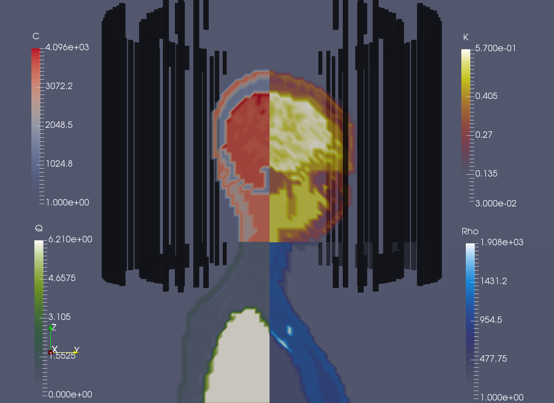
An example of Specific Heat Capacity (C), Heat generated by metabolic rate (Q), Thermal Conductivity (K), and Material Density (R) maps on Ella of the virtual family model. The black structure surrounding the images is the birdcage coil.

An example of the Temperature map estimated by TESS overlayed on the blood perfusion map (W)
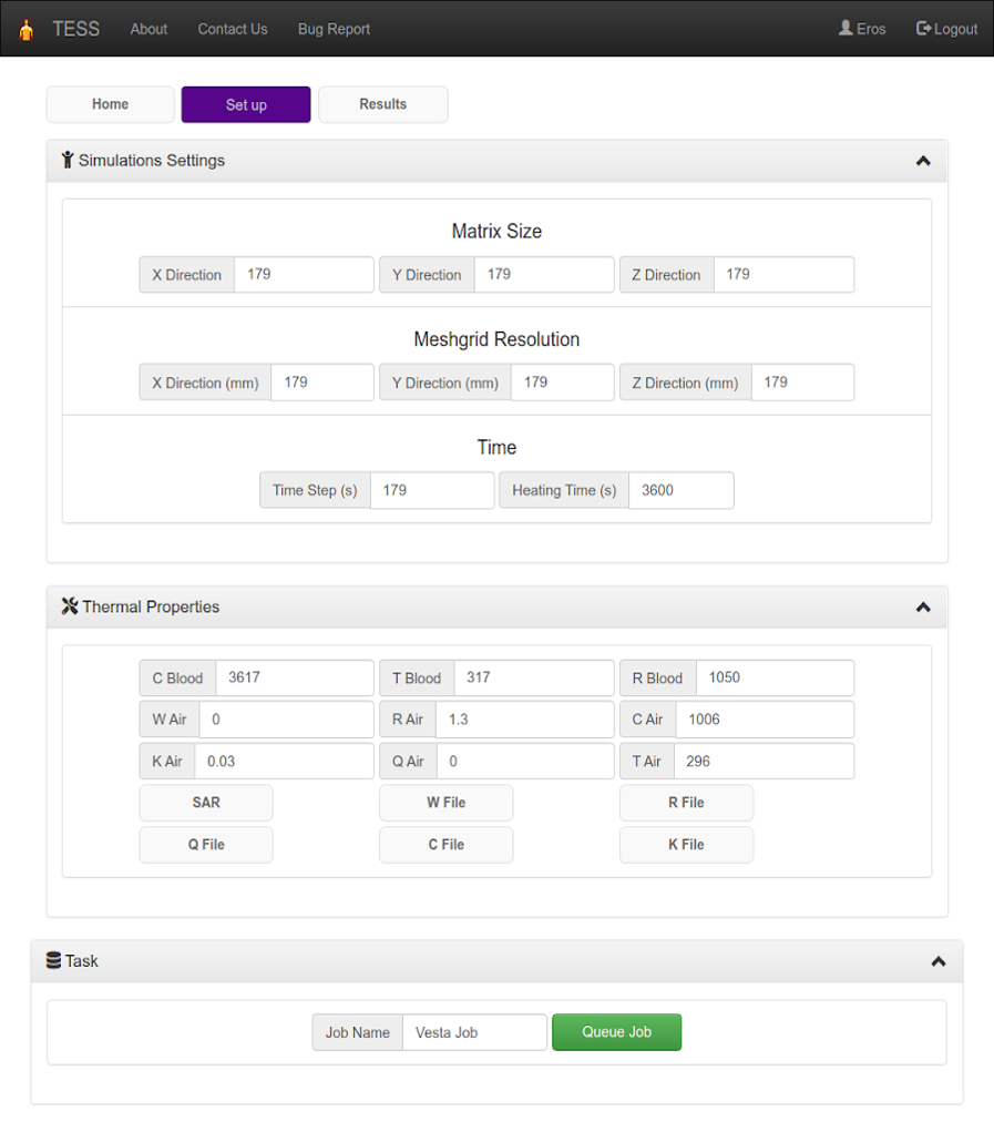
The Set Up tab of TESS’s web GUI. The web GUI is implemented in HTML and uses several javascripts libraries (angularjs, jquery, bootstrap, plotly.js, math.js and fabric.js). The Set Up tab is divided in panels that enable customizing different aspects of the simulations.

The Results tab of TESS’s web GUI. The Results tab is organized in two panels. From the “Job Queue” panel users can visualize the status of the requested jobs and manage simulation results. A completed job will be highlighted in green, pending jobs in yellow, etc. Users can download the results as JSON or mat files using restful API’s. The “Plots” panel allows users to visualize simulation results (e.g., coil performance map, coil SNR, etc.) and analyze them by drawing regions of interest.
DOI: https://doi.org/10.58530/2022/0582