0547
MR Molecular Imaging of Extradomain-B Fibronectin for Non-invasive Active Surveillance of Prostate Cancer1Department of Biomedical Engineering, Case Western Reserve University, Cleveland, OH, United States
Synopsis
C-X-C motif chemokine receptor 2 (CXCR2) expression is associated with high-grade and advanced prostate cancer. LNCaP-CXCR2+ prostate cancer cells with stable expression of CXCR2 exhibited mesenchymal features and became more aggressive with elevated expression of an ECM oncoprotein EDB-FN as compared to the slow-growing parental cell line, LNCaP. MRMI of EDB-FN with a targeted contrast agent MT218 resulted in significant increase of T1-weighted contrast enhancement in LNCaP-CXCR2+ prostate tumors as compared to the LNCaP tumors in mice. The results suggest that MRMI of EDB-FN has the potential to provide non-invasive active surveillance of low-grade prostate cancer.
Introduction
Prostate cancer is the most common noncutaneous cancer in men and the second leading cause of cancer deaths in the United States. An estimated quarter million men are annually diagnosed with prostate cancer in the United States 1. Prostate cancer is a highly heterogeneous disease with significant variations in molecular and pathological trajectory. Accurate early detection, risk-stratification, and active surveillance of primary prostate cancer are critical to identify the high-risk disease for timely treatment at a treatable stage for disease-free outcomes and to monitor the status and progression of low-risk tumors. C-X-C motif chemokine receptor 2 (CXCR2) neuroendocrine tumor cells are enriched in high-grade and advanced prostate cancer 2. CXCR2 expression is associated with therapy resistance and progression of the disease. It may also play a role in epithelial-mesenchymal transition (EMT) and extracellular matrix (ECM) remodeling. MRI is routinely used for diagnostic imaging and active surveillance of prostate cancer 3,4. We have recently developed small peptide targeted GBCAs specific to extradomain B fibronectin (EDB-FN) for cancer MR molecular imaging (MRMI). A lead targeted macrocyclic GBCA, ZD2-N3-Gd(HP-DO3A), otherwise known as MT218, has been identified for clinical translation 5. Our previous studies have shown that EDB-FN overexpression is associated with cancer invasiveness and MRMI with MT218 is effective to differentiate high-risk prostate cancer from low grade cancer 6,7. In this study, we aimed to determine potential correlation of EDB-FN overexpression with CXCR2 acquisition in the LNCaP and LNCaP-CXCR2+ prostate cancer cells and to assess the potential of MRMI of EDB-FN with MT218 for non-invasive monitoring of the progression of low-grade prostate cancer after establishing stable expression CXCR2 in mouse models.Methods
LNCaP prostate cancer cell were purchased from ATCC (Manassas, VA). LNCaP-CXCR2 cells were obtained from the lab of Dr. Jiaoti Huang (Duke University, Durham, NC). The prostate cancer cell lines were cultured in RPMI1640 medium (Sigma-Aldrich), supplemented with 10% fetal bovine serum (Gibco) and 100 Units/mL Penicillin/ Streptomycin (Gibco). The EDB-FN expression levels in LNCaP and LNCaP-CXCR2+ prostate cancer cells were determined at the mRNA and protein levels using qRT-PCR and Western blotting, respectively. Transwell assays, with and without Matrigel, were performed to assess the invasive and migratory properties of the different PCa cells. LNCaP or LNCaP-CXCR2, cells suspended in Matrigel-PBS mixture (1:1) were subcutaneously injected in the left flanks of male nude mice to develop prostate cancer models. MRMI was conducted on a 3T MRS 3000 scanner (MR Solutions, Surrey, UK) with a mouse short quad coil when the tumors reached volumes of 50-75 mm3. Images were acquired before and after i.v. injection of 0.04 mmol/kg dose of MT218. T1-weighted MR images were obtained with respiratory gating using a axial fast spin echo (FSE) sequence (TR = 305 ms, TE = 11 ms, FA = 90°, FOV = 40 mm x 40 mm, slice thickness = 1 mm, slice number = 15, Nav = 2, matrix = 256 x 256) and a coronal FSE sequence (TR = 305 ms, TE = 11 ms, FA = 90°, FOV = 90 mm x 90 mm, slice thickness = 1 mm, slice number = 20, Nav = 1, matrix = 248 x 512). Contrast-to-noise ratios (CNRs) were calculated using the formula: (average tumor intensity – average muscle intensity)/standard deviation of noise. The animals were euthanized at the end of imaging acquisition, and immunohistochemistry (IHC) was performed using an anti-EDB-FN G4 antibody to assess EDB-FN expression in the tumors. All the animal experiments were performed according to the protocol and guidelines laid down by the IACUC of CWRU.Results and discussion
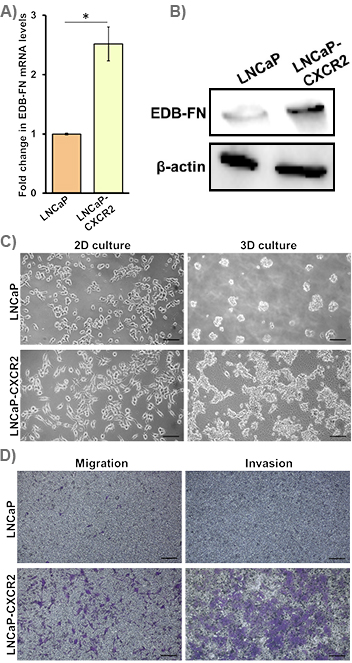
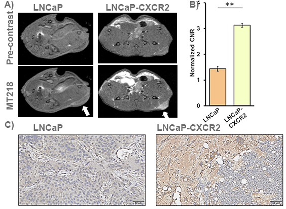
LNCaP-CXCR2+ prostate cancer cells exhibited significantly increased EDB-FN expression at both mRNA and protein levels as compared to the slow-growing parental LNCaP cells. LNCaP-CXCR2+ cells exhibited mesenchymal features and increased invasion and migration abilities, Figure 1. The results indicate that EDB-FN expression is significantly increased in LNCaP-CXCR2+ cancer cells in correlation with their acquired invasiveness, as compared to non-invasive parental cells. Figure 2 shows the representative MRI images of LNCaP and LNCaP-CXCR2+ prostate cancer xenografts established in athymic nu/nu mice before and 25 minutes after intravenous injection of MT218. Strong signal enhancement was observed in the invasive LNCaP-CXCR2+ tumors, while only slight signal enhancement was observed in the low-grade LNCaP tumors (Fig. 2A). Quantitative analyses of the signal intensities showed over 3-fold increased CNRs in the LNCaP-CXCR2+ tumors with MT218, while the LNCaP tumors only had about 50% CNR increase (Fig. 2B). Post-mortem IHC analysis of the tumor tissues with G4 monoclonal antibody showed strong staining EDB-FN in the LNCaP-CXCR2+ tumors as compared to the low EDB-FN in the LNCaP tumor sections, indicating increased expression of EDB-FN protein in the LNCaP-CXCR2+ tumors after the conversion to an aggressive phenotype with acquired CXCR2 expression (Fig. 2C). IHC data corroborated the MRMI resultsConclusion
Acquired CXCR2 expression in LNCaP-CXCR2+ converts the previously non-invasive prostate cancer cells into an aggressive phenotype with increased EDB-FN expression. MRMI of EDB-FN is effective to monitor the progression of prostate cancer based on acquired CXCR2 expression and increased EDB-FN expression. MRMI with MT218 has the promise for non-invasive risk-stratification and active surveillance of prostate cancer.Acknowledgements
This research was supported by the National Cancer Institute of the National Institutes of Health grant under award number R01CA211762.References
1. Siegel, R.L., Miller, K.D., Fuchs, H.E. & Jemal, A. Cancer Statistics, 2021. CA Cancer J Clin 71, 7-33 (2021).
2. Li, Y., et al. Targeting cellular heterogeneity with CXCR2 blockade for the treatment of therapy-resistant prostate cancer. Sci Transl Med 11(2019).
3. Tosun, M. & Uslu, H. Prebiopsy multiparametric MRI and PI-RADS version 2.0 for differentiating histologically benign prostate disease from prostate cancer in biopsies: A retrospective single-center comparison. Clin Imaging 78, 98-103 (2021).
4. Maggi, M., et al. SelectMDx and Multiparametric Magnetic Resonance Imaging of the Prostate for Men Undergoing Primary Prostate Biopsy: A Prospective Assessment in a Multi-Institutional Study. Cancers (Basel) 13(2021).
5. Ayat, N.R., et al. Optimization of ZD2 Peptide Targeted Gd(HP-DO3A) for Detection and Risk-Stratification of Prostate Cancer with MRI. ACS Med. Chem. Lett. 9, 730-735 (2018).
6. Han, Z., et al. Targeted Contrast Agent Specific to an Oncoprotein in Tumor Microenvironment with the Potential for Detection and Risk Stratification of Prostate Cancer with MRI. Bioconjug Chem 28, 1031-1040 (2017).
7. Han, Z., et al. Targeted gadofullerene for sensitive magnetic resonance imaging and risk-stratification of breast cancer. Nature communications 8, 692 (2017).
Figures