0537
FAI Bone Morphology Correlates with Increased T2 Relaxations Times in the Hip Cartilage of Female Water-Treading Athletes1Radiology, Stanford University, Stanford, CA, United States, 2Stanford University, Stanford, CA, United States, 3University of Easy Anglia, Norwich, United Kingdom, 4University of British Columbia, Vancouver, BC, Canada
Synopsis
Water polo players and synchronized swimmers have previously been found to have an increased prevalence of femoroacetabular impingement morphology compared to the general population. In this study, we used MRI to identify regional patterns in the microstructure of the hip cartilage and joint of high-level female water polo players and synchronized swimmers. Compared to healthy controls, the water-treading athletes had significantly higher T2 relaxation times in the right hip and a significantly higher prevalence of cam morphology in both hips. This study suggests that microstructural and morphological differences are present in these athletes’ hip cartilage compared to controls.
Introduction
Femoroacetabular impingement (FAI) is a syndrome diagnosed by variation in bony anatomy of the femur and/or acetabulum resulting in cam/pincer morphologies and hip pain. Sporting activity during adolescence has been strongly associated with the development of cam morphology1. Water polo and synchronized swimming require extensive eggbeater kicking, which involves a continuous rotational motion in the hip joint. Water polo players and synchronized swimmers at the National Collegiate Athletic Association (NCAA) Division 1 (D1) level have been found to have a higher incidence of cam morphology than the general population2. In addition, it has been previously reported that NCAA Division 3 (D3) female athletes (basketball, cross-country running, soccer, swimming, tennis, track, and water polo) report a higher rate of hip injuries than male athletes3. Quantitative MRI methods offer a way to non-invasively study early changes in hip joint cartilage microstructure4 that may be associated with FAI morphologies. This study hypothesizes that (1) quantitative MRI will identify different regional T2 patterns in the hip cartilage of NCAA D1 female water polo players and synchronized swimmers compared to female non-athlete controls and (2) that the female water-treading athletes’ increased cam and pincer morphology prevalence will correlate with increased T2 relaxation times in the hip cartilage.Methods
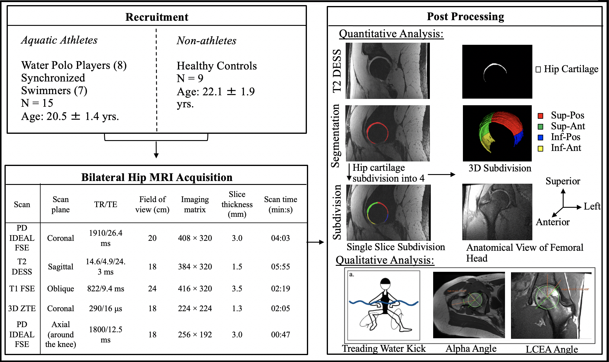
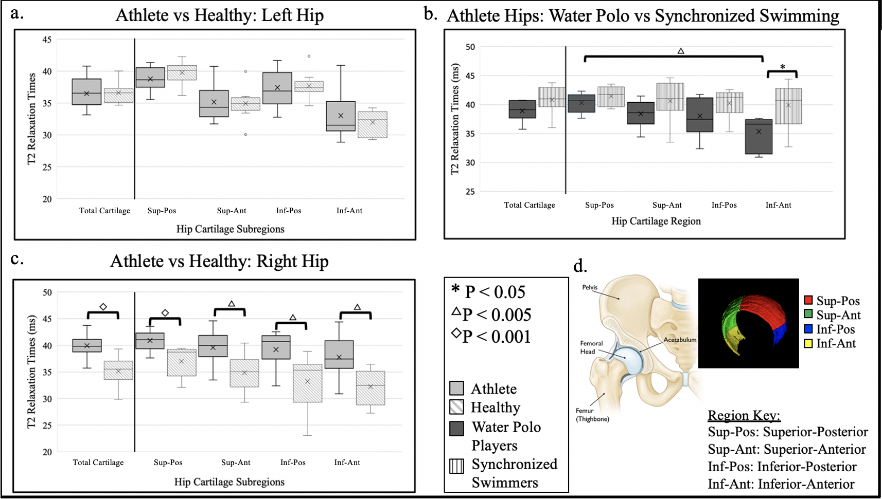
This Institutional Review Board approved study enrolled fifteen NCAA D1 athletes (20.5±1.4 yrs.), including 8 water polo players and 7 synchronized swimmers, and 9 healthy controls (22.1±1.9 yrs.), with informed consent. Athletes were excluded if they had previous hip surgery. On the right and left hip, each participant underwent a non-contrast unilateral hip MRI scan using a whole-body 3 Tesla MRI scanner (GE Healthcare, Waukesha, WI, USA) with a 16-channel flexible phased-array-receive-only coil (NeoCoil, Pewaukee, WI, USA). A quantitative double-echo in steady-state (qDESS) sequence (5 minutes/hip) was acquired to calculate T2 relaxation times4. Hip cartilage was manually segmented to define the region of interest for T2 maps. The segmentation included both acetabular and femoral cartilage as a single unit due to the close proximity of the two cartilage surfaces5. We manually subdivided the segmentation, using a circle fitted around the femoral head, into four quadrants: superior-posterior (sup-pos), superior-anterior (sup-ant), inferior-posterior (inf-pos), and inferior-anterior (inf-ant) (Figure 1). The four quadrants were created using vertical and horizontal planes parallel to the coronal and axial imaging planes. A Generalized Linear Model with a Bonferroni correction and α=0.05 was used to test for T2 differences between athletes vs. controls and between hip cartilage subregions. A Pearson correlation was used to compare T2 relaxation times with previously determined cam and pincer morphology measures2. Cam and pincer morphologies are clinically defined respectively by alpha angle (> 55°) and lateral central edge angle (LCEA) (> 39°)(Figure 2b,c).Results
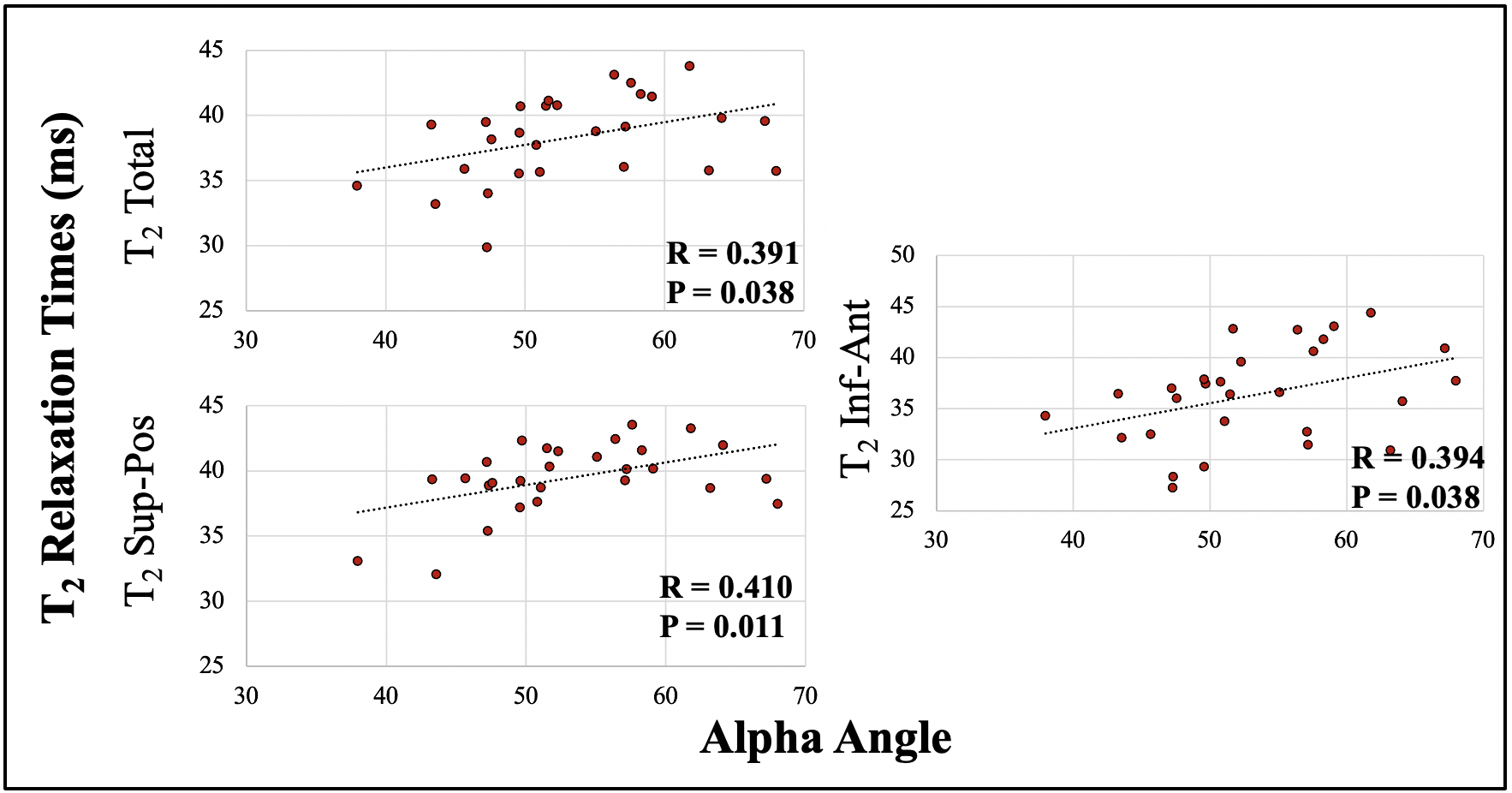
In all four regions of the right hip cartilage, the female aquatic athletes had significantly higher T2 relaxation times compared with the healthy controls (Total Cartilage: (39.8±2.4, 35.1±2.8, p < 0.001), Sup-Pos: (40.6±1.8, 37.0±2.8, p < 0.001), Sup-Ant: (39.5±3.1, 34.9±3.5, p <0.005), Inf-Pos: (38.7±3.8, 33.3±5.0, p < 0.005), Inf-Ant: (38.3±3.9, 32.2±3.3, p < 0.005) (Fig 2). In both the right and left hips, the aquatic athletes had significantly higher alpha angles compared to the healthy controls (p < 0.001). Cam morphology prevalence, respectively in the right and left hips, was 60% and 75% in the water-treading athletes and 0% and 0% in the controls. In both athlete and control populations, the right hip Total Cartilage, Sup-pos region, and Inf-ant regions, there was a significant positive moderate correlation between T2 relaxation times and alpha angle/cam morphology (p = 0.038, p = 0.011, p = 0.038) (Fig 3).Discussion
Significantly higher T2 relaxation times and alpha angles in the female aquatic athletes compared to the controls indicate that treading water could be affecting hip cartilage microstructure and joint health. Right hip T2 relaxation time elevation in every subregion of the athletes compared to the controls suggests that these sports globally affect the joint cartilage microstructure. The significant differences in just the right hip could indicate a dominant leg that has increased stress to its hip cartilage, but further investigation is required. The correlation between T2 relaxation times and alpha angle indicates that quantitative MRI could measure early cartilage degeneration that occurs with morphologic bone changes prevalent in the FAI disease progression. Further study is required to identify if treading water results in higher T2 relaxation times or if the abnormal morphology from treading water is causing the early cartilage damage. The ability to quantify cartilage changes could allow for monitoring of training and rehabilitation that may help mitigate the future need for FAI surgery.Conclusion
This work demonstrates that quantitative and morphologic MRI can detect differences in water polo players’ and synchronized swimmers’ hip cartilage composition compared to healthy controls. Additionally, that quantitative MRI could be a useful tool in both longitudinal studies of these athletes’ cartilage health and in the monitoring of training and rehabilitation. Knowledge of injury patterns and early detection of cartilage damage could help inform strategies for keeping athletes healthy when they present with hip pain.Acknowledgements
This work was supported by a Major Grant from the Stanford University Vice Provost for Undergraduate Education as part of the Human Biology Honors Program. Support also came from GE Healthcare, and the National Institutes of Health (R01EB002524, R01AR077604).References
1. Palmer, A., Fernquest, S., Gimpel, M., et al. 2018. Physical activity during adolescence and the development of cam morphology: a cross-sectional cohort study of 210 individuals. British Journal of Sports Medicine 52(9), pp. 601–610.
2. Langner, J.L., Black, M.S., MacKay, J.W., et al. 2020. The prevalence of femoroacetabular impingement anatomy in Division 1 aquatic athletes who tread water. Journal of hip preservation surgery 7(2), pp. 233–241.
3. Sallis, R.E., Jones, K., Sunshine, S., Smith, G. and Simon, L. 2001. Comparing sports injuries in men and women. International journal of sports medicine 22(6), pp. 420–423.
4. Sveinsson, B., Chaudhari, A.S., Gold, G.E. and Hargreaves, B.A. 2017. A simple analytic method for estimating T2 in the knee from DESS. Magnetic Resonance Imaging. 38, pp. 63–70.
5. Nishii, T., Nakanishi, K., Sugano, N., Masuhara, K., Ohzono, K. and Ochi, T. 1998. Articular cartilage evaluation in osteoarthritis of the hip with MR imaging under continuous leg traction. Magnetic Resonance Imaging 16(8), pp. 871–875.
6. Neumann, J., Zhang, A.L., Schwaiger, B.J., et al. 2019. Validation of scoring hip osteoarthritis with MRI (SHOMRI) scores using hip arthroscopy as a standard of reference. European Radiology 29(2), pp. 578–587.
Figures