0483
Noninvasive assessment of unilateral ureter obstruction using dynamic contrast-enhanced MR-CEST urography
Julia Stabinska1, Aruna Singh1,2, Farzad Sedaghat2, Max Kates3, Yuguo Li1,2, and Michael T. McMahon1,2
1F.M. Kirby Research Center for Functional Brain Imaging, Kennedy Krieger Institute, Baltimore, MD, United States, 2The Russell H. Morgan Department of Radiology and Radiological Science, The Johns Hopkins University School of Medicine, Baltimore, MD, United States, 3The James Buchanan Brady Urological Institute, Department of Urology, The Johns Hopkins University School of Medicine, Baltimore, MD, United States
1F.M. Kirby Research Center for Functional Brain Imaging, Kennedy Krieger Institute, Baltimore, MD, United States, 2The Russell H. Morgan Department of Radiology and Radiological Science, The Johns Hopkins University School of Medicine, Baltimore, MD, United States, 3The James Buchanan Brady Urological Institute, Department of Urology, The Johns Hopkins University School of Medicine, Baltimore, MD, United States
Synopsis
The extent of recovery of renal function in patients with urinary tract obstructions depends on early diagnosis and prompt intervention. In the present study, we employed dynamic contrast-enhanced MR-CEST urography to noninvasively assess kidney function in mice with unilateral ureter obstruction (UUO) at two time points (day 1 and 2) after UUO surgery. Using a dynamic CEST MRI protocol, we were able to detect a slight increase in pH values (pH 6.74 ± 0.08 and 6.62 ± 0.04 in the UUO and contralateral kidney, respectively) as well as iopamidol retention in the UUO kidney.
Introduction
Urinary tract obstructions (UTOs) are blockages in the urinary tract that impede normal urine flow, causing urinary retention and increased retrograde pressure1. Because the extent of recovery of renal function in patients with UTO depends on severity and duration of the obstruction, early diagnosis and prompt intervention are crucial for preventing irreversible kidney damage1.While effective in characterizing upper tract function, ultrasonography and traditional CT are limited in quantifying kidney function2. Renal scintigraphy can be applied for assessment of differential kidney function but its anatomical resolution is poor2. In contrast, iopamidol-enhanced chemical exchange saturation transfer (CEST) MRI has been shown to provide functional information by generating spatially localized renal pH maps in addition to time-activity curves which are similar to standard renograms3,4,5. In this study, we explore the potential of dynamic contrast-enhanced MR-CEST urography for assessing renal function in mice with UUO by simultaneous pH and renal filtration measurements.
Methods
For the in vivo study, the right ureter of six mice was obstructed via suture ligation. The animals were imaged at day 1 (3 mice) and 2 (3 mice) post-obstruction on an 11.7 T Bruker MRI animal scanner. A total of 204 CEST images, including twelve M0 images and 96 sets of saturated images at 4.3 and 5.5 ppm, were collected with the following parameters: B1 = 3.6 µT, tsat = 3 sec, TE/TR = 3.49/5125 msec, matrix: 64x64, slice thickness: 1.5 mm, RARE factor: 32. Iopamidol was injected via tail-vein catheter. Renal time-course signal enhancement curves were measured as a percentage change in the post-injection CEST-prepared signal at 4.3 ppm relative to the average pre-injection signal.For pH mapping, post-injection magnetization transfer ratio (MTR) at 4.3 and 5.5 ppm was quantified from three averaged images collected at the peak enhancement time. Averaged pre-injection MTR maps were then subtracted from the post-injection MTR images to remove endogenous CEST signals. Subsequently, renal pH values were obtained by calculating the signal ratio6 for these two frequencies and using the pH calibration curve as described previously3.
Results
The time-activity curves which reflect the uptake and excretion of iopamidol by the kidneys showed clear differences between the healthy and UUO kidneys. For the healthy mice, the dynamic CEST curves of both kidneys were nearly identical and displayed rapid excretion of contrast. In the UUO mice, the time-activity curve for the obstructed kidneys displayed prolonged contrast excretion with a decreased STenh% values with time after ligation compared with the contralateral kidney (Fig. 1).A representative renal pH map and histogram obtained in a healthy mice displayed similar acidity for both kidneys (pH 6.71 ± 0.06 and 6.68 ± 0.08 in the right and left kidney, respectively). At one day after UUO, there was an increase in pH values in the obstructed kidney, especially in the inner and outer medulla, compared with the contralateral (pH 6.77 ± 0.05 and 6.66 ± 0.05 in the UUO and CL kidney, respectively). The same trend could be observed at day 2 after obstruction (pH 6.69 ± 0.05 and 6.56 ± 0.04 in the UUO and CL kidney, respectively) (Fig. 2). The box-plot analysis for all six UUO mice revealed slightly increased pH values in the UUO kidneys compared with the native kidney (Fig. 3).
Discussion
In this study, we have applied DCE-MR CEST urography to a murine model of obstructive nephropathy and non-invasively assessed kidney function over 2 days after UUO. Our results demonstrate that a dynamic CEST acquisition combined with a single injection of iopamidol allows simultaneous measurements of both renal perfusion and pH, as opposed to the conventional diagnostic techniques, which provide only one type of metric to characterize kidney function. As early as one day after UUO, we observed both increased pH values, which are likely due to tubular defects in urinary acidification7,8, and retention of iopamidol in the renal parenchyma of the UUO kidney, which suggests a decline in renal function5,9. Overall, these results suggest that CEST imaging might be particularly useful for detecting and monitoring the progression of renal injury caused by UUO. On the basis of these findings, we believe that our dynamic CEST MRI protocol is promising for early assessment of upper UTOs and could be translated to patients with obstructive nephropathy.Conclusion
Our findings indicate that DCE-MR-CEST urography can detect changes in renal filtration and pH homeostasis and distinguish between obstructed and unobstructed kidney as early as one day after UUO.Acknowledgements
This work has been supported by NIH grant 5R01DK121847-02.References
- Brenner BM, Rector FC. Brenner&Rector’s the Kidney. 8th ed. Saunders Elsevier; 2008
- Bo S, Sedaghat F, Pavuluri K et al. Dynamic Contrast Enhanced-MR CEST Urography: An Emerging Tool in the Diagnosis and Management of Upper Urinary Tract Obstruction. Tomography. 2021;7(1):80-94
- Pavuluri K, Manoli I, Pass Al et al. Noninvasive monitoring of chronic kidney disease using pH and perfusion imaging. Sci Adv. 2019;5(8):eaaw8357
- Irrera P, Consolino L, Cutrin JC et al. Dual assessment of kidney perfusion and pH by exploiting a dynamic CEST-MRI approach in an acute kidney ischemia-reperfusion injury murine model. NMR Biomed. 2020 Jun;33(6):e4287
- Tantawy MN, Jiang R, Wang F et al. Assessment of renal function in mice with unilateral ureteral obstruction using 99mTc-MAG3 dynamic scintigraphy. BMC Nephrol 13, 168 (2012).
- Longo DL, Dastru W, Digilio G et al. Iopamidol as a responsive MRI-chemical exchange saturation transfer contrast agent for pH mapping of kidneys: In vivo studies in mice at 7 T. Magn. Reson. Med.2011, 65: 202-211.
- Harris RH, Yarger WE. Renal function after release of unilateral ureteral obstruction in rats . Am J Physiol . 1974; 227:806–15.
- Thirakomen K, Kozlov N, Arruda JAL et al. Renal hydrogen ion secretion after release of unilateral ureteral obstruction . Am J Physiol . 1976; 231:1233–9.
- Schlotmann A, Clorius JH, Rohrschneider WK, Clorius SN, Amelung F, Becker K. Diuretic renography in hydronephrosis: delayed tissue tracer transit accompanies both functional decline and tissue reorganization. J Nucl Med. 2008 Jul;49(7):1196-203.
Figures
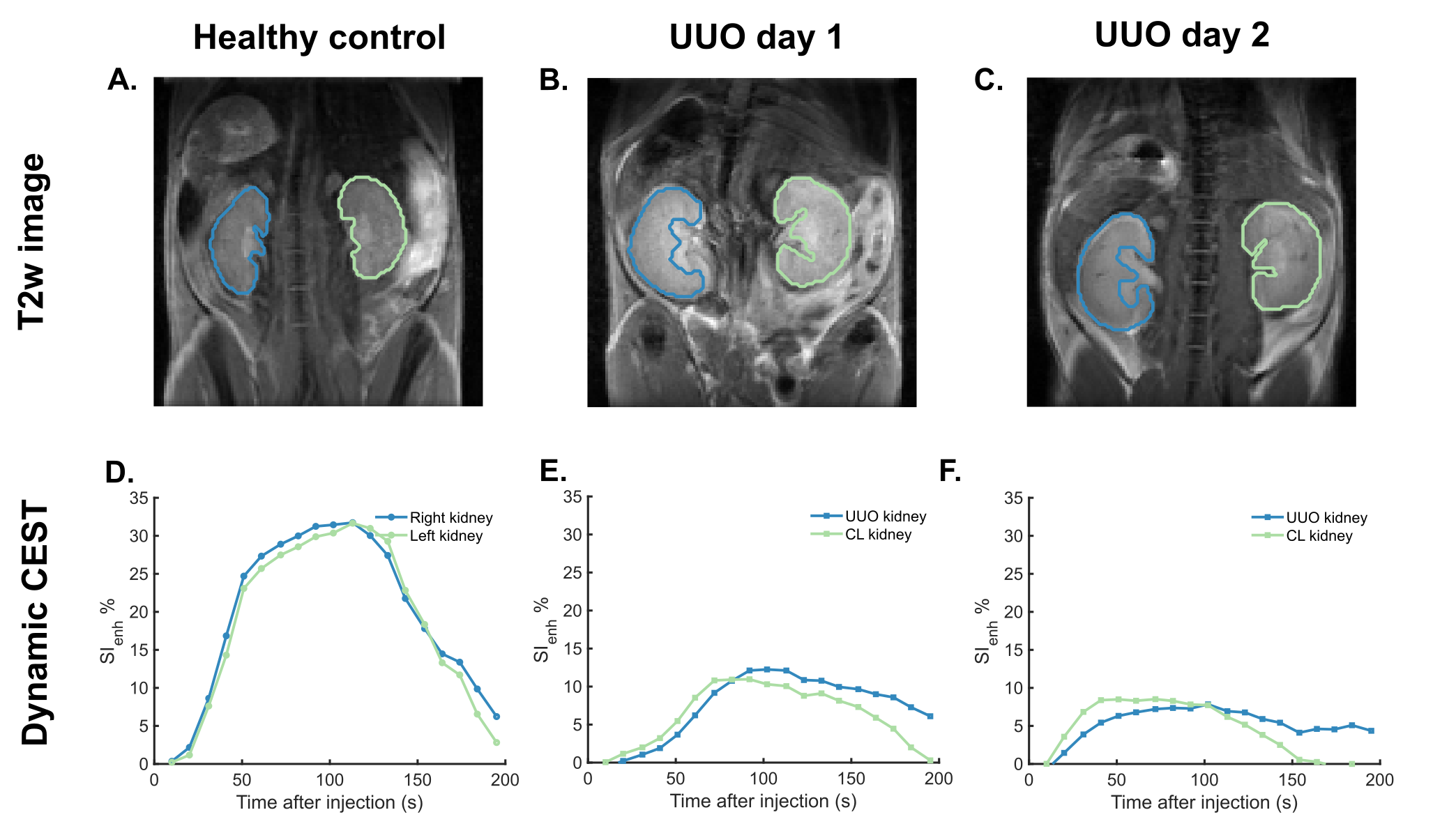
Figure 1: T2w images and dynamic CEST signal enhancement curves measured in (A, D) a control mouse, (B, E) UUO mice at day 1 and (C, F) day 2 after obstruction.
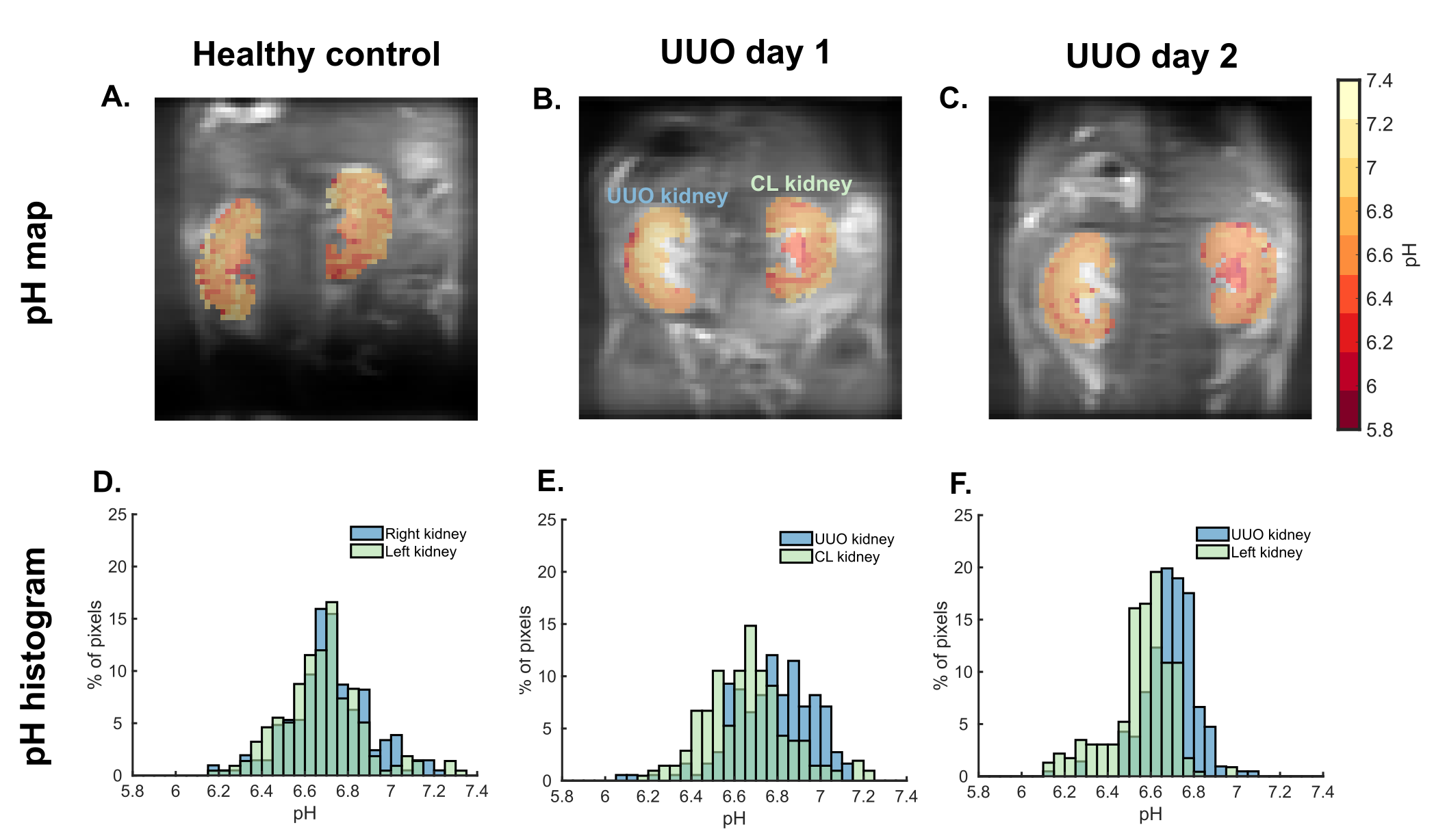
Figure 2: pH maps and pH histograms measured in (A, D) a control mouse, (B, E) UUO mice at day 1 and (C, F) day 2 after obstruction.
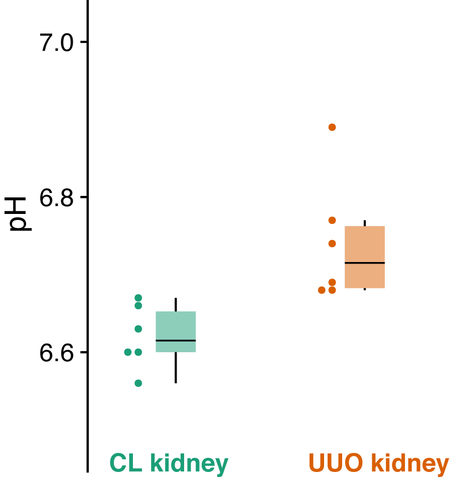
Figure 3: pH
distribution in contralateral kidney and UUO kidney measured in six animals.
DOI: https://doi.org/10.58530/2022/0483