0476
Detection of kidney cysts using a Convolutional Neural Network on 11,000 MRI data sets from the German National Cohort1University Medical Center Freiburg, Freiburg, Germany
Synopsis
We implemented a processing pipeline for kidney cyst segmentation using a hierarchical patch-based stack of U-nets and applied it to abdominal MRI images of the German National Cohort (GNC) study. The training data set included 300 cases, and the final net was applied to the dataset of 11,207 MRIs. Kidney cysts could be segmented with 98% sensitivity above a lower detection threshold of 1ml. The relation of first parameters based on the cyst segmentation with age and sex are presented. This result presents an optimal starting point to identify more advanced bimarkaers and their correlates, especially with kidney function parameters.
Introduction
Kidney cysts are a frequent incidental finding in normal kidneys[1]. The number and size of kidney cysts that are detected increases with age, but so far there is no automatically generated data on the occurrence and size of kidney cysts originating from a large, population-based dataset. Moreover, the genetic disorder polycystic kidney disease (PKD) is among the leading genetic causes for chronic kidney disease (CKD)[2], and an early detection of PKD would be of great clinical relevance. We set out to develop a robust image processing pipeline for the segmentation of kidneys and detection of kidney cysts and to apply it to abdominal MRI images from a large, population-based cohort study, the GNC.Methods
Within the GNC, 30,000 participants underwent the MRI protocol described in [3]. For the first 11,207 available data sets, 3D gradient echo and 2D haste images of the abdominal part of the data were imported into the imaging platform, NORA (www.nora-imaging.org) for analysis. The platform provides tools to manage, annotate and perform training of deep-learning convolutional neural networks (CNNs) on a large scale. For comparison, a subsample of 200 cases were initially manually investigated and segmented for the occurrence and size of kidney cysts by two expert raters. As a result, a lower detection limit was chosen to achieve at least 98% detection of manually detected cysts above that threshold volume with the available images and contrasts.A requirement analysis was performed to design a CNN, and a U-Net model was configured using the “patchwork”-framework, developed in-house at the University Medical Center Freiburg (https://bitbucket.org/reisert/patchwork/wiki/Home). This hierarchical patch-based stack of U-nets was trained to segment the cysts automatically. At this stage, only the 3D gradient echo data was used for analysis. Data was randomized, balanced, and mildly augmented on the fly. After a first training round, the model was iteratively improved by a loop of prediction, manual correction, and re-training in four rounds. The final model was trained on 300 cases and then applied to the dataset of 11,207 abdominal MRI. A manual quality control reading was performed to determine problematic cases and to select a valid dataset for statistical analysis. Based on the resulting segmentation and detection of kidney cysts, prevalence and volumetric parameters (total cystic volume) were calculated. Values were correlated with age and sex.Results
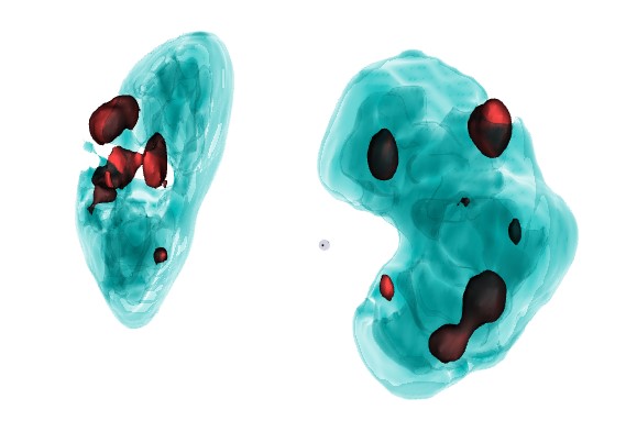
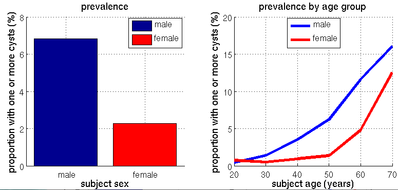
The cysts were detected and segmented with a detection rate of 98% within the kidneys when a lower detection limit of (>1 mL) was chosen (Fig. 1). Manual quality control revealed that the image quality was insufficient for automated segmentation in 10% of the cases. This was predominantly due to initial imaging artifacts or bad image quality and these cases were excluded from the statistical analysis. For the remaining data sets, the prevalence of cysts (>1 mL) was strongly related to age, as was expected, and to male sex (Fig. 2). The overall prevalence was 7% in men compared to 2.5% in women. We also found that the mean total cystic volume was related to male sex with 17ml in men compared to 14ml in women.Discussion and Conclusions
The developed framework allows for robust detection and segmentation of kidney cysts in abdominal MRI data from anon-specific clinical routine protocol of a large cohort study. Basic parameters such as prevalence of cysts and total cyst volume show correlations with participants' age and sex that are consistent with prior knowledge. The correlation of prevalence to male sex was even higher than expected, which could be due to the fact that women have reportedly [1] smaller cysts, that could stay below the detection threshold. Even though the used data was acquired on multiple sites within Germany, the scanner setup and used MR protocol was identical. Compared to most other multicentric studies, this leads to a high uniformity in data quality. Not only to better understand the requirements for the quality of the input data for such a workflow, it will be of interest to apply it also to data acquired on other scanners with similar but at least slightly variant protocols and therefore less uniform data. In conclusion, in our study we used a CNN to assess the prevalence and size of renal cysts MRI in a large number of MRI data sets from the German National Cohort (NAKO/GNC). Older age and male sex could be confirmed as risk factors for the occurrence and volume of renal cysts. This result appears as an optimal starting point to identify more advanced biomarkers for the cyst formation and classification and their correlates, especially kidney function parameters. This will help to better understand the prevalence of kidney cysts in the general population and how established kidney disease markers relate to imaging-based biomarkers of kidney function and disease.Acknowledgements
This work funded by the Deutsche Forschungsgemeinschaft (DFG, German Research Foundation) – DFG KE 2513/1-1, / DFG KO 3598/6-1 / SPP 2177 within the DFG Priority Program SPP2177 „Radiomics: Nächste Generation der medizinischen Bildgebung“.References
[1] Mensel, B., Kühn, JP., Kracht, F. et al. Prevalence of renal cysts and association with risk factors in a general population: an MRI-based study. Abdom Radiol 43, 3068–3074 (2018). doi: 10.1007/s00261-018-1565-5
[2] Eckardt KU, Coresh J, Devuyst O, et al. Evolving importance of kidney disease: from subspecialty to global health burden. Lancet 2013;382(9887):158-69. doi: 10.1016/S0140-6736(13)60439-0
[3] Bamberg et. al. Whole-Body MR Imaging in the German National Cohort: Rationale, Design, and Technical Background. Radiology 2015, doi 10.1148/radiol.2015142272
Figures