0439
Anisotropic Mechanical Properties of White Matter Tracts in Aging via Transversely Isotropic MR Elastography1University of Delaware, Newark, DE, United States, 2Dartmouth College, Hanover, NH, United States, 3Université de Sherbrooke, Sherbrooke, QC, Canada, 4Washington University in St. Louis, St. Louis, MO, United States
Synopsis
This study combined multi-excitation MR Elastography (ME-MRE) with a transversely isotropic inversion nonlinear inversion algorithm and diffusion tensor imaging (DTI) fiber directions to investigate anisotropic white matter tract integrity in aging. Participants were 5 older adults and 17 young adults. MRE outcomes include substrate shear modulus, μ, shear anisotropy, φ, tensile anisotropy, ζ, and DTI measures were also examined. Shear and tensile anisotropies were significantly different in the Corona Radiata and Superior Longitudinal Fasciculus. Diffusion measures were significantly different between groups in most tracts. These results suggest sensitivity of anisotropic properties to biological changes along white matter tracts in aging.
Introduction
Magnetic resonance elastography (MRE) produces quantitative maps of tissue mechanical properties, such as shear stiffness, in the human brain1. These mechanical properties capture information regarding the composition and organization of the brain tissue microstructure. Previous MRE studies have shown that brain mechanical properties decline with age and in neurological diseases2, and that they relate to cognitive outcomes3. However, there is limited data on how properties change in white matter (WM) tracts owing to limitations in MRE approaches that assume isotropic behavior of tissue, and thus exhibit uncertainty in recovering properties of anisotropic WM tracts4. Diffusion tensor imaging (DTI) studies have reported radial diffusivity (RD), mean diffusivity (MD), and fractional anisotropy (FA) changes in healthy aging white matter5. However, DTI primarily captures measures related to intra-axonal integrity and provides limited information about changes to the surrounding tissues.Hence, we have recently developed an approach for estimating anisotropic properties in WM tracts by combining a three-parameter model of nearly incompressible transversely isotropic behavior6, multi-excitation MRE data acquisition (ME-MRE)4,7 and a finite element-based inversion that estimates anisotropic parameters while accounting for material heterogeneity8. ME-MRE has been shown to generate sufficient waves for repeatable anisotropic inversions in a group of young healthy volunteers9,10. In this study, we perform ME-MRE on a group of normal older adult volunteers and estimate the substrate shear modulus, shear anisotropy, and tensile anisotropy across major WM tracts. MRE outcomes are compared between older and young adults to investigate the age-related changes of these properties. We expect tissue anisotropy to decline with age, based on previous works that demonstrated softening of the brain11 and loss of WM integrity in normal aging12.
Methods
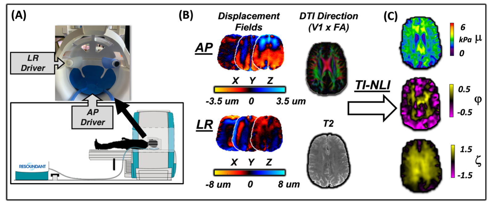
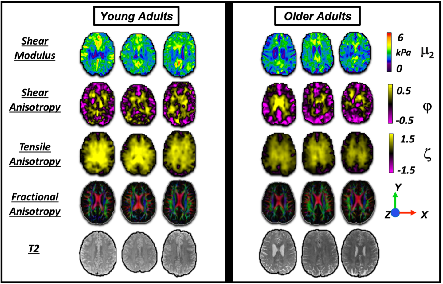
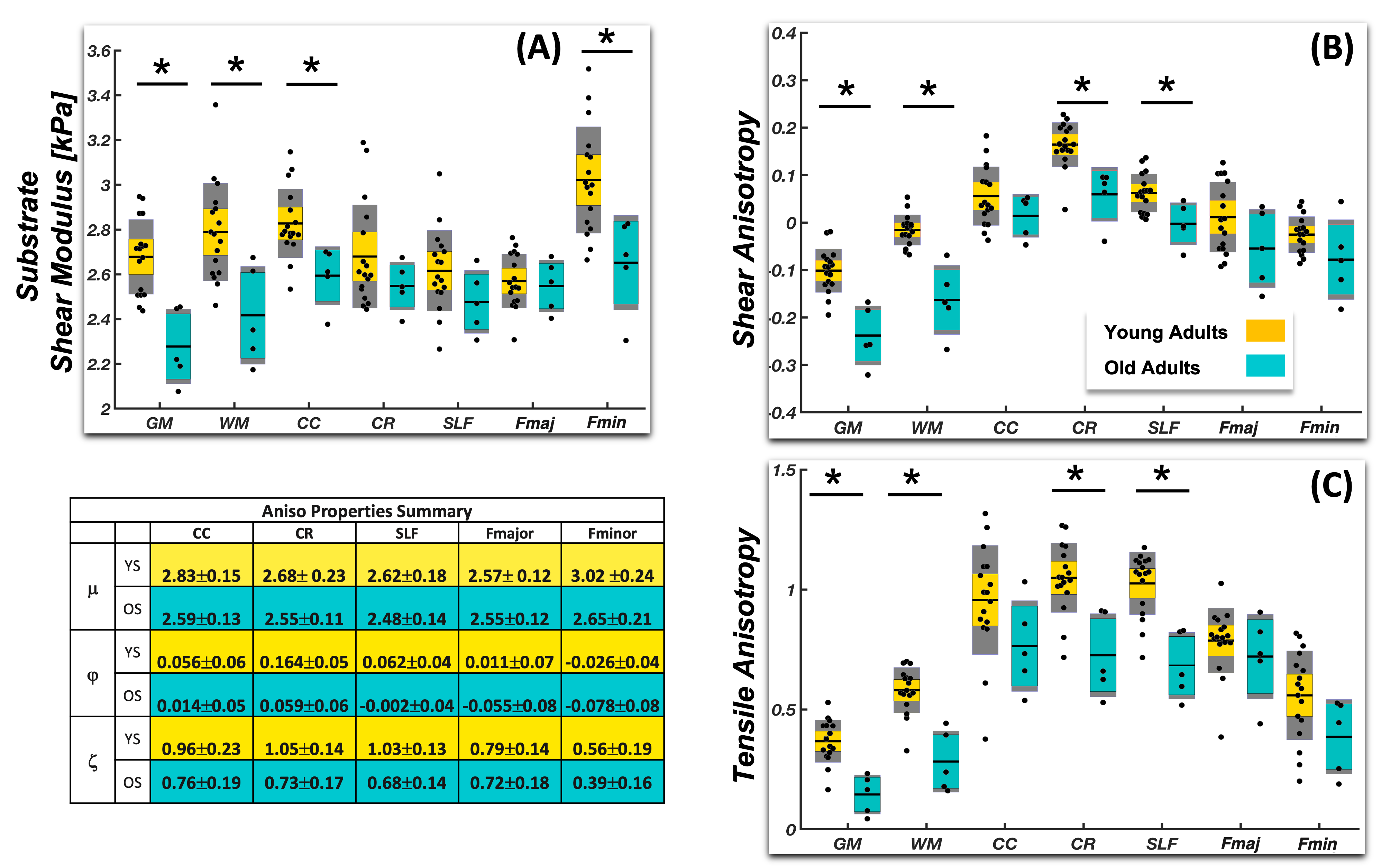
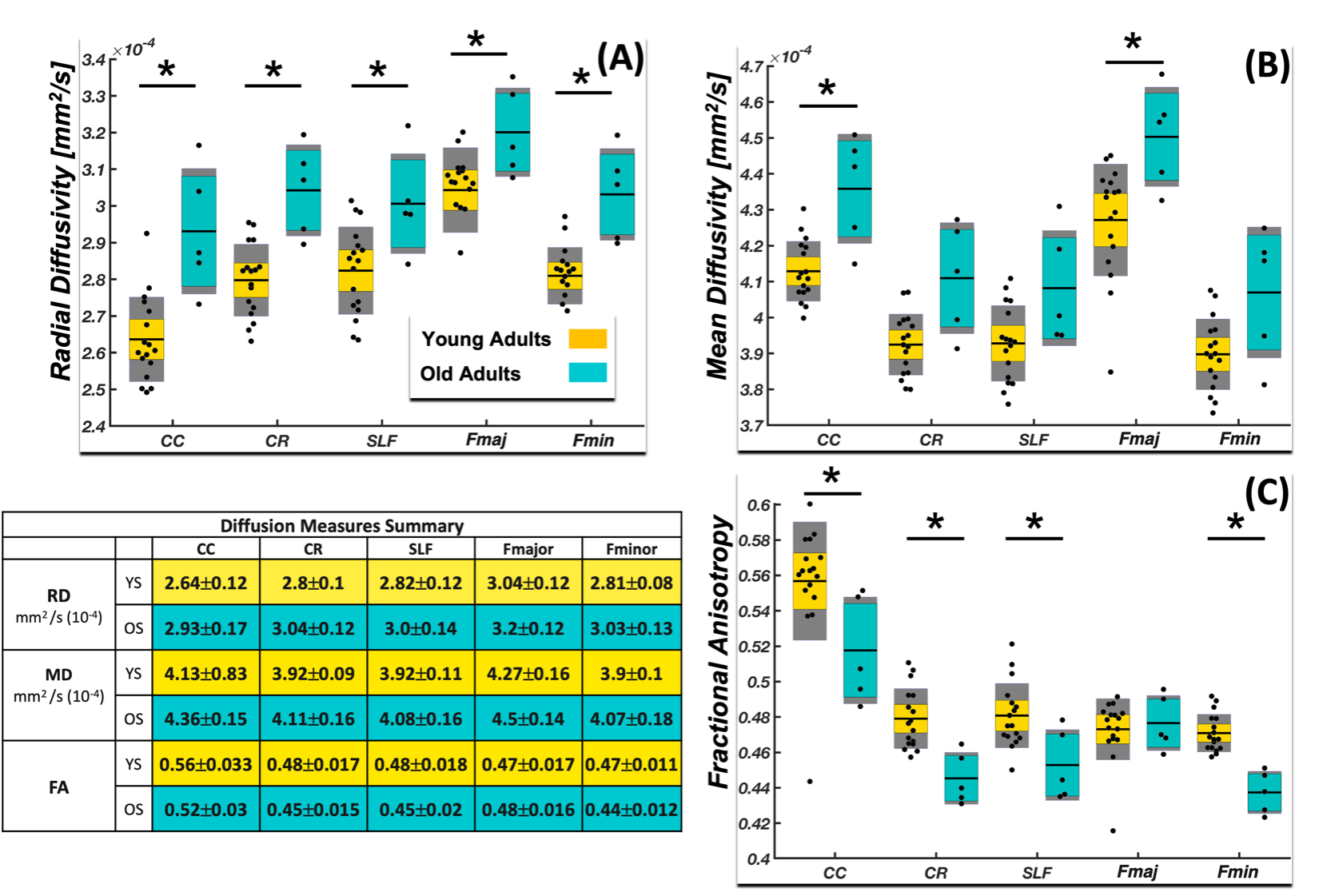
A group of 5 normal older subjects (4M/1F, 65.87.1 years), and a group of 17 normal young subjects (7M/10F, 25.22.1 years) with no history of neurological conditions. Participants completed the ME-MRE protocol on a 3T Siemens Prisma scanner using a 20-channel head coil. In ME-MRE we generate steady state harmonic vibrations in the anterior-posterior (AP) direction using a pillow driver and, in the left-right (LR) direction using a lateral bottle driver affixed to the head coil as seen in Fig.1.9 Two MRE scans were acquired separately for LR and AP excitations at 50 Hz vibration frequency. A 3D multiband, multishot spiral MRE sequence13 was used to image whole-brain displacements at 2 mm isotropic resolution (240x240x128 mm FOV, 64 slices, TR/TE = 2240/76 msec). Auxiliary scans included DTI at 1.5 mm isotropic resolution (210x240x138 FOV, 92 slices, TR/TE = 3520/95.2 msec) and T1-weighted MPRAGE at 0.9 mm isotropic resolution for segmentations. LR and AP displacement fields were combined and used together with fiber direction from DTI in TI-NLI8,14 to estimate substrate shear modulus, μ2, shear anisotropy (φ=μ1/μ2-1) and tensile anisotropy (ζ=E1/E2–1).MRE data was skull stripped15 and segmented into whole grey and white matter using the MNI template, and registered to MRE space via FSL-FLIRT16,17 and WM tract masks were determined via registration from the JHU white matter atlas18. MRE properties and DTI measures were examined in the Corpus Callosum (CC), Corona Radiata (CR), Superior Longitudinal Fasciculus (SLF), Forceps Major (Fmaj) and Forceps Minor (Fmin) and mechanical properties were averaged across each region. Two sample t-tests assuming unequal variances were performed to compare young subjects and older subjects for all the MRE properties and the DTI measures.
Results and Discussion
Anisotropic mechanical properties decrease in normal aging within white matter and are summarized in Fig.3. We found that μ is lower in older adults in most regions. φ significantly differed between groups in the CR (YS 0.164±0.05, 0.059±0.06) and the SLF (YS 0.062±0.04, OS -0.002±0.04). Similarly, ζ was lower in the older adults in the CR (YS 1.05±0.14, OS 0.73±0.17) and the SLF (YS 1.03±0.13, OS 0.68±0.14). Estimated negative values of φ may stem from model-data mismatch at the grey/white matter boundaries where tissue properties abruptly change from anisotropic to isotropic and there is a lack of strong fiber direction. Figure 4 compares DTI measures between groups and shows that fractional anisotropy (FA) decreases with age, while radial diffusivity (RD) and mean diffusivity (MD) both increase with age. These findings suggest that TI-NLI properties are changing with age and are sensitive to individual tracts.Conclusion
Anisotropic mechanical properties of white matter tracts were estimated in a group of older adults and compared to young adults. Shear and tensile anisotropy were lower for all regions in the older group suggesting that these properties reflect microstructural changes, such as degeneration, demyelination, and cell death. A previous study examined anisotropic properties in aging but showed no differences in WM anisotropy19, potentially due to methods that don’t model tissue heterogeneity. This novel approach can detect changes in tissue integrity for individual white matter tracts that reflect differences between young and old participants, confirming that this method is sensitive to biological changes in the white matter microstructure that occur in normal aging.Acknowledgements
This research was supported by NIH grants R01-EB027577 and R01-AG058853.References
1. Hiscox, L. V et al. Magnetic resonance elastography (MRE) of the human brain: technique, findings and clinical applications. Phys. Med. Biol. 61, R401–R437 (2016).
2. Murphy, M. C., Huston, J. 3rd & Ehman, R. L. MR elastography of the brain and its application in neurological diseases. Neuroimage 187, 176–183 (2019).
3. Schwarb, H., Johnson, C. L., McGarry, M. D. J. & Cohen, N. J. Medial temporal lobe viscoelasticity and relational memory performance. Neuroimage 132, 534–541 (2016).
4. Anderson, A. T. et al. Observation of direction-dependent mechanical properties in the human brain with multi-excitation MR elastography. J. Mech. Behav. Biomed. Mater. 59, 538–546 (2016).
5. Bennett, I. J., Madden, D. J., Vaidya, C. J., Howard, D. V & Howard, J. H. J. Age-related differences in multiple measures of white matter integrity: A diffusion tensor imaging study of healthy aging. Hum. Brain Mapp. 31, 378–390 (2010).
6. Tweten, D. J., Okamoto, R. J., Schmidt, J. L., Garbow, J. R. & Bayly, P. V. Estimation of material parameters from slow and fast shear waves in an incompressible, transversely isotropic material. J. Biomech. 48, 4002–4009 (2015).
7. Smith, D. et al. Multi-Excitation MR Elastography of the Brain: Wave Propagation in Anisotropic White Matter. J. Biomech. Eng. 142, 710051–710059 (2020).
8. McGarry, M. D. J. et al. Model-Based Heterogeneous Transverse Isotropic MR Elastography Inversion for Brain Tissue with Aligned Fiber Tracts. 28th Annu. Meet. Int. Soc. Magn. Reson. Med. (2020).
9. Caban-Rivera, D. A., Smith, D., Kailash, K., Okamoto, R., McGarry, M., Williams, L., Guertler, C., McIlvain, G., Sowinski, D., Houten, E. Van, Paulsen, K., Bayly, P., Johnson, C. Multi-Excitation Actuator Design for Anisotropic Brain MRE. 29th Annu. Meet. Int. Soc. Magn. Reson. Med. May 15-20, (2021).
10. Smith, D. R. et al. Anisotropic mechanical properties of white matter tracts in the healthy brain estimated with multi-excitation MR elastography and transversely isotropic nonlinear inversion. 29th Annu. Meet. Int. Soc. Magn. Reson. Med. May 15-20, 2021 (2021).
11. Hiscox, L. V., Schwarb, H., McGarry, M. D. J. & Johnson, C. L. Aging brain mechanics: Progress and promise of magnetic resonance elastography. Neuroimage 232, 117889 (2021).
12. Davis, S. W. et al. Assessing the effects of age on long white matter tracts using diffusion tensor tractography. Neuroimage 46, 530–541 (2009).
13. Johnson, C. L., Holtrop, J. L., Anderson, A. T. & Sutton, B. P. Brain MR Elastography with Multiband Excitation and Nonlinear Motion-Induced Phase Error Correction. 24th Annu. Meet. Int. Soc. Magn. Reson. Med. (2016)
14. McGarry, M. D. J. et al. A heterogenous, time harmonic, nearly incompressible transverse isotropic finite element brain simulation platform for MR elastography. Phys. Med. Biol. (2020) doi:10.1088/1361-6560/ab9a84.
15. Smith, S. M. Fast robust automated brain extraction. Hum. Brain Mapp. 17, 143–155 (2002).
16. Jenkinson, M., Bannister, P., Brady, M. & Smith, S. Improved optimization for the robust and accurate linear registration and motion correction of brain images. Neuroimage 17, 825–841 (2002).
17. Jenkinson, M. & Smith, S. A global optimisation method for robust affine registration of brain images. Med. Image Anal. 5, 143–156 (2001).
18. Hua, K. et al. Tract probability maps in stereotaxic spaces: analyses of white matter anatomy and tract-specific quantification. Neuroimage 39, 336–347 (2008).
19. P. Kalra, B. Raterman, X. Mo, and A. Kolipaka, “Magnetic resonance elastography of brain: Comparison between anisotropic and isotropic stiffness and its correlation to age.,” Magn. Reson. Med., vol. 82, no. 2, pp. 671–679, Aug. 2019, doi: 10.1002/mrm.27757.
Figures