0388
Image acceleration with an 8-channel local B0 coil array compatible with parallel imaging in a 9.4T human MR scanner
Rui Tian1, Alexander Loktyushin1, Kai Buckenmaier1, Theodor Steffen1, and Klaus Scheffler1,2
1High-Field MR center, Max Planck Institute for Biological Cybernetics, Tübingen, Germany, 2Department for Biomedical Magnetic Resonance, University of Tübingen, Tübingen, Germany
1High-Field MR center, Max Planck Institute for Biological Cybernetics, Tübingen, Germany, 2Department for Biomedical Magnetic Resonance, University of Tübingen, Tübingen, Germany
Synopsis
A recent idea of spread spectrum MRI utilizes rapid modulations of localized magnetic fields to speed up image acquisitions. To push this concept further, we experimentally demonstrated 2D FLASH scans at 9.4T accelerated by 3-fold with modulation of a newly built 8-channel local B0 coil array, and 6-fold together with parallel imaging. Moreover, for FLASH and single shot spiral acquisitions, we designed and simulated another two modulation schemes for the B0 coil array to further investigate the advantages of independent control over each local coil for imaging acceleration.
Introduction
Among several concepts to utilize nonlinear gradients to accelerate MRI acquisitions1–5, we push a recent idea of spread spectrum MRI forward, which is based on rapid modulation of localized magnetic fields superimposed with the linear gradients, to further reduce the scan time. The modulation is produced by a set of current loops, and thus imposes unique and localized spin phase evolution during signal readout. Consequently, the k-space data is sampled by a “stamp” instead of a point at a time6 and therefore, can be fully collected within a shorter time duration.With a recently built 8-channel local B0 coil array compatible with our parallel imaging setup at 9.4T, highly accelerated imaging can be achieved jointly with SENSE7. Therefore, the goal of this study is to experimentally test the feasibility of imaging acceleration with our newly built system, and further investigate the advantages of the local modulation by simulation, as significant steps towards highly accelerated in-vivo scans.
Methods
A new local B0 coil array (8 square loops, each 10cm x 10cm/14 windings) was built with geometry compatible with a shielded 18/32 transmit/receive RF coil for in-vivo head imaging at 9.4T8. With the temperature constantly monitored, each coil has its current independently driven and monitored by the power amplifiers (IECO, Finland), which were controlled by a high-speed signal generator/monitor device (ADwin, Germany) synchronized with the scanner. The coil setup was filled with epoxy to increase the weight for reducing the mechanical vibration. The experiments (Figure 1) were carried out on a 9.4T whole-body MR scanner (Siemens Healthineers, Erlangen, Germany).An algorithm was proposed to estimate the phase modulations by local B0 coils with monitored currents in real-time (Figure 2). First, eight high-resolution maps of accumulated phase offsets, contributed from each coil per unit current and time, were estimated with the ESPIRiT algorithm9, by comparing each of the eight FLASH scans with an additional current blip in a local coil and a reference scan, all in low-resolution. Second, the phase evolution of spins was calculated from these unit phase maps and the monitored currents in the coils during later modulation experiments.
Experimentally, 2D FLASH scans of a head-shoulder phantom with 10x readout oversampling were performed with a basic sinusoidal modulation scheme of the local B0 coils (10kHz/40Apk in all coils, 45° phase shift between neighboring coils)5. The receiver sensitivity maps were estimated by the ESPIRiT9. The final images were reconstructed similar to the Wave-CAIPI10, with and without SENSE.
In one simulation, a 2D FLASH scan with similar modulation but slower frequency (5kHz/40Apk) was investigated, which should introduce less current distortions and slower magnetic field switching with our amplifiers. In addition to the sinusoidal modulation used above, a phase shift of π was applied for currents in all channels during even phase encoding steps, given 2-fold undersampling.
In the other simulation, the local coils modulation was combined with a single shot spiral-out trajectory acquisition(~50 ms)11. The basic sinusoidal modulation (7.5kHz/45Apk, 45° phase shift between coils)5 was tested, and compared with another one, in which the sinusoidal amplitude was reduced (7.5kHz/30Apk) but DC currents randomly switched between ±15A every 3ms were added in each coil. The latter aims to oscillate the Larmor frequency of local regions of the object in a randomly shifted bandwidth to increase the orthogonality of spin signals from voxels.
Both simulations share the same FOV/matrix size (i.e., 220mm/128) and the unit phase maps with the experiments, and were mainly examined by calculating the G-map7 and the noise amplification map in k-space12. However, in simulation, the reconstruction matrix and the noise maps were directly calculated from the encoding matrix on a high memory compute node (1T-RAM) for simplicity.
Results
In experiments with the basic sinusoidal modulation, a rotating magnetic field was superimposed on the linear gradient during readout, similar to the Wave-CAIPI/FRONSAC but with a local-coils setup (equiv. 6.6 and 7.7 mT/m approx. linear gradient). The images were successfully reconstructed from undersampled (i.e., reduced phase encoding steps) k-space data (Figure 3). With local coil modulation alone, 3-fold acceleration can be reached with negligible artifacts. Furthermore, 6-fold acceleration can be jointly achieved with SENSE, which provides higher SNR (i.e., lower G-factor) and less residual artifacts than the SENSE reconstruction without modulation.In the simulated FLASH scan (Figure 4), the additional π shift of modulation currents during even phase encoding steps can achieve substantially noise reduction (i.e., lower noise maps values) by sampling the k-space more uniformly (i.e., based on k-space noise amplification patterns).
In the simulated spiral acquisition (Figure 5), the reconstructed image in all sampling schemes can approximate the object well, but only with the sinusoidal modulation with random DC offsets, the object can be sampled uniformly in k-space and thus has much lower noise amplification.
Conclusion/Discussion
The experimental results have validated the feasibility of our new system to perform local phase modulation for image acceleration jointly with SENSE at 9.4T. In the simulation, the two newly designed schemes provide much more uniform and efficient k-space sampling, showing the advantages of having B0 modulation in a local manner. These results make our system promising for the highly accelerated in-vivo experiments in the near future.Acknowledgements
The first author thanks Praveen Iyyappan Valsala, Felix Glang, and Dr. Jonas Bause for helpful discussions.
This study is supported by ERC Advanced Grant No 834940.
References
1. Patz S, Hrovat MI, Pulyer YM, Rybicki FJ. Novel encoding technology for ultrafast MRI in a limited spatial region. Int J Imaging Syst Technol. 1999;10(3):216–24.2. Hennig J, Welz AM, Schultz G, Korvink J, Liu Z, Speck O, et al. Parallel imaging in non-bijective, curvilinear magnetic field gradients: a concept study. Magn Reson Mater Phys Biol Med. 2008 Mar;21(1–2):5.
3. Stockmann JP, Ciris PA, Galiana G, Tam L, Constable RT. O ‐space imaging: Highly efficient parallel imaging using second‐order nonlinear fields as encoding gradients with no phase encoding. Magn Reson Med. 2010 Aug;64(2):447–56.
4. Wang H, Tam LK, Constable RT, Galiana G. Fast rotary nonlinear spatial acquisition (FRONSAC) imaging. Magn Reson Med. 2016 Mar;75(3):1154–65.
5. Scheffler K, Loktyushin A, Bause J, Aghaeifar A, Steffen T, Schölkopf B. Spread‐spectrum magnetic resonance imaging. Magn Reson Med. 2019 Apr 26;mrm.27766.
6. Galiana G, Stockmann JP, Tam L, Peters D, Tagare H, Constable RT. The role of nonlinear gradients in parallel imaging: A k-space based analysis. Concepts Magn Reson Part A. 2012 Sep;40A(5):253–67.
7. Pruessmann KP, Weiger M, Scheidegger MB, Boesiger P. SENSE: Sensitivity encoding for fast MRI. Magn Reson Med. 1999;42(5):952–62.
8. Avdievich NI, Giapitzakis I-A, Bause J, Shajan G, Scheffler K, Henning A. Double-row 18-loop transceive-32-loop receive tight-fit array provides for whole-brain coverage, high transmit performance, and SNR improvement near the brain center at 9.4T. Magn Reson Med. 2019 May;81(5):3392–405.
9. Uecker M, Lai P, Murphy MJ, Virtue P, Elad M, Pauly JM, Vasanawala SS, Lustig M. ESPIRiT-an eigenvalue approach to autocalibrating parallel MRI: Where SENSE meets GRAPPA. Magn Reson Med. 2014 Mar;71(3):990–1001.
10. Bilgic B, Gagoski BA, Cauley SF, Fan AP, Polimeni JR, Grant PE, Wald LL, Setsompop K. Wave-CAIPI for highly accelerated 3D imaging: Wave-CAIPI for Highly Accelerated 3D Imaging. Magn Reson Med. 2015 Jun;73(6):2152–62.
11. Lustig M, Kim S-J, Pauly JM. A fast method for designing time-optimal gradient waveforms for arbitrary k-space trajectories. IEEE Trans Med Imaging. 2008 Jun;27(6):866–73.
12. Athalye V, Lustig M, Martin Uecker. Parallel magnetic resonance imaging as approximation in a reproducing kernel Hilbert space. Inverse Probl. 2015 Apr 1;31(4):045008.
Figures
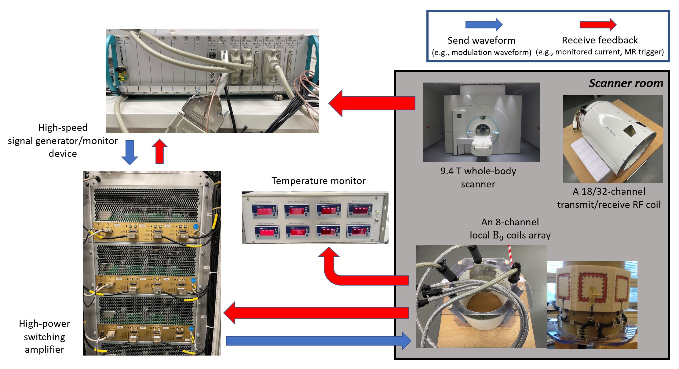
Figure 1. Our newly built 8-channel local B0 coil array system. During MR scans, where a 18/32-channel transmit/receive RF coil is inserted into the cylinder of the B0 coil array, the scanner sends triggers to the signal generator device, which transmits the designed waveform to the power amplifier and outputs to the B0 coil array. The modulation current in the B0 coils is monitored by the amplifier and the signal monitoring device for off-line reconstruction, and the temperature of each coil is continuously monitored during experiments for safety.
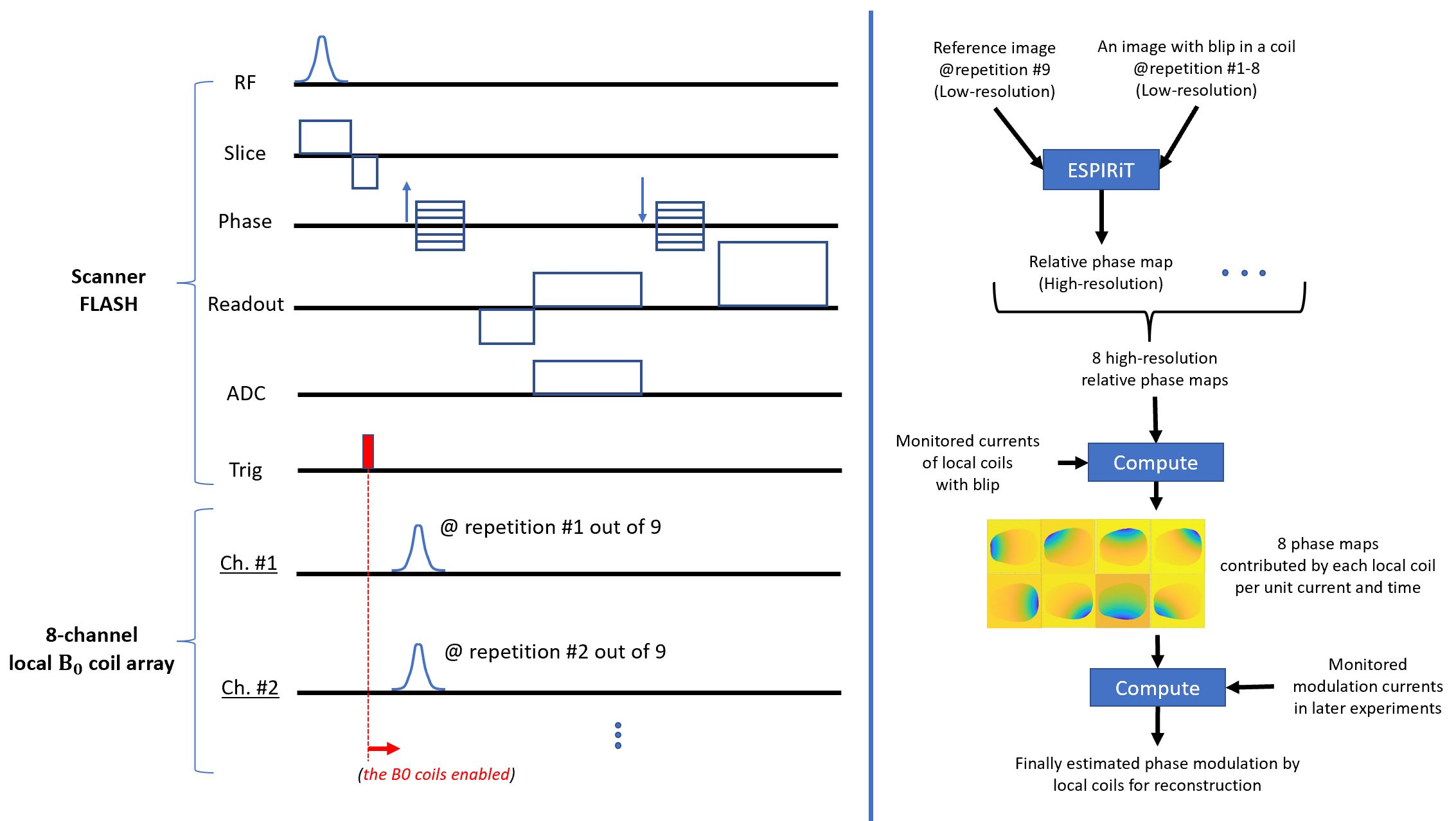
Figure 2. The pulse sequence diagram and the algorithm used to estimate the phase modulations by local B0 coils with monitored currents in real-time. Left: the pulse sequence diagram for nine FLASH scans, without or with an additional current blip in a channel of the local coils. Right: the signal processing flowchart to calculate the phase modulation, with the unit phase maps computed from the scans on the left, and the monitored modulation current recorded in later experiments.
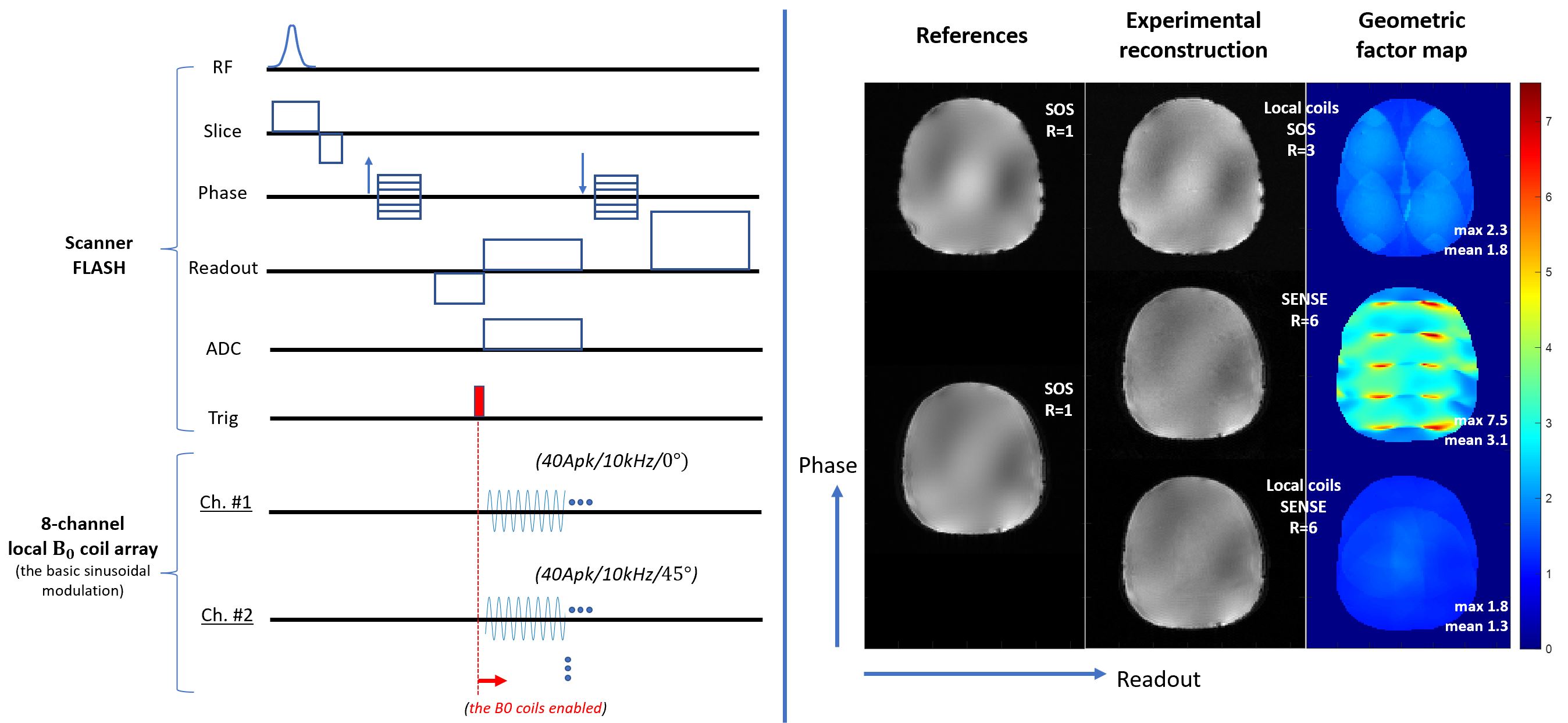
Figure 3. Experiments of accelerated scans with local B0 coils modulation only, and jointly with SENSE. Left: the pulse sequence diagram for the FLASH scans with the basic sinusoidal modulation of B0 coils. Right: The reconstructed images and the G maps. Columns left to right: the reference sum of square images, the reconstruction given reduction factor R, the G maps. Rows upper to bottom: B0 modulation alone and the sum of square coil combination given R=3, SENSE reconstruction without B0 modulation given R=6, and joint reconstruction of SENSE and B0 modulation given R=6.
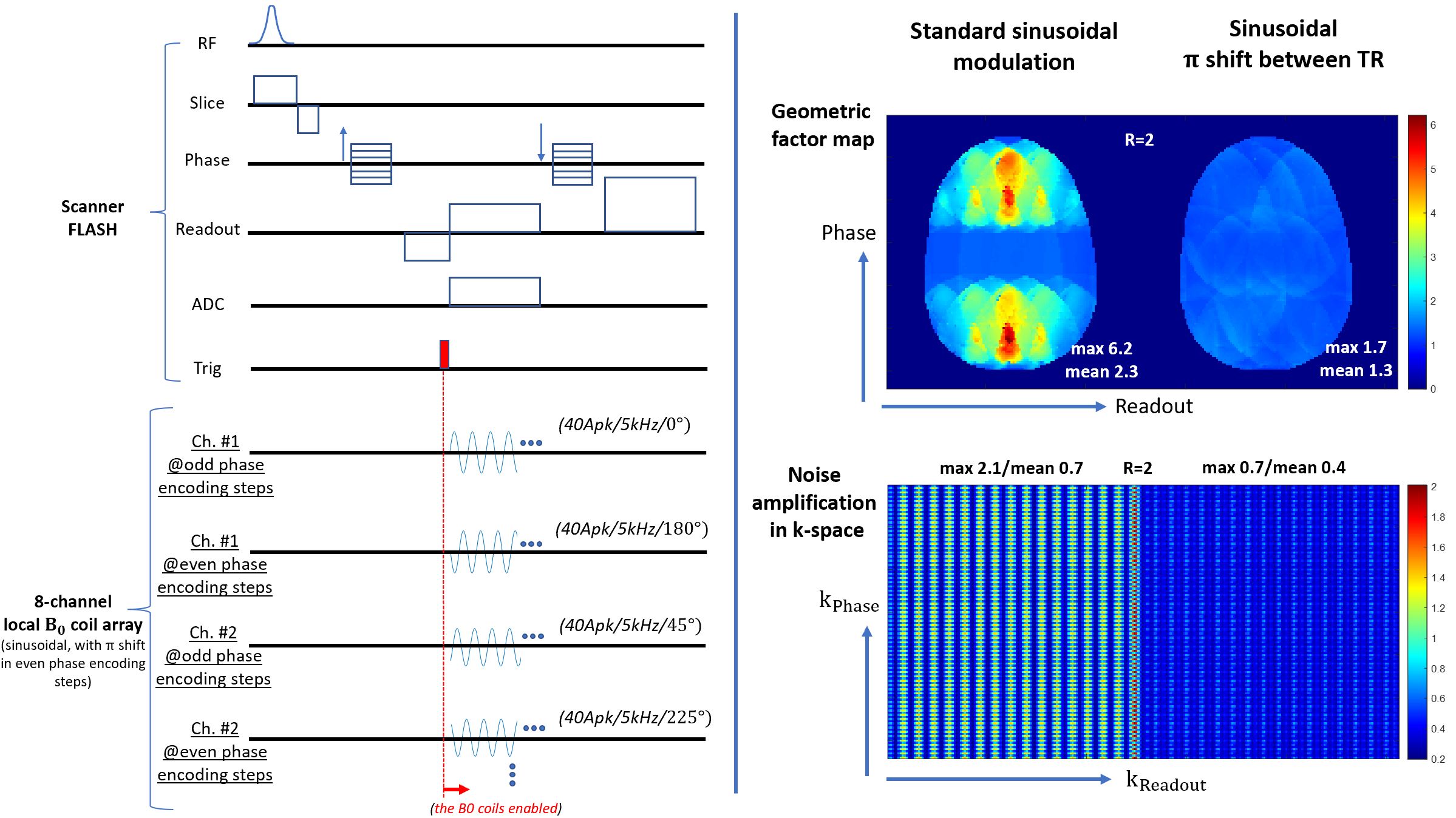
Figure 4. Simulated FLASH scans with 5kHz modulation of B0 coils. Left: The pulse sequence diagram for the scans with the basic sinusoidal modulation of B0 coils and the additional π shift for all coils between consecutive phase encoding steps. Right: The G maps (upper) and the noise amplification maps in k-space (bottom) for the basic sinusoidal modulation, and for the modulation with the π shift between consecutive phase encoding steps.
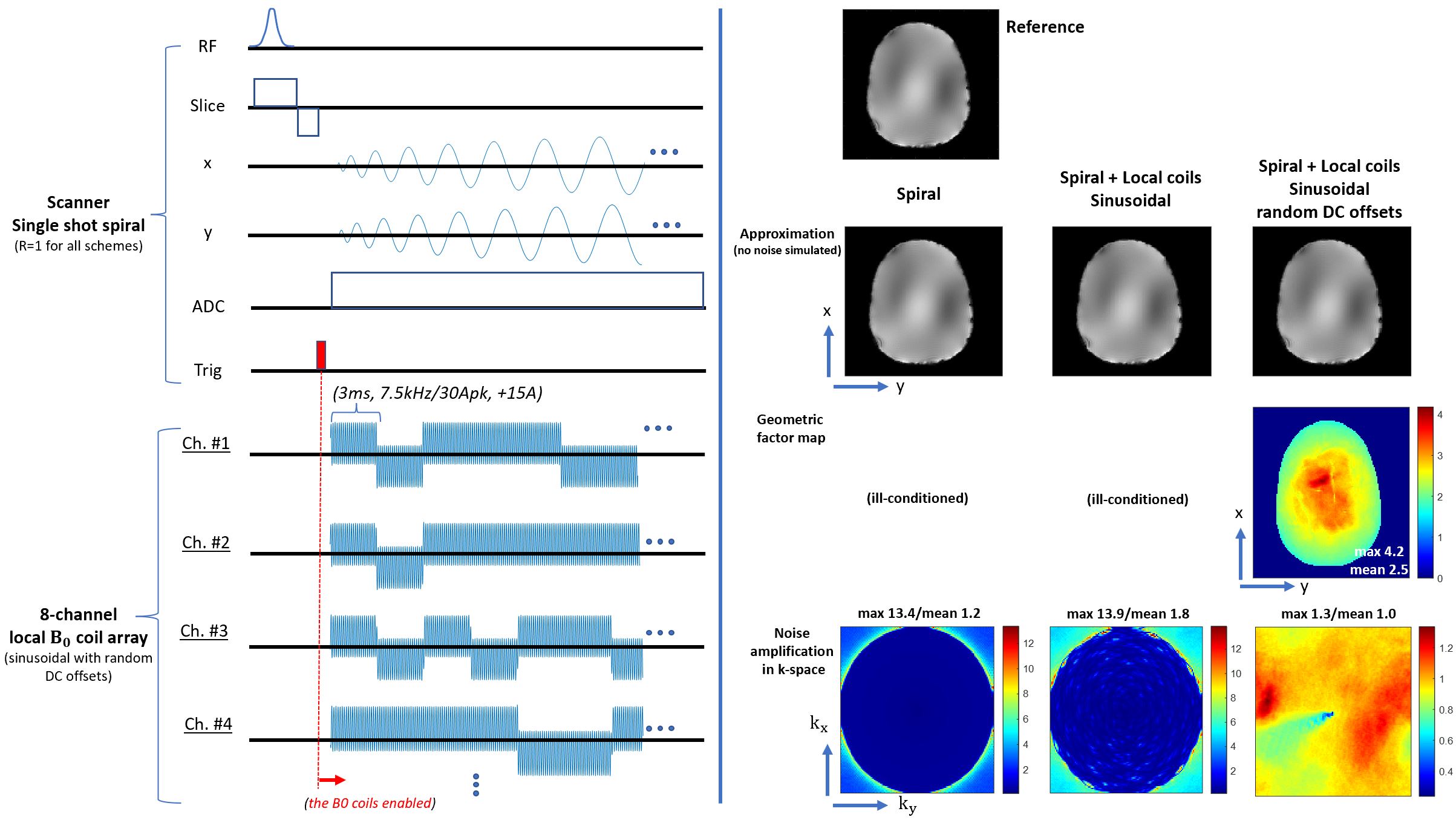
Figure 5. Simulated spiral acquisition with and without the modulations of B0 coils. Left: the pulse sequence diagram for a single shot scan of a spiral-out trajectory, with the sinusoidal modulation and random DC offsets of B0 coils. Right: the reconstructed images which approximate the object (i.e., without simulated noise), the G maps and the noise amplification maps in k-space, for the spiral acquisition alone, the spiral with the basic sinusoidal modulation of B0 coils, and the spiral with the sinusoidal modulation and random DC offsets, respectively.
DOI: https://doi.org/10.58530/2022/0388