0382
Progress on the commissioning of the Iseult 11.7T whole-body MRI: first images1University of Paris-Saclay, CEA, Irfu, Gif sur Yvette, France, 2University of Paris-Saclay, CEA, CNRS, BAOBAB, NeuroSpin, Gif sur Yvette, France, 3Siemens Healthcare GmbH, Erlangen, Germany, 4Siemens Healthcare SAS, Saint-Denis, France, 5Ensimtech, Buckenhof, Germany, 6Sorbonne University, Institut du Cerveau - Paris Brain Institute - ICM, INSERM, CNRS, Paris, France, 7ICM, Centre de NeuroImagerie de Recherche – CENIR, Paris, France
Synopsis
The Iseult project started in 2001 and is a collaborative effort involving CEA and the University of Freiburg in academics, Guerbet, Siemens Healthineers and Bruker Biospin as industrial partners. One central aspect of the project has been the design by CEA of a whole-body magnet of 11.7T field strength with a 90-cm wide bore. After nearly 20 years of research and development, prototyping, integration and tests, first images have been acquired with this unique MRI scanner. Some key validation results are presented here.
Introduction
MRI at ultra-high field (UHF) offers great promises in SNR and CNR and has fueled the race to achieve ever higher fields, the highest field used on humans to date being 10.5T at the University of Minnesota (CMRR). With higher fields however come greater challenges (e.g. field homogeneity and stability) which must be overcome to enable proper exploitation. Here we review the key results of the tests performed in the last year to finalize the commissioning of this unique 11.7T whole-body MRI scanner1, and we present its first high resolution images acquired on a pumpkin and on an ex-vivo brain.Methods
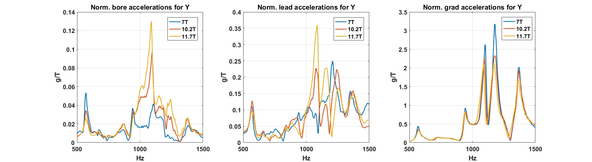
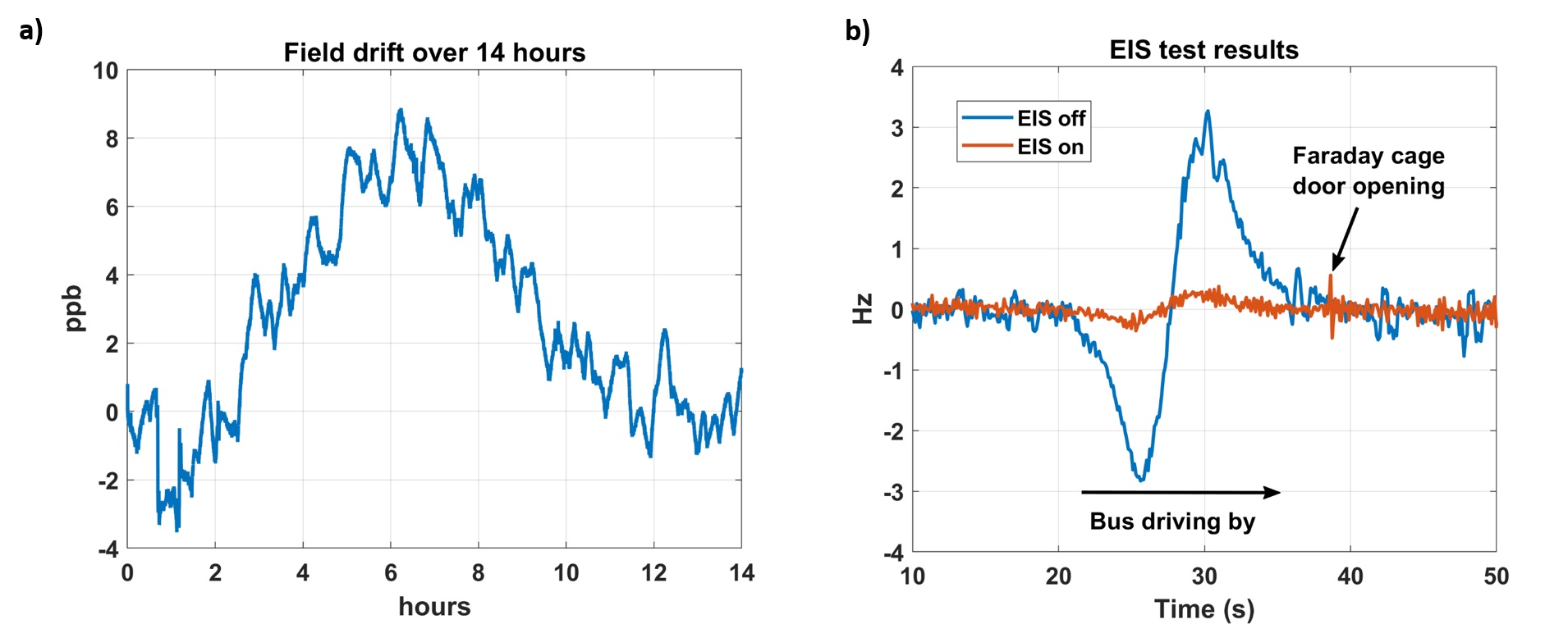
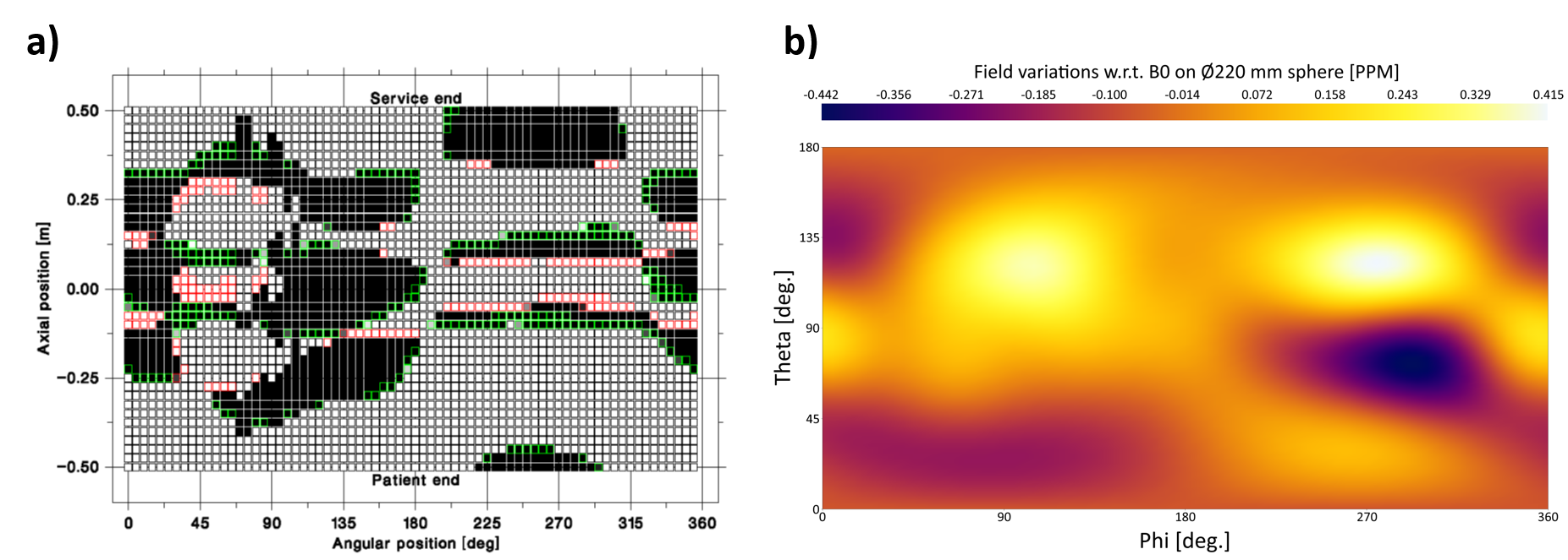
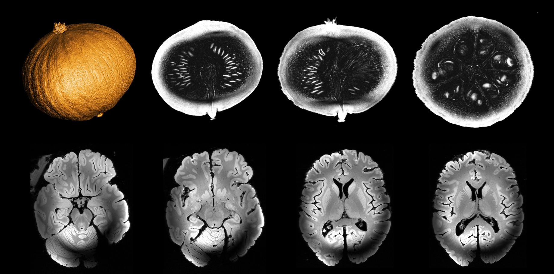
The main superconducting coil of the Iseult magnet is made of NbTi and is immerged into a 7000-liter He vessel at a temperature of 1.8 K (below the 2.17 K transition from liquid to superfluid He). Following a double pancake design, it sustains a current of 1483 A and has an inductance of 308 H, allowing to ramp-up the field from 0 to 11.7T in approximately 5 hours. This magnet is actively shielded by two 5-m diameter superconducting coils also immerged in superfluid He. The whole magnet assembly weighs 132 tons with external diameter and length of 5 m. After the cool down of the magnet and the first ramp-up at 11.7T in 2019, the imaging equipment (of the MAGNETOM 11.7T MRI series) has been integrated around the magnet. Gradient coil-magnet interaction tests were performed at 3T, 7T, 10.2T and 11.7T, the gradient being the Siemens Healthineers SC72 whole-body gradient. They included acoustics, vibration and power deposition measurements during variable speed frequency sweeps of the three gradient coils independently. Figure 1 reports some vibration data measured on the gradient, the lead tube (designed by CEA to screen gradient-magnet interactions at specific frequencies2) and the magnet cryostat. Temporal field stability (spec of 0.05 ppm/hour max at rest) was measured at 11.7T with an FID sequence over 14 hours on a phantom. The External Interference Shield (EIS) designed to screen external field perturbations was tested with a Skope (Skope MRT, Zurich, Switzerland) field camera with measurements performed at an intermediate field of 7T every 105 ms while a bus was driving on the road nearby. Field homogeneity (spec of 0.5 ppm peak-to-peak over the surface of a 22 cm diameter sphere with passive and cryo shimming only) was mapped by a Metrolab (Metrolab, Geneva, Switzerland) field camera. Tune-up and Quality Assessment of the imaging equipment, in quadrature mode only for the moment, was performed by Siemens Healthineers with support of CEA. First images were acquired on a pumpkin and an ex-vivo brain using a Gradient Echo sequence with a 0.4 mm isotropic spatial resolution (TR=20 ms, TE = 1.82/2.5 ms, Matrix = 512 × 512 × 448, 4 NEX, TA = 5 H 05 min), using the single channel service quadrature coil (QED) delivered by Siemens Healthineers.Results
Field stability measurement is shown in Fig. 2a revealing a fluctuation of less than 0.01 ppm over 14 hours. The EIS also reduces by 10 fold the field disturbance caused by a bus driving on the road 50 m from the magnet (Fig. 2b). Field homogeneity relies on passive shim iron (optimized schematics of Al/Iron blocks), cryoshims and the Siemens Healthineers active shim. The last measurement returns a peak-to-peak variation of 0.88 ppm and a RMS deviation of 0.15 ppm with the passive and (partial) cryo shim only (Fig. 3), the nominal frequency being 499.415 MHz. Despite a severe but expected3 RF field inhomogeneity at 500 MHz with a quadrature coil, these experimental conditions led to the visualization of exquisite details on high spatial resolution images of the pumpkin (e.g. vegetal fibers) and of the ex-vivo brain (e.g. internal structure of the thalamus), thus validating up to this stage the commissioning of the 11.7T Clinical MRI system.Discussion and conclusion
The results reported in this work reflect and validate the research and developments performed at CEA for nearly 20 years and now make MRI at 11.7T and with a 90 cm diameter bore a reality. The safety assessment of this new medical device will be started soon to seek authorization to scan human volunteers. Parallel transmission also will be deployed in the near future to mitigate the RF field inhomogeneity problem. Small refinements on the passive shim configuration are also needed to reach the final specifications.Acknowledgements
The Musée National d’Histoire Naturelle (Marc Herbin) in Paris is thanked for lending CEA the post-mortem brain for scanning. This part of the Iseult project has been funded by CEA and BPIfrance. NB is funded by the European Union’s Horizon 2020 research and innovation program under grant agreement No 885876 (AROMA project).References
[1] D. Le Bihan, T. Schild, Human Brain MRI at 500MHz, scientific perspectives and technological challenges, Supercond. Sci. Techno. 2017;30:1-19.
[2] G. Aubert. NMR imaging system with reduced cryogenic losses and reduced acoustic noise. US patent no 8,410,777 B2.
[3] A. Webb. Concepts in Magnetic Resonance Part A, 2011; 38A:148-184.
Figures