0364
High resolution diffusion-weighted MRI combining markerless prospective motion correction and locally low-rank constrained reconstruction1School of Biomedical Engineering, Shanghai Jiao Tong University, Shanghai, China, 2United Imaging Healthcare, Shanghai, China, 3The Weizmann Institute of Science, Rehovot, Israel
Synopsis
High-resolution diffusion-weighted MRI is susceptible to head movement artifacts and background phase variations. We investigate a crucial combination of prospective motion correction and Spatial-angular local low rank (SPA-LLR) constrained reconstruction to obtain robust multi-shot high-resolution diffusion-weighted MRI under substantial motion. Prospective motion correction with optical markerless motion tracking is used to remove artifacts and blurring due to bulk motion whereas locally low-rank regularization is used to correct remaining artifacts due to the background phase variations from scan to scan. We present the results in healthy adult volunteers, demonstrating high-resolution diffusion-weighted images and FA images under different motion patterns with PMC.
Introduction
Multi-shot diffusion-weighted imaging (MS-DWI) is a promising approach for enabling high spatial resolution and high-fidelity diffusion imaging [1]. Unfortunately, during the inherent diffusion-encoding process with strong diffusion gradients, subject tiny bulk motion and physiological effects cause background phase variations, resulting in aliasing artifacts and signal cancellation [2]. Some retrospective reconstruction schemes, such as SPA-LLR and MUSSELS, have been proposed to correct the artifacts but become invalid if there is subject head motion during the acquisition that leads to mismatches of images [3, 4]. This causes edge artifacts, severe blurring and even signal dropouts in the calculated diffusion maps, as well as errors in the tensor estimation in the case of diffusion tensor imaging [5]. Recently, robust prospective motion correction techniques are applied in DWI for real-time motion correction [6]. Here, we investigate a crucial combination of prospective motion correction (PMC) and Spatial-angular local low rank constrained reconstruction for multi-shot diffusion-weighted MRI under substantial motion. Prospective motion correction with optical markerless motion tracking is used to remove artifacts and image blurring due to bulk motion whereas locally low-rank regularization (LLR) is used to correct remaining artifacts due to the background phase variations from scan to scan. We demonstrate the performance in healthy adult volunteers on 3 Tesla MRI systems performing different motion patterns, especially with the ability of PMC to preserve the orientational information in DTI images.Methods
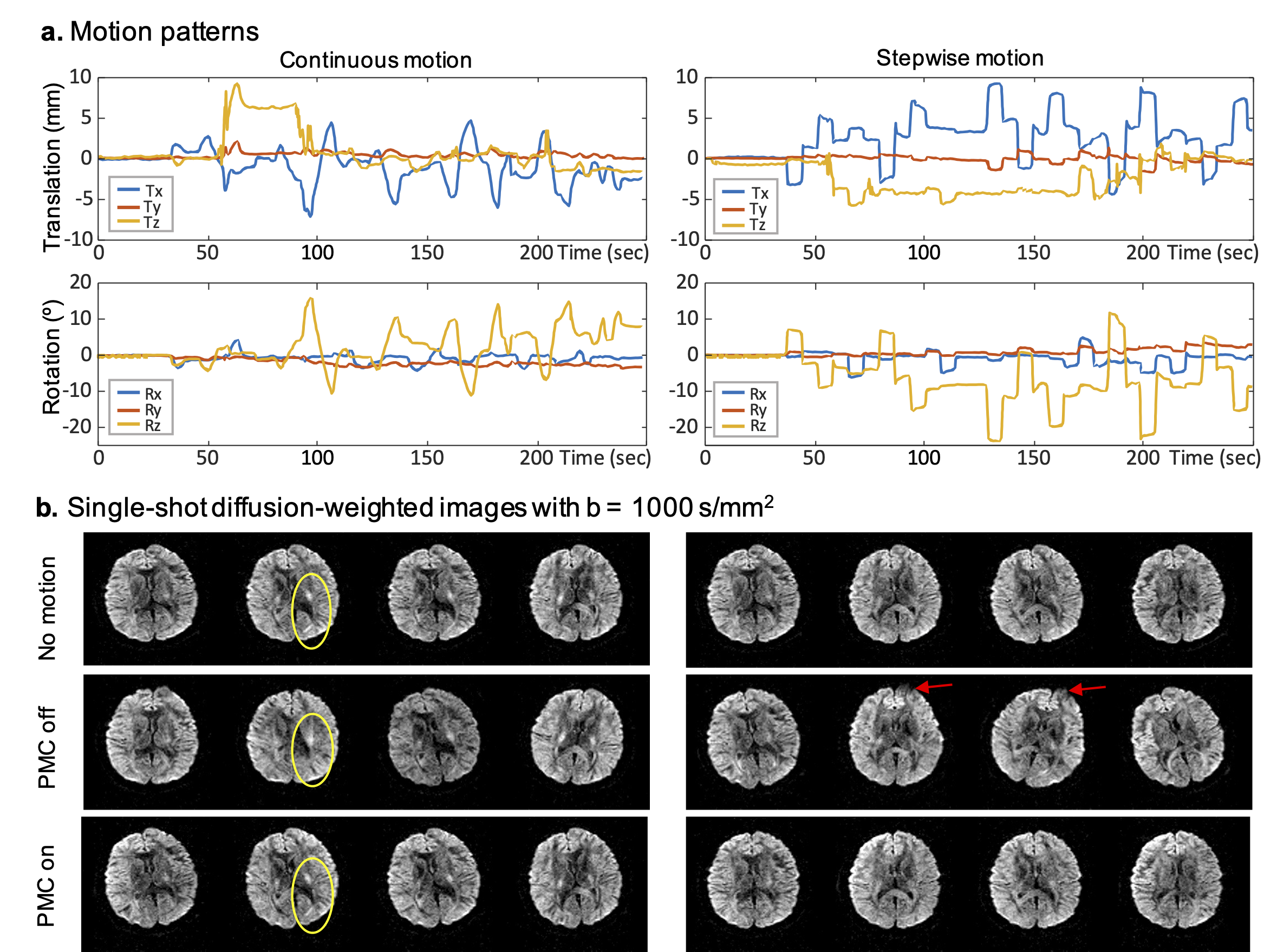
PMC camera and sequence control system: The PMC camera and sequence control system are shown in Fig. 1a. The camera records the images in stereo and transfers the data streaming to an FPGA-based computation implementation of fast 3D point cloud modeling of the face. Then, head motion is estimated at six degrees of freedom by a real-time 3D registration referring to an initial point cloud model. The RF, gradient and acquisitions are synchronous prospective updated with the 6-DOF to make the imaging volume “follows” the object in “real-time”. A threshold operation on the 6-DOF that the translation larger than 100 µm and rotation larger than 0.1º was implemented in the sequence.Imaging experiments: Single-shot and multi-shot DTI with PMC on/off under three different motion patterns (still, stepwise and continuous) was performed on a 3.0T United Imaging uMR890 system with ethical approval. Parameters of SS-DTI include 4000ms TR, 92ms TE, 220×220 mm2 FOV, 96×96 matrix size, 4mm slice thickness, 32 slices, 32 uniformly distributed diffusion directions, 1000 s/mm2 b-value. MS-DTI sequence includes 82ms TE, 160×160 matrix size and 4 shots.
Data analysis: The reconstruction pipeline is shown in Fig. 1b. Even odd ghost correction with inline three-echo navigations, SVD-based coil compression [7] and coil sensitivity estimation are done in the pre-processing step. A Spatial-Angular LLR regularization (SPA‐LLR) with SENSE reconstruction initialization was applied to the magnitude images from all diffusion-encoding directions to exploit the spatial and angular correlation substantially improves multi-direction DWI reconstruction, simultaneously removes motion-induced phase artifacts, and denoises images. The diffusion tensor model was then fitted using FSL’s “dtifit” function to derive the fractional anisotropy (FA) and the primary eigenvector (V1).
Results and Discussion
Figure 2 shows examples of the motion data that the camera captures and how they affect the single-shot EPI images with/without using the PMC system. The diffusion-weighted images from representative diffusion encoding directions acquired with PMC off (the second row in Fig. 2b) are moved in-plane and through-plane compared to the no motion reference (the first row in Fig. 2b) while the images stay well aligned across the multiple scanning with PMC on (the third row in Fig. 2b). Furthermore, the PMC can maintain the diffusion-weighted contrast sensitized to a fixed diffusion encoding gradient as marked by the yellow ellipses. Please note there are some distortion variations due to field inhomogeneity manifested in the front of the brain from different scans. However, the distortion variations with PMC on are smaller than the counterparts with PMC off as indicated by the red arrows.Figure 3 shows the results of single-shot DTI with and without PMC under the two above-mentioned motion patterns. The images magnitude-averaged and color FA across all the diffusion directions of a representative slice are severely blurred and deteriorated with PMC off but greatly improved with PMC on.
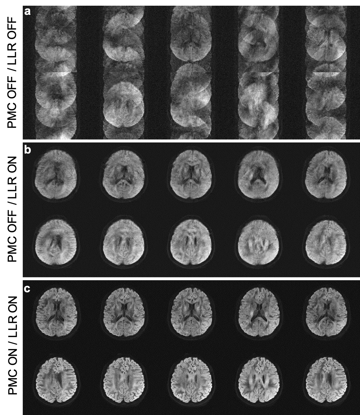
Figure 4 shows the necessity and feasibility to combine the PMC and LLR reconstruction in multi-shot diffusion-weighted MRI under substantial motion under continuous motion. Nyquist ghosts are severe due to the inconsistent background phases along the shot and diffusion direction dimensions without LLR regularized reconstruction. The aliasing is markedly reduced with LLR regularized reconstruction while the images are grievously blurred due to the motion effects without PMC. With PMC and LLR reconstruction, we obtained high-resolution and high-fidelity diffusion-weighted images.
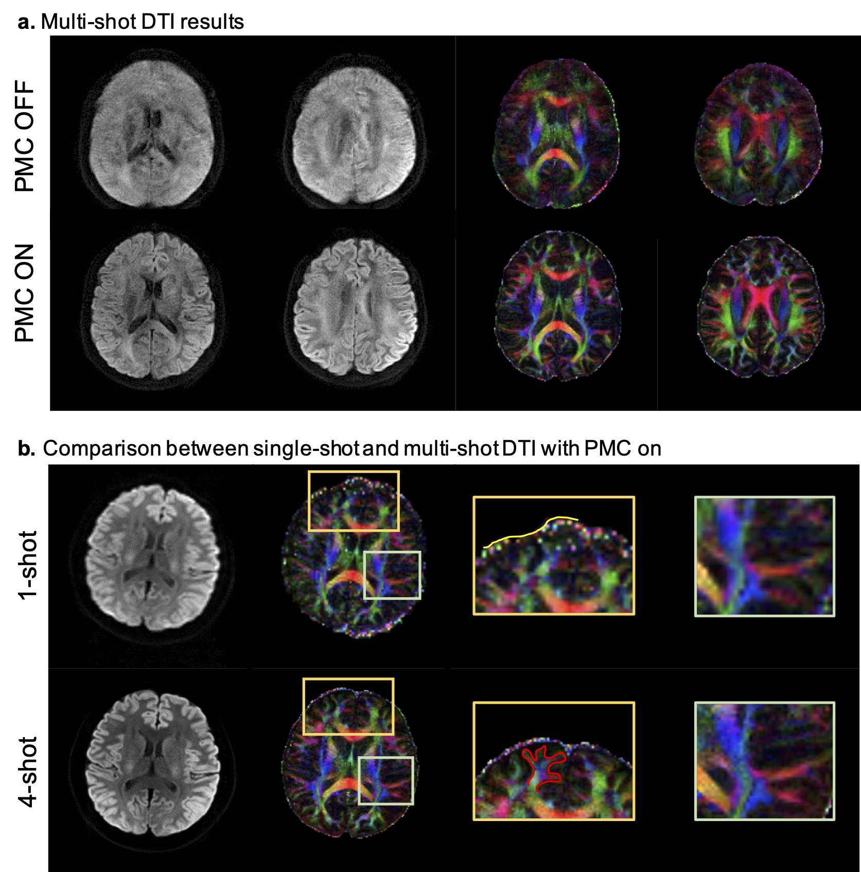
Figure 5a shows 4-shot diffusion-weighted images and color FA are severely blurred and degraded without PMC but improved a lot with PMC. Figure 5b shows comparisons of single-shot and multi-shot DTI under continuous motion. Small structures such as branches are clearly identified and the distortion is mitigated in multi-shot DTI.
Conclusions
We propose a crucial combination of prospective motion correction and spatial-angular local low rank constrained reconstruction to obtain robust high-resolution diffusion-weighted MRI under substantial motion.Acknowledgements
This work is supported by National Natural Science Foundation of China National Science Foundation of China (No. 62001290) and sponsored by Shanghai Sailing Program (20YF1420900). We thank Dr. Yuxin Hu for sharing reconstruction code, and Dr. Jon Tamir for helpful discussions.References
[1] Mani, M., Jacob, M., Kelley, D., Magnotta, V., 2017. Multi-shot sensitivity-encoded diffusion data recovery using structured low-rank matrix completion (MUSSELS). Magnetic Resonance in Medicine 78, 494-507.
[2] Eichner, C., Cauley, S.F., Cohen-Adad, J., Möller, H.E., Turner, R., Setsompop, K., Wald, L.L., 2015. Real diffusion-weighted MRI enabling true signal averaging and increased diffusion contrast. NeuroImage 122, 373-384.
[3] Hu, Y., Wang, X., Tian, Q., Yang, G., Daniel, B., McNab, J., Hargreaves, B., 2020. Multi-shot diffusion-weighted MRI reconstruction with magnitude-based spatial-angular locally low-rank regularization (SPA-LLR). Magnetic Resonance in Medicine 83, 1596-1607.
[4] Mani, M., Aggarwal, H.K., Magnotta, V., Jacob, M., 2020. Improved MUSSELS reconstruction for high-resolution multi-shot diffusion weighted imaging. Magnetic Resonance in Medicine 83, 2253-2263.
[5] Hoinkiss, D.C., Porter, D.A., 2017. Prospective motion correction in diffusion-weighted imaging using intermediate pseudo-trace-weighted images. NeuroImage 149, 1-14.
[6] Berglund, J., van Niekerk, A., Rydén, H., Sprenger, T., Avventi, E., Norbeck, O., Glimberg, S.L., Olesen, O.V., Skare, S., 2021. Prospective motion correction for diffusion weighted EPI of the brain using an optical markerless tracker. Magnetic Resonance in Medicine 85, 1427-1440.
[7] Zhang, T., Pauly, J.M., Vasanawala, S.S., Lustig, M., 2013. Coil compression for accelerated imaging with Cartesian sampling. Magnetic Resonance in Medicine 69, 571-582.
Figures