0359
Multi-shot DW-EPI using a modified interleaving scheme and second-order motion-compensated diffusion sensitization1Institute for Biomedical Engineering, ETH Zurich and University of Zurich, Zurich, Switzerland
Synopsis
Multi-shot techniques for diffusion MRI are desirable because of the associated improvement potential in image resolution but are inhibited by phase variability across interleaves, which results from subject motion and causes ghosting. Here, an EPI-based acquisition strategy is devised which diminishes the ghosting pattern by employing a modified interleaving scheme and mitigates motion sensitivity by nulling the first- and second-order moments of the diffusion-sensitizing gradient waveforms. This solution yielded unperturbed, high-resolution images in in vivo human brain scans and clear improvements in image quality compared to acquisitions incorporating standard interleaving and uncompensated diffusion sensitization.
Introduction
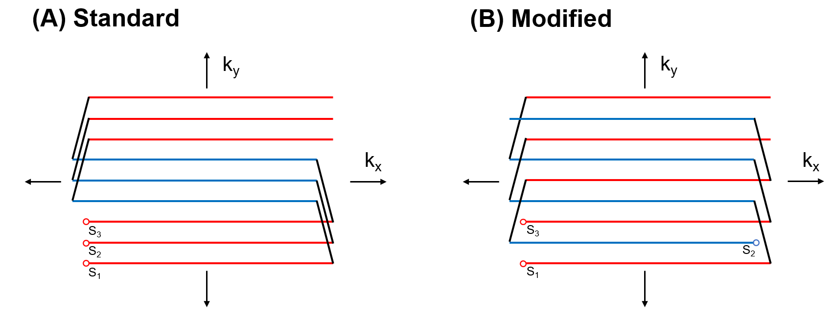
Diffusion-weighted imaging (DWI) is a powerful diagnostic tool but is predominantly combined with single-shot EPI acquisitions, thereby limiting the attainable resolution. Segmented acquisitions can provide improved resolution but are hindered by motion-induced shot-to-shot phase variability, which leads to ghosting in reconstructed images when neglected.1 One promising approach for resolving this issue is to suppress phase variations by utilizing diffusion-sensitizing gradient sequences with nulled moments (i.e., motion-compensation); this method yielded high quality images for interleaved spiral acquisitions.2 Translation of this approach to EPI, however, may be nontrivial because interleaved EPI acquisitions suffer trains of ghosts for standard interleaving (i.e., when all interleaved k-space trajectories begin at the same corner of k-space) even in the absence of phase fluctuations.3 That said, ghosting can be reduced by employing a modified interleaving scheme (particularly, for acquisitions with an odd number of interleaves) in which even and odd interleaves begin at opposite corners of k-space, thereby returning the sampling pattern to that of single-shot EPI.4In light of these considerations, the aim of this work is to evaluate the feasibility of multi-shot DW-EPI in conjunction with second-order motion-compensated diffusion gradients and the modified EPI interleaving scheme.
Methods
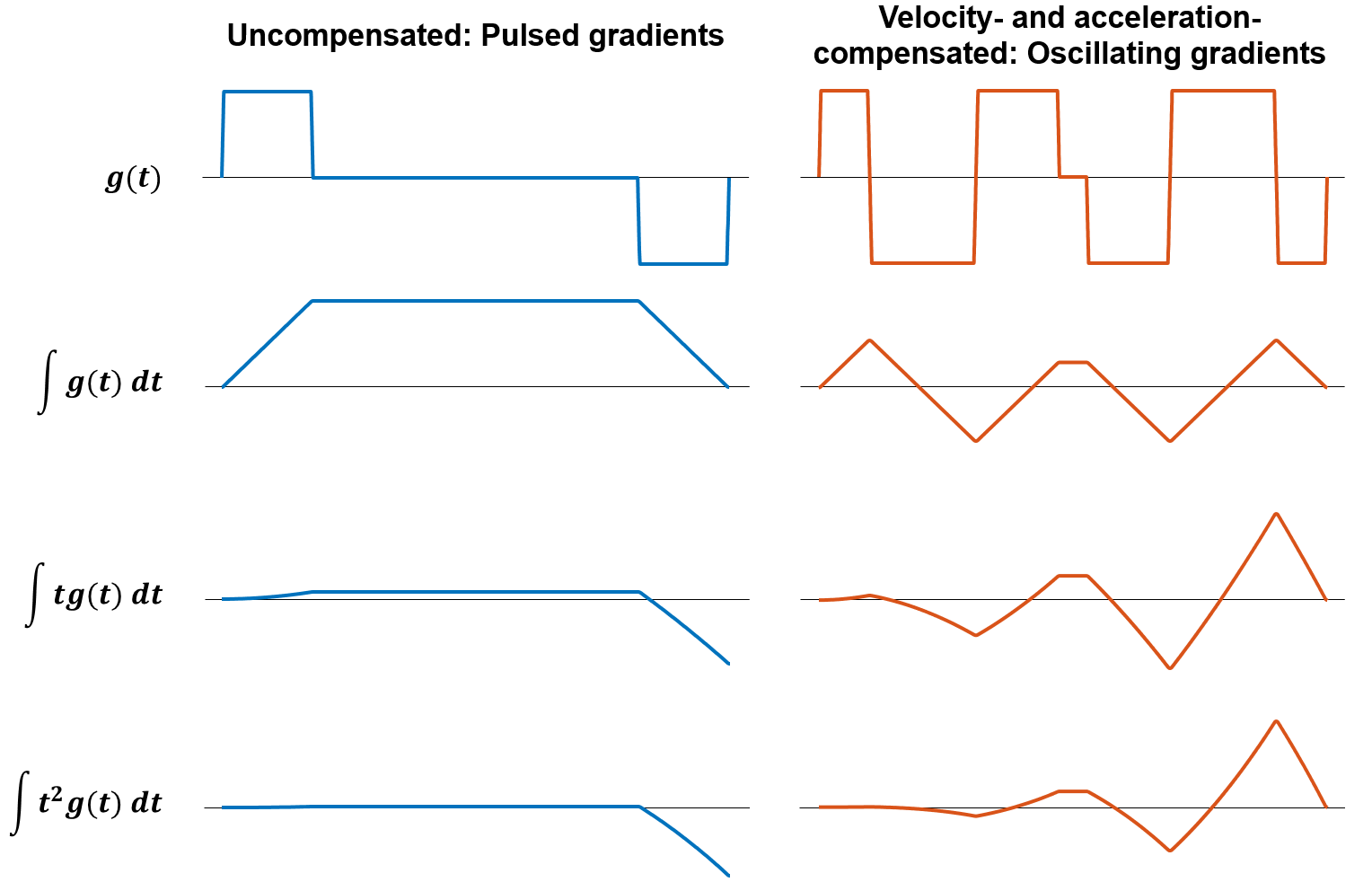
Scanning was performed using a 3T Philips Achieva system (Philips Healthcare, Best, the Netherlands) with a high-performance gradient insert that could reach gradient amplitudes up to 100 mT/m and slew rates up to 1200 mT/m/ms at 100% duty cycle.5 To capture the dependence of imaging performance on the interleaving scheme, a stationary silicon oil phantom was scanned using pulsed gradient spin-echo (PGSE) sequences, which are uncompensated, combined with three-shot EPI readouts with standard and modified interleaving schemes, which are depicted in Figure 1. Subsequently, to examine the utility of motion-compensated diffusion gradients in conjunction with modified interleaving, two healthy adult volunteers were scanned using the modified three-shot EPI acquisitions combined with pulsed gradients and velocity- and acceleration-compensated oscillating diffusion gradients2 derived from improved oscillating gradient spin-echo (OGSE) waveforms.6 Figure 2 depicts the two gradient shapes as well as nulled higher-order moments of the oscillating gradients, which corresponds to the desired motion compensation. Identical imaging parameters were used across all acquisitions: 1.5 mm in-plane resolution, 10 slices, 3 mm slice thickness, 2 mm slice gap, b = 800 s/mm2, two b = 0 acquisitions and 3 DWI directions (each aligned with a Cartesian coordinate axis) with 2 repetitions each, and TR/TE = 4000/103 ms.All scans were repeated using a Dynamic Field Camera7 (Skope Magnetic Resonance Technologies, Zurich, Switzerland) to monitor the spatiotemporal field dynamics associated with the imaging gradients; third-order spherical harmonic models were fitted to these data and were utilized in an algebraic reconstruction algorithm,8 which also incorporated off-resonance maps. Reconstructed images were smoothed with Hamming filters to reduce Gibbs ringing, after which maps of mean diffusivity (MD) were computed as the average diffusivity of the three diffusion gradient directions.
Results
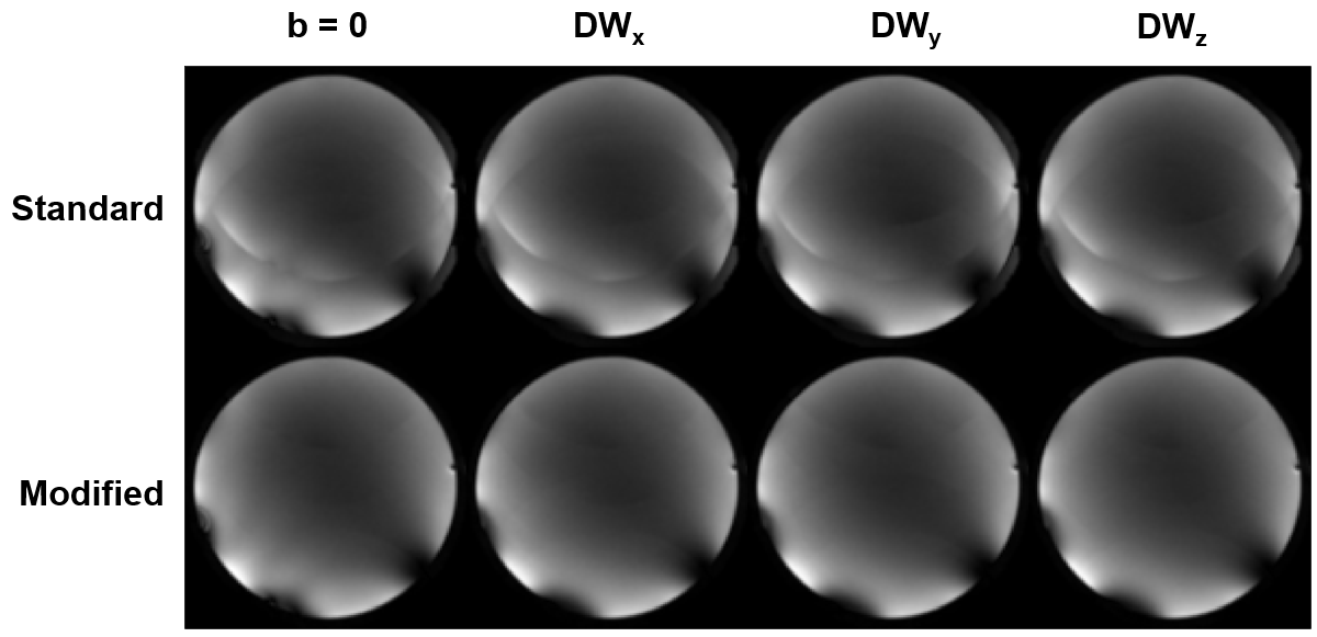
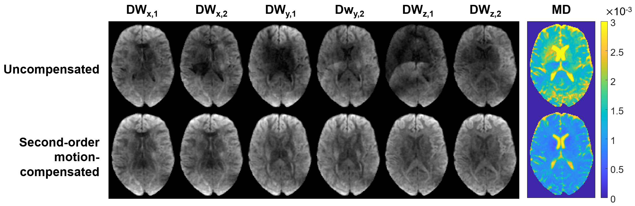
Figure 3 depicts phantom images acquired with standard and modified interleaving schemes for b = 0 and for each diffusion gradient direction; multiple ghosts are visible for standard interleaving but become highly suppressed for the modified interleaving. The combination of the modified scheme with pulsed and second-order motion-compensated diffusion gradients is shown in Figure 4 for a slice of one volunteer, which depicts repetitions of diffusion-weighted images for each gradient direction as well as mean diffusivity (MD) maps. In the acceleration-compensated case, all diffusion-weighted images lack discernible artifacts and yield a high quality MD map, whereas all but one uncompensated image have some level of ghosting or signal dropout, which deteriorate the MD map.Discussion
The reduction in the number of visible ghosts for acquisitions with modified interleaving compared to those with standard interleaving can be attributed to the favorable k-space sampling pattern of the former,4 which yields only very faint ghosts at FOV/2, like single-shot EPI. The suppression of this residual ghost is likely a byproduct of the higher-order field model, without which stronger ghosting would have occurred.In the subsequent diffusion-weighted in vivo images, the visible ghosting for the uncompensated diffusion gradients again only appears at FOV/2, whereas the signal dropouts, which appear near CSF in the ventricles and lead to falsely elevated MD values, do not appear patterned and are likely caused by phase variability induced by pulsatile motion.9 In the motion-compensated case, for which constant velocity and acceleration motion is not encoded into phase, the repeated acquisitions do not contain discernible forms of either artifact, thereby obviating the need for a reconstruction method dedicated to accounting for shot-to-shot phase variability.1,10
Compensating for motion of increasing orders inherently reduces the encoding efficiency and is responsible for the relatively long TEs. However, more efficient methods for motion compensation exist;11 future work should investigate such approaches with this implementation, which may enable a sufficient b-value/TE combination for translation to a standard gradient system.
Conclusion
This study demonstrates the feasibility of the multi-shot DW-EPI implementation utilized here, which combined acceleration-compensated diffusion sensitization and a modified interleaving scheme and provided images of high quality without a reconstruction method dedicated to the typical issues of interleaved DWI.Acknowledgements
No acknowledgement found.References
1. Chen N-k, Guidon A, Chang H-C, Song AW. A robust multi-shot scan strategy for high-resolution diffusion weighted MRI enabled by multiplexed sensitivity-encoding (MUSE). Neuroimage. 2013;72:41-47.
2. Michael ES, Hennel F, Pruessmann KP. Multi-shot diffusion MRI of the human brain with motion-compensated oscillating gradients. In Proceedings of the 2021 ISMRM & SMRT Annual Meeting, 2021. Abstract #1324.
3. McKinnon GC. Ultrafast interleaved gradient-echo-planar imaging on a standard scanner. Magn Reson Med. 1993;30(5):609-616.
4. Hennel F. Image-based reduction of artifacts in multishot echo-planar imaging. J Magn Reson. 1998;134(2):206-213.
5. Weiger M, Overweg J, Rösler MB, et al. A high-performance gradient insert for rapid and short-T2 imaging at full duty cycle. Magn Reson Med. 2018;79(6):3256-3266.
6. Hennel F, Michael ES, Pruessmann KP. Improved gradient waveforms for oscillating gradient spin-echo (OGSE) diffusion tensor imaging. NMR Biomed. 2021;34(2):e4434.
7. Dietrich BE, Brunner DO, Wilm BJ, et al. A field camera for MR sequence monitoring and system analysis. Magn Reson Med. 2016;75(4):1831-1840.
8. Wilm BJ, Barmet C, Pavan M, Pruessmann KP. Higher order reconstruction for MRI in the presence of spatiotemporal field perturbations. Magn Reson Med. 2011;65(6):1690-1701.
9. Miller KL, Pauly JM. Nonlinear phase correction for navigated diffusion imaging. Magn Reson Med. 2003;50(2):343-353.
10. Mani M, Jacob M, Kelley D, Magnotta V. Multi-shot sensitivity-encoded diffusion data recovery using structured low-rank matrix completion (MUSSELS). Magn Reson Med. 2017;78(2):494-507.
11. Aliotta E, Wu HH, Ennis DB. Convex optimized diffusion encoding (CODE) gradient waveforms for minimum echo time and bulk motion–compensated diffusion-weighted MRI. Magn Reson Med. 2017;77(2):717-729.
Figures