0319
Maxwell Field Compensation for 2D Spiral-Ring Turbo Spin-Echo Imaging at 0.55T and 1.5T1Biomedical Engineering, University of Virginia, Charlottesville, VA, United States, 2Cardiovascular Branch, Division of Intramural Research, National Heart, Lung, and Blood Institute, National Institutes of Health, Bethesda, MD, United States, 3Radiology & Medical Imaging, University of Virginia, Charlottesville, VA, United States
Synopsis
Concomitant (Maxwell) fields are problematic for TSE imaging when the readout waveforms vary along the echo train, leading to severe signal dropouts and image blurring. These effects are exaggerated when using prolonged, high-gradient amplitude readouts at lower magnetic-field strengths. The purpose of this work was to implement 2D TSE imaging using annular spiral rings with compensation of self-squared concomitant-gradient terms at the echo time and across echo spacings via gradient waveform modifications, and with compensation of the residual Maxwell- and B0-field induced phase accruals along the readout via image reconstruction. This method reduced artifacts at both 0.55T and 1.5T.
Introduction:
Spiral-based TSE techniques have shown advantages over conventional Cartesian TSE at high fields (3T), in terms of SNR efficiency, improved image contrast, and reduced SAR1,2. However, for TSE imaging at lower field strengths, concomitant-gradient effects may induce artifacts because Maxwell-terms scale inversely with the field strength. Maxwell-field phase errors induced by differences in spiral readouts along the echo-train may disturb the TSE signal pathway and violate the CPMG condition3, thus resulting in severe signal loss that cannot be recovered in image reconstruction.Researchers have investigated Maxwell compensation for interleaved-spiral TSE imaging1,4, and recently Mugler et al. proposed a general method to achieve the compensation of self-squared Maxwell-field terms by modifying gradient-waveforms along the entire echo-train for interleaved-spiral T2-weighted 2D-TSE imaging with different trajectory types4. Promising results show this approach enables Maxwell compensation for spiral trajectory types that are temporally asymmetric, such as spiral-out.
In this work, 2D-TSE imaging using annular-spiral-rings2 (SPRING-RIO TSE) was developed that incorporates gradient-waveform modifications to compensate the self-squared Maxwell-terms at the echo time and over echo spacings, with an image reconstruction method to correct for residual Maxwell and B0-inhomogeneity induced phase accruals along the trajectory.
Methods:
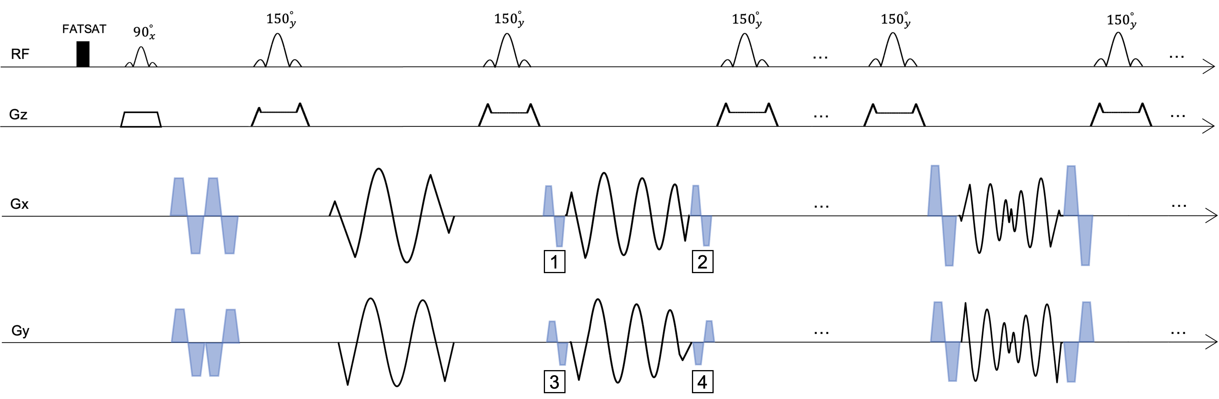
Sequence-based Maxwell field-corrections: The goal of gradient-waveform modifications was to eliminate the phase shift from self-squared terms at the k-space center, and to reduce the difference in phase shifts between echoes targeting a constant phase shift at the end of every echo. The implementation was accomplished (Fig.1) as follows:1) For each shot, the maximum Maxwell integral $$$M$$$ for each gradient axis was determined from the spiral-ring with the highest gradient amplitude.
2) Bipolar-gradient pairs were added at both the beginning and end of each remaining echo spacing (i.e., two pairs for each axis) to increase the Maxwell integrals. The gradient amplitudes were determined by subtraction of the Maxwell integral $$$M_{i}$$$ for the current $$$i$$$th ring from $$$M$$$.
3) The polarity of one of the four bipolar-gradient pairs in each echo spacing was set to be the opposite of the others for self-balancing the quadratic cross-terms5 induced by the added bipolar gradients. Additional time was added to the echo spacing as needed to achieve compensation, and the final Maxwell integral at the end of each echo spacing was designed to be a constant value of $$$\frac{M}{2}$$$.
Reconstruction-based Maxwell field-corrections: The goal of the image reconstruction method was to further reduce the residual phase errors from Maxwell-gradients6 and off-resonance effects accrued during the readout.
1) For the axial scan case, the data from each ring was demodulated by multiplying its own time-dependent Maxwell phase shift by the factor of $$$e^{-\phi_c{(z,t)}}$$$, where $$$\phi_c{(z,t)}=\frac{\gamma z^2}{2 B_0}\int(g_x^2+g_y^2)\ dt$$$.
2) For sagittal and coronal orientations, the data was first demodulated as indicated for the axial plane, followed by multi-frequency interpolation7 to mitigate the in-plane blurring caused by spatial- and time-dependent Maxwell phase error.
3) A semi-automatic deblurring method8 was used for off-resonance correction.
Experiments were performed on 1.5T (MAGNETOM Avanto) and 0.55T9 (prototype MAGNETOM Area) MR scanners (Siemens Healthcare, Erlangen, Germany). For phantom and human studies, images were acquired using SPRING-RIO TSE with and without compensation, and Cartesian TSE as a reference.
Results and Discussion:
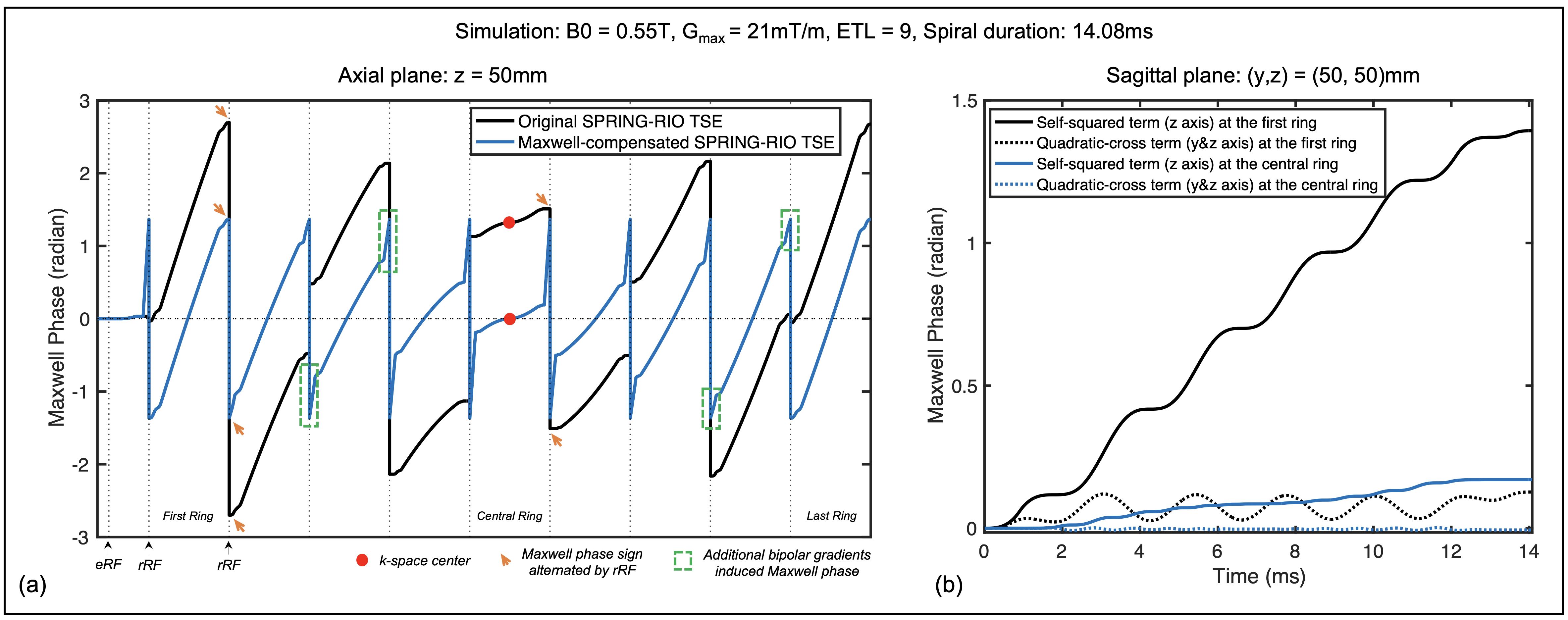
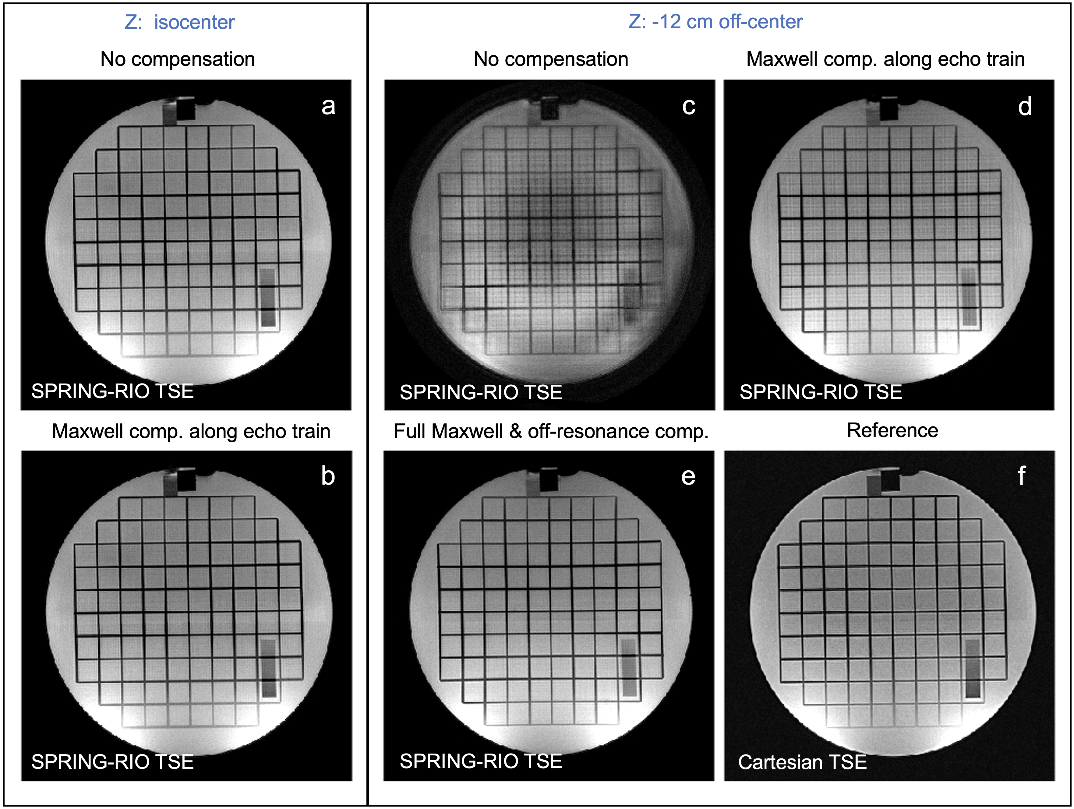
Fig. 2a shows the simulation of the Maxwell phase pathway from self-squared terms along the echo-train for SPRING-RIO TSE with (blue) and without (black) sequence modifications (axial plane). After adding compensation gradients, the accrued phase for each echo spacing starts at $$$-\phi$$$ and ends at $$$\phi$$$, where $$$\phi$$$ is a constant value, and the phase at the k-space center (spin echo) is zero. The green dashed boxes indicate examples of increased Maxwell phase by added bipolar gradients. Fig. 2b shows that the first ring produces the largest Maxwell phase accrual while the central ring has the smallest value. For the sagittal scan example, the self-squared phase terms are substantially larger than the quadratic-cross phase terms.Fig. 3 illustrates the effectiveness of the proposed Maxwell compensation via sequence modification and image reconstruction in a phantom study at 1.5T. While both uncompensated and compensated axial images show no significant artifacts at isocenter (Fig.3a-b), the uncompensated image (Fig.3c) shows substantial artifacts at z = -12 cm. With Maxwell compensation along the echo-train, no signal loss and reduced artifacts are seen (Fig.3d). Performing further Maxwell and off-resonance phase correction during image reconstruction removes residual artifacts (Fig.3e).
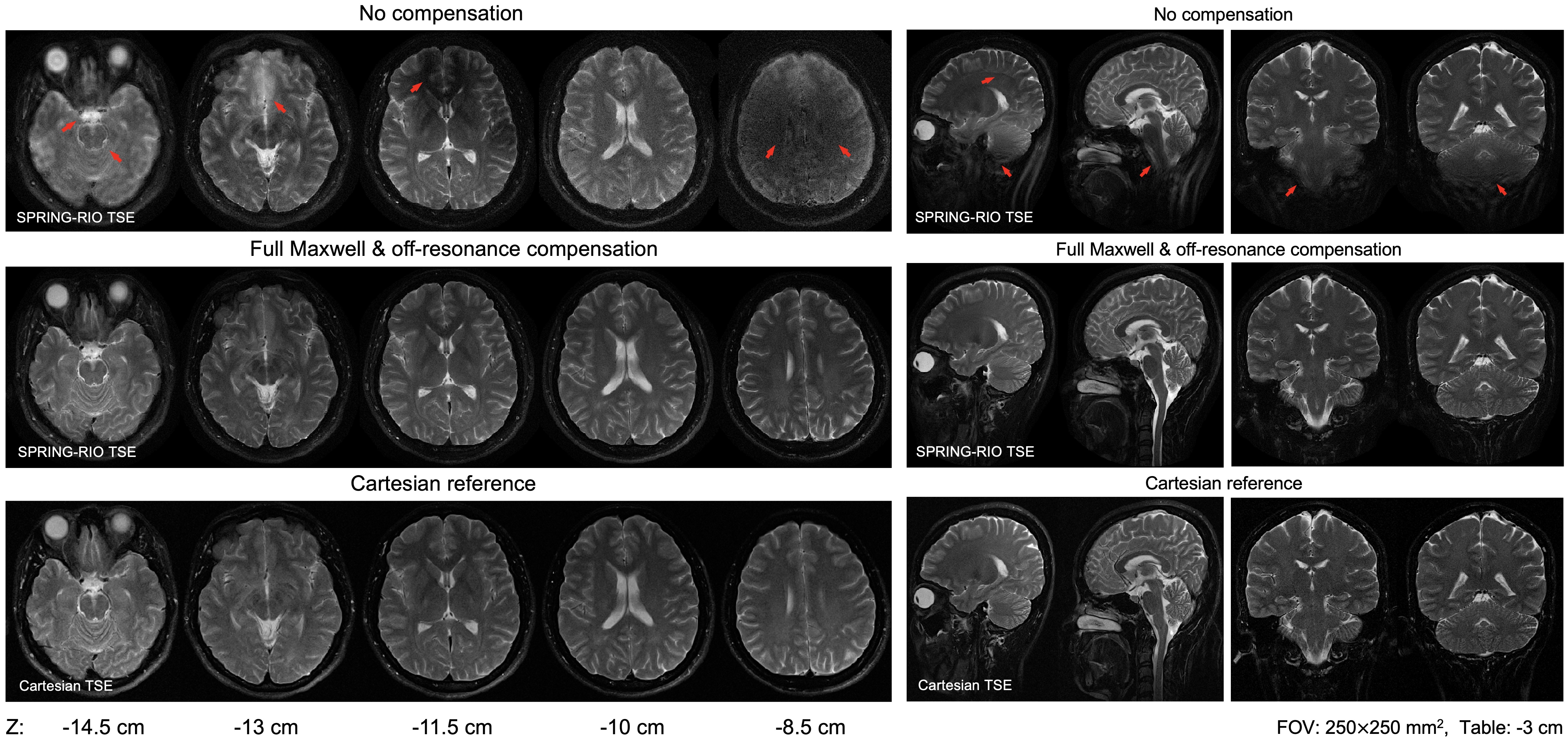
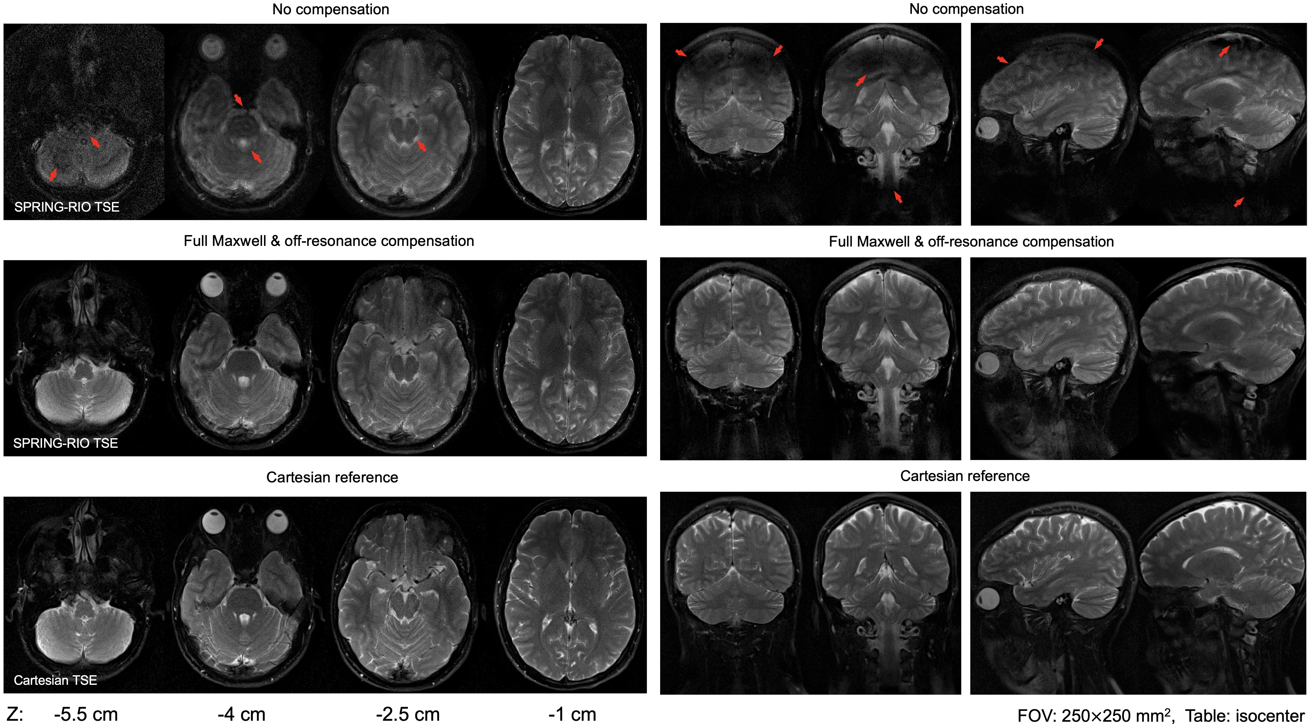
Fig. 4 and Fig. 5 show the image quality obtained in-vivo from SPRING-RIO TSE, with no compensation, or with full Maxwell and off-resonance compensation, compared to that for Cartesian TSE at 1.5T (Fig.4) and 0.55T (Fig.5), respectively. The uncompensated images at different off-center locations and slice orientations show image degradation, while the fully compensated images show similar image quality when compared to Cartesian TSE.
Conclusion and future work:
We demonstrated a spiral-ring T2-weighted 2D-TSE pulse sequence that incorporates sequence modification and image reconstruction to mitigate the image degradation associated with concomitant-gradient effects at 0.55T and 1.5T. This approach can be extended to compensate Maxwell-gradient-induced effects for TSE imaging with other asymmetric trajectory types. Further studies are needed to determine whether quadratic-cross Maxwell-terms will also require full compensation to obtain high image quality, and to optimize the sequence parameters to improve acquisition efficiency when including Maxwell compensation.Acknowledgements
The authors would like to acknowledge the assistance of Siemens Healthcare in the modification of the MRI system for operation at 0.55T under an existing cooperative research agreement (CRADA) between NHLBI and Siemens Healthcare. This work was supported in part by Siemens Healthcare.References
[1] Li Z, Karis JP, Pipe JG. A 2D spiral turbo-spin-echo technique. Magn Reson Med. 2018;80:1989–1996.
[2] Wang Z, Allen S, Feng X, Mugler JP, Meyer CH. SPRING-RIO TSE: 2D T2-Weighted Turbo Spin-Echo Brain Imaging using SPiral RINGs with Retraced In/Out Trajectories. Proceedings of the ISMRM 29th Annual Meeting, Virtual Conference. 2021:0837.
[3] Zhou XJ, Tan SG, Bernstein MA. Artifacts induced by concomitant magnetic field in fast spin-echo imaging. Magn Reson Med 1998; 40:582-591.
[4] Mugler JP, Campbell-Washburn AE, Ramasawmy R, Pfeuffer J, Meyer, CH. Maxwell Compensation for Spiral Turbo-Spin-Echo Imaging. Proceedings 29th Annual Meeting ISMRM, Virtual meeting. 2021:0003.
[5] Bernstein MA, Zhou XJ, Polzin JA, et al. Concomitant gradient terms in phase contrast MR: analysis and correction. Magn Reson Med 1998; 39:300-308.
[6] King KF, Ganin A, Zhou XJ, Bernstein MA. Concomitant gradient field effects in spiral scans. Magn Reson Med 1999; 41:103-112.
[7] Man LC, Pauly JM, Macovski A. Multifrequency interpolation for fast off-resonance correction. Magn Reson Med 1997; 37:785–792.
[8] Chen W, Meyer CH. Semiautomatic off‐resonance correction in spiral imaging. Magn Reson Med. 2008;59:1212–1219.
[9] Campbell-Washburn AE et al. Opportunities in interventional and diagnostic imaging by using high-performance low-field-strength MRI. Radiology 2019; 293:384-393.
Figures