0318
Fat-Separated Radial 3D bSSFP Imaging at Low Field Using Frequency-Sweep RF Saturation and XD-GRASP Reconstruction1Center for Biomedical Imaging, Department of Radiology, NYU School of Medicine, New York, NY, United States
Synopsis
Low-field systems have recently gained interest because of the potential to make MRI more accessible, but the field reduction comes at expense of low SNR and challenges with fat suppression. This work describes a new concept for fat separation at low field, based on radial acquisition during frequency-sweep RF saturation and combined with frequency-resolved XD-GRASP reconstruction. The approach is demonstrated for free-breathing abdominal imaging using a radial stack-of-stars 3D bSSFP sequence at 0.55T, showing that fat and water can be reliably distinguished based on the response to saturation at different frequencies. Fat-suppressed composite images can be calculated as final step.
Introduction
MRI systems with low magnetic field <1T have recently gained strong interest due to lower total cost of ownership and reduced facility requirements, potentially enabling broader accessibility of MRI1,2. However, lowering the field strength leads to reduction of SNR, especially in T2-weighted sequences, which translates into longer scan times and diminishes some of the cost savings2. A sequence class that has potential to counter such SNR losses are bSSFP sequences, which can be used without SAR constraint at low field. To utilize them clinically as replacement for established sequences, an option for fat separation is needed, which enables categorizing lesions. However, fat suppression is challenging at low field strength. Due to small spectral distance of water and fat, conventional saturation approaches require long RF pulses and can lead to loss of water signal or insufficient fat suppression, while Dixon-type approaches cannot be utilized for rapid scanning due to highly prolongated in-/opposed-phase conditions. Here, we describe a new concept for fat separation at low field using continuous radial acquisition with frequency sweep of an RF saturation pulse, combined with frequency-resolved compressed-sensing reconstruction. The approach is demonstrated for free-breathing abdominal imaging at 0.55T using a radial stack-of-stars 3D bSSFP sequence.Methods
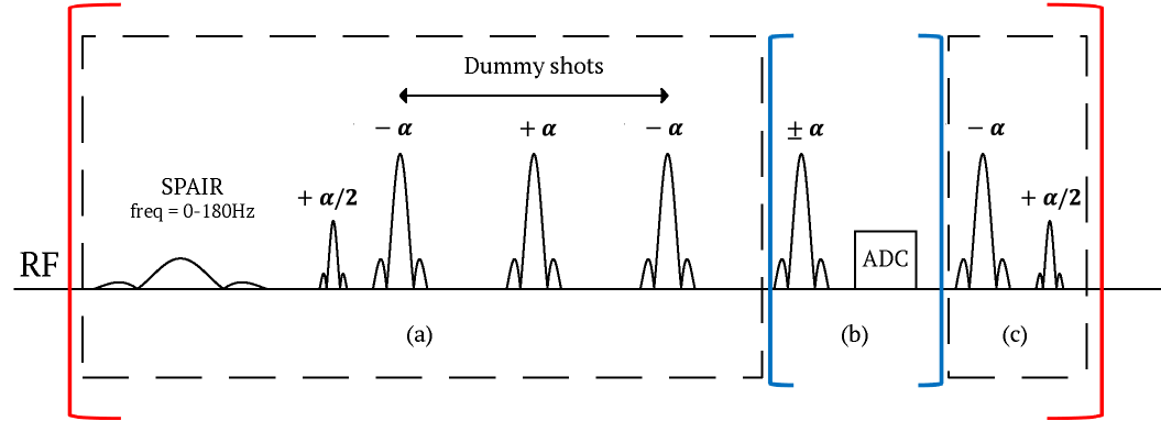
Sequence DesignA radial stack-of-stars 3D bSSFP sequence with golden-angle ordering has been implemented following the scheme described for GRE acquisition3. Prior to acquiring a stack of projections for each angle, a fat saturation module is executed (Figure 1a), consisting of an adiabatic spectrally-selective inversion pulse (SPAIR) and spoiler gradient, followed by one α/2 preparation and multiple dummy pulses to establish steady state. After the bSSFP readout train, a flip-back module (Figure 1c) is played to store the transverse magnetization into the longitudinal plane4 before repeating the fat saturation module. During the scan, the frequency offset of the SPAIR pulse is swept from 0Hz to 180Hz with an increment of 20Hz for each step. 100 projection angles are acquired for each frequency offset, resulting in 1000 spokes for the whole scan. Due to radial sampling, separate images can be reconstructed for the frequency offsets.
Experiments
Exemplary datasets were acquired in healthy volunteers after obtaining informed consent using a prototypic 0.55T scanner (MAGNETOM Aera, Siemens). For free-breathing abdominal imaging, 64 slices were acquired in axial orientation with 50% slice oversampling. Relevant parameters included: FOV 350x350x192mm3, FA 70°, TR/TE 3.74/1.87ms, BW 890Hz/px, resolution 1.37x1.37x3.0mm3, 1000 radial projections, total duration 7:26min. For comparison, scans without fat saturation were collected with 400 projections (total duration 2:24min).
Image Reconstruction
To obtain separate images for the frequency offsets, eXtra-Dimensional GRASP (XD-GRASP) reconstruction5 is performed, which exploits correlations along the frequency-sweep dimension to recover images from the undersampled data. Total variation is used as sparsity transform.
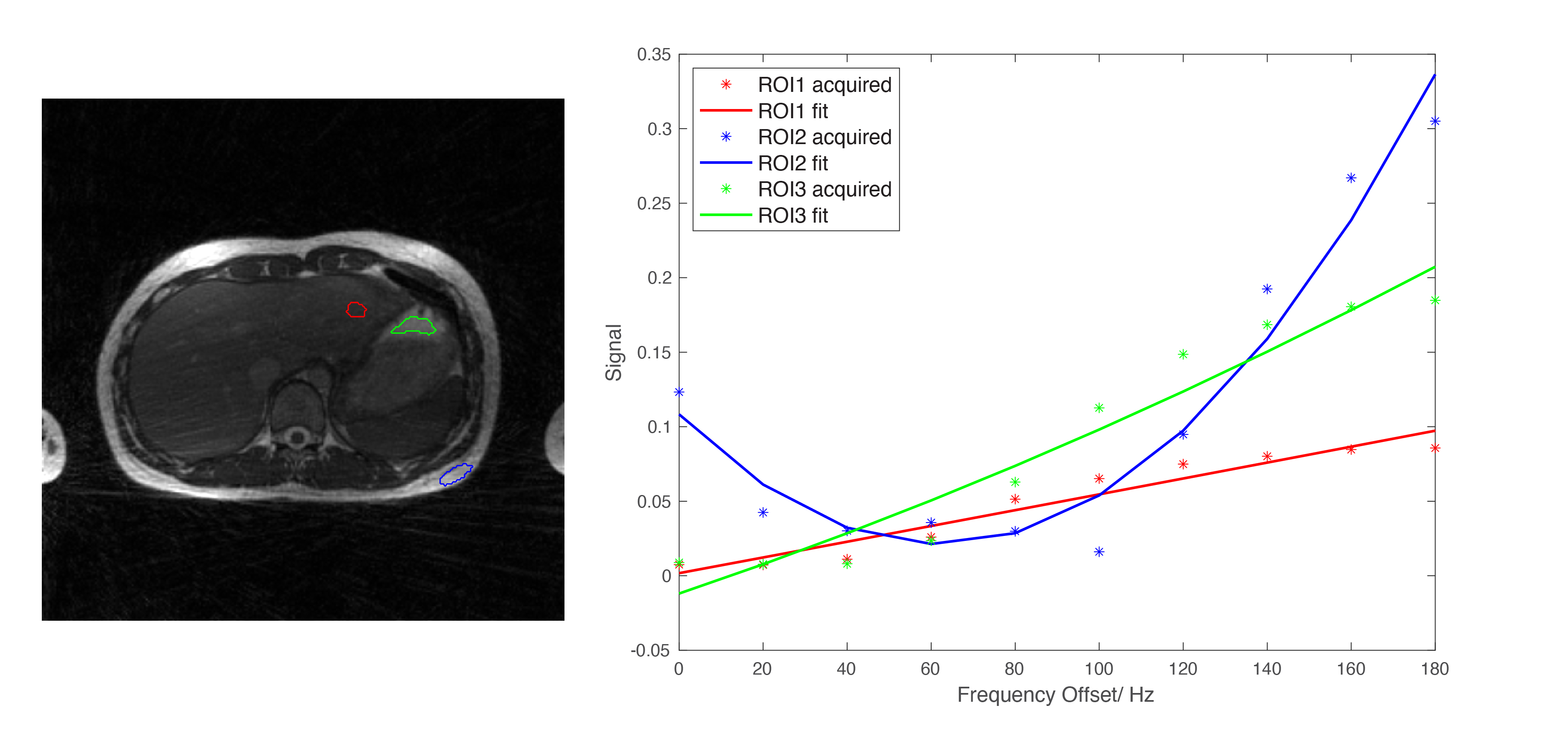
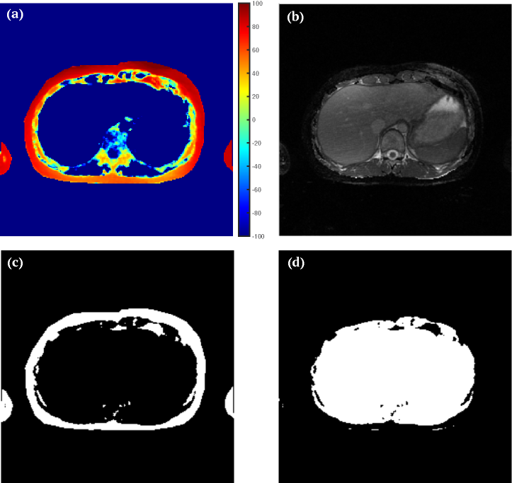
The image series is then used for voxel-by-voxel fitting of a second-degree polynomial $$$y=a(x-b)^{2}+c$$$, which allows classifying voxels into fat and water based on the frequency-dependent response to the saturation pulse (in the simplest case, the frequency shift $$$b$$$). For illustration purpose, a fat-suppressed image was generated by combining the minimum value along the frequency dimension for fat-containing and the maximum value for water-containing voxels.
Results
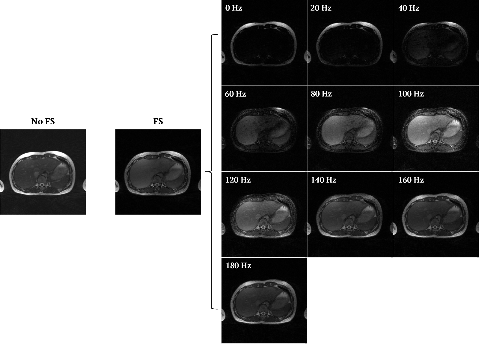
Figure 2 compares a scan without fat saturation to the proposed frequency-sweep acquisition. By performing XD-GRASP reconstruction, images are obtained that reflect the response to RF saturation at different frequency offsets. As noticeable, a single frequency offset is insufficient to suppress fat uniformly, while the frequency offset with strongest fat suppression (80Hz) also considerably weakens the water signal (due to the small spectral distance at 0.55T).By obtaining images for a range of saturation frequencies, fat and water can be clearly distinguished based on the response profile to the RF frequency sweep. This can be seen from the curves shown in Figure 3 for three exemplary ROIs: While the water signal increases monotonously, the fat signal shows a distinct parabolic shape. Figure 4 shows the map of the fitted frequency shift, which can be used for fat/water classification and for creating a fat-suppressed composite image.
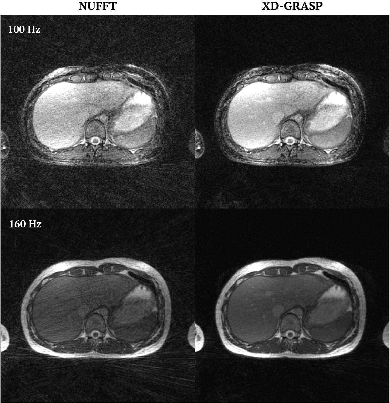
Figure 5 shows how XD-GRASP reduces streaking artifacts compared to standard gridding reconstruction. In the present case, also the gridding images are of acceptable quality, indicating that higher acceleration should be possible.
Discussion
In this work, we describe a new idea for fat separation at low magnetic field where conventional fat-saturation techniques fail. By acquiring a spectral footprint of the response to RF saturation, fat can be reliably distinguished from water without relying on an “ideal” saturation profile. Thus, instead of requiring a long, optimized RF pulse, the proposed method allows using short, imperfect pulses and exploiting the spectral footprint instead. Due to use of radial k-space sampling, such footprint can be obtained in a time-efficient manner since complementary projections can be acquired and combined using XD-GRASP. Moreover, it allows for free-breathing acquisition. While a simple classification approach was used in a preliminary attempt for composing fat-suppressed images, more advanced strategies could be employed that handle coexistence of fat and water and provide reliability in presence of significant B0 inhomogeneities. Further work is warranted to identify best experimental parameters for different applications, including frequency range, step width, acceleration, and pulse shape.Acknowledgements
We thank Thomas Benkert for providing the underlying radial 3D bSSFP sequence and for helpful discussions.References
1. Campbell-Washburn AE, Ramasawmy R, Restivo MC, et al. Opportunities in Interventional and Diagnostic Imaging by Using High-Performance Low-Field-Strength MRI. Radiology. 2019;293(2):384-393.
2. Chandarana H, Bagga B, Huang C, et al. Diagnostic abdominal MR imaging on a prototype low-field 0.55 T scanner operating at two different gradient strengths. Abdom Radiol (NY). 2021;10.1007/s00261-021-03234-1.
3. Block KT, Chandarana H, Milla S, Bruno M, Mulholland T, Fatterpekar G, et al. Towards Routine Clinical Use of Radial Stack-of-Stars 3D Gradient-Echo Sequences for Reducing Motion Sensitivity. Vol. 18, Journal of the Korean Society of Magnetic Resonance in Medicine. 2014;p.87.
4. Scheffler K, Heid O, Hennig J. Magnetization preparation during the steady state: fat-saturated 3D TrueFISP. Magn Reson Med. 2001;45(6):1075-1080.
5. Feng L, Axel L, Chandarana H, Block KT, Sodickson DK, Otazo R. XD-GRASP: Golden-angle radial MRI with reconstruction of extra motion-state dimensions using compressed sensing. Magn Reson Med. 2016;75(2):775-788.
Figures