0263
The relation between brain anatomy, functional connectivity, and emotional behavior in 3- and 9-month old infants1University of Pittsburgh, Pittsburgh, PA, United States
Synopsis
Uncinate fasciculus, forceps minor, and cingulum bundle volumes measured in infants at 3 months of age via DWI tractography were found to predict infant emotionality at 3 months and 9 months of age. These relations were found to be mediated and/or suppressed by functional connectivity from the orbitofrontal cortex, dorsolateral prefrontal cortex, and posterior default mode. Therefore, modulating connectivity among regions in the default mode, central executive, and salience networks might be promising future approaches for interventions to maintain infant emotionality and reduce risk of future psychopathology later in childhood and adolescence.
Introduction
Lower infant positive emotionality (PE) and greater negative emotionality (NE) predispose to future mental health problems later in childhood and adolescence1,2,3. Here we investigate influences of regional WM volume on relationships among functional connectivity in large-scale neural networks and infant emotionality at 3 and 9 months of age.Materials and Methods
Participants: fcMRI and DWI scans were successfully collected from 52 infants (age = 15.2 +/- 2.9 wks) during natural sleep. Caregiver-infant dyads were recruited from the community. All infants were full-term and physiologically and neurologically normal.MRI Scans: All scans were acquired on a 3T Siemens Skyra scanner at UPMC Children’s Hospital of Pittsburgh (CHP). fcMRI: TR = 800ms, TE = 30ms, MB factor = 4, voxel resolution = 3 mm isotropic, 2 scan runs of 5 minutes each. DTI: Two-shell acquisition (100 acquisitions at b = 2000 s/mm2, 50 acquisitions at b = 750 s/mm2, 9 b=0 acquisitions), MB factor = 3, voxel resolution = 2 mm isotropic.
Temperament/Emotionality: Infant temperament was measured using the Infant Behavior Questionnaire Revised. Positive Emotionality (PE) and Negative Emotionality (NE) were determined using composite scores. Caregiver depression and affective instability were assessed at 3 and 9 months using the Edinburgh Postnatal Depression (EPDS) and the Personality Assessment Inventory–Borderline Features Scale (PAIBOR). A summary measure of public assistance was used as a proxy for socioeconomic status (SES).
Pre-processing: Standard pre-processing4 was used for fcMRI. Seed regions were delineated using the neonatal parcellation atlas5. Seed-based connectivity maps were computed from the bilateral orbitofrontal cortex (OFC), dorsolateral prefrontal cortex (dlPFC), and posterior cingulate gyrus (PCC), as representative regions in central executive network (CEN), default mode network (DMN), and salience (SN) network, connected by the cingulum bundle (CB) forceps minor (FM), and uncinate fasciulus (UF). DWI data were preprocessed using fMRI Software Library (FSL). Using DSI Studio, tractography was performed for each tract (CB, FM, UF); tract volume and FA were extracted from each tract and averaged across hemispheres.
WM-emotionality: Elastic net regressions were followed by General Linear Models with elastic net-selected variables using a robust estimator and bootstrapping (1000 samples). We included 6 variables representing white matter integrity (FA and volume from each of the 3 tracts) as predictors. Additionally, we included the infant’s sex and age at time of scan, and the caregiver’s age, SES, EPDS and PAIBOR scores at the time of the infant scan (3 months). Outcome variables included NE and PE scores at 3 months. Prospective analyses were performed in a similar fashion with 9 months NE/PE scores as outcomes and with the 3-month emotionality scores, infant age, caregiver EPDS and PAI-BOR scores at the time of the 9-month assessment added as predictors.
FcMRI Mediation: Voxelwise mediation analyses were conducted with white matter structural volumes the independent variable, functional connectivity the mediator, and infant emotionality the outcome with the same sets of covariates. Bootstrapping (1000 repetitions) was used to assess statistical significance. Additional GLMs were conducted for each arm (WM volume – connectivity and connectivity-outcome). Standard methods were used to correct for multiple comparisons across voxels; results were deemed significant at FWE-corrected p < 0.05.
Results
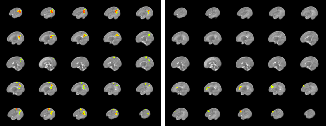
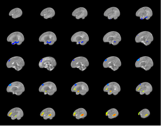
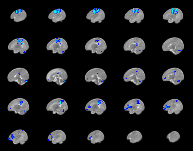
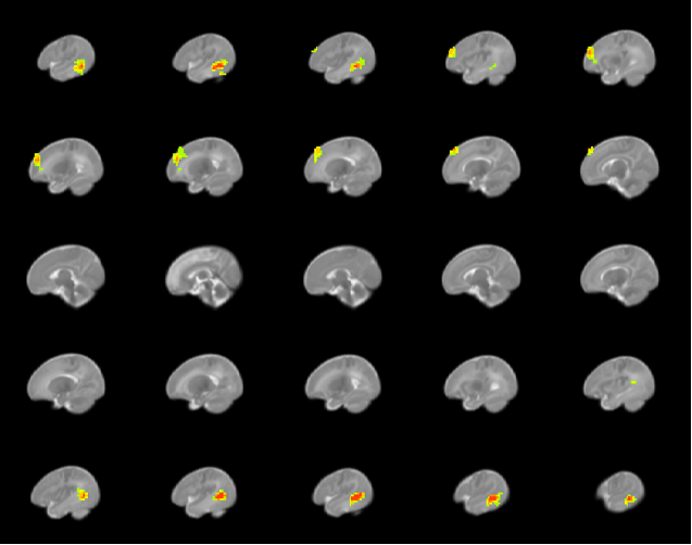
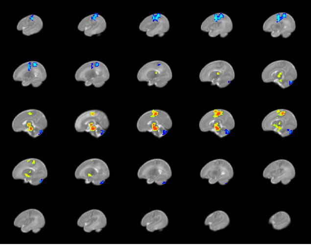
3 month emotionality relationships: Greater CB volume at 3 mo was associated with less PE (p = .031). Mediation analyses revealed significant positive indirect effects for dlPFC connectivity with regions in the CEN and DMN, and significant positive indirect effects for PCC connectivity with prefrontal cortical regions in the CEN, (Figure 1). Greater UF volume at 3 mo was associated with less NE (p = .022). Mediation analyses revealed significant negative indirect effects for OFC connectivity with regions in the temporal cortex, and significant positive indirect with prefrontal cortical regions in the CEN (Figure 2).Prospective 9-month emotionality relationships: Greater CB volume at 3 mo was associated with less PE at 9 mo (p = .031). Mediation analyses revealed significant negative indirect effects for dlPFC connectivity with regions in the CEN, SN and DMN (Figure 3). Greater FM volume at 3 mo predicted less PE at 9 mo (p = 0.003). Mediation analyses revealed significant positive indirect effects for OFC connectivity with temporal and prefrontal cortical regions (Figure 4); positive indirect effects for dlPFC connectivity with thalamus and supplemental motor area, and negative indirect effects with parietal cortical regions in the CEN and DMN (Figure 5).
Discussion
Greater CB and FM volumes predicted worse emotional outcomes (PE) at 3 and 9 months. This may reflect greater axonal dispersion. Our mediation findings show that relationships in infants among white matter microstructure in tracts supporting emotional regulation are mediated or suppressed by opposite patterns of resting state connectivity in the DMN, CEN, and SN. In particular, greater connectivity among “task positive” network (CEN, SN) and “task negative” networks (DMN) is associated with lower concurrent and future positive emotionality.Conclusion
Our results show that white matter structure – emotionality relationships in infants are mediated or suppressed by functional connectivity in the DMN, CEN, and SN and indicate that modulating connectivity among regions in these networks might be promising future approaches for interventions to maintain PE and reduce risk of future psychopathology later in childhood and adolescence.Acknowledgements
No acknowledgement found.References
1. Phillips ML, Schmithorst VJ, Banihashemi L, Taylor M, Samolyk A, Northrup JB, English GE, Versace A, Stiffler RS, Aslam HA, Bonar L, Panigrahy A, Hipwell AE. Patterns of Infant Amygdala Connectivity Mediate the Impact of High Caregiver Affect on Reducing Infant Smiling: Discovery and Replication. Biol Psychiatry. 2021 Sep 1;90(5):342-352. doi: 10.1016/j.biopsych.2021.03.026. Epub 2021 Mar 26. PMID: 34130856; PMCID: PMC8364485.
2. A. Sanson, S.A. Hemphill, D. Smart. Connections between temperament and social development: A review. Soc Dev, 13 (2004), 142-170.
3. E. Anderson, D.A. Hope A review of the tripartite model for understanding the link between anxiety and depression in youth. Clin Psychol Rev, 28 (2008), 275-287.
4. Power JD, Mitra A, Laumann TO, Snyder AZ, Schlaggar BL, Petersen SE. Methods to detect, characterize, and remove motion artifact in resting state fMRI. Neuroimage. 2014;84:320-41.
Figures