0233
Field-Map Combination Method for Phase-Cycled bSSFP using Inherent Bo Mapping1Stanford University, Stanford, CA, United States, 2Western University, London, ON, Canada
Synopsis
Phase-cycled bSSFP enables off-resonance-robust bSSFP imaging. Conventional phase-cycle combination methods weight brighter phase-cycles more, assuming that passband signal is brighter than off-resonant signal. However, near-band signal can be hyperintense, most notably for flowing spins, so traditional methods fail to mitigate these artifacts. Incorporating knowledge of Bo enables inclusion of only passband signal, and exclusion of dark bands and near-band hyperintense artifacts. Using a golden-angle radial trajectory for a free-breathing, phase-cycled acquisition enables reconstruction of a Bo-map time series without any additional scans. This facilitates field-map-based combination throughout the respiratory and cardiac cycles, resulting in substantially reduced hyperintense artifacts than root-sum-of-squares.
Introduction
Balanced SSFP (bSSFP) is susceptible to banding artifacts in the presence of off-resonance. For banding-artifact removal, multiple bSSFP acquisitions with different RF phase-cycling can be combined into a final image. Traditional combination methods, such as root-sum-of-squares (RSOS), implicitly assume that, for any given voxel, the phase-cycle with the highest signal is the desired on-resonant signal, and weight the component images accordingly. For some tissue and sequence parameters, however, off-resonant signal is brighter than the center of the passband. In the heart, where bSSFP is often used, flow in near-band regions results in hyperintense artifacts, which can be an order-of-magnitude brighter than the desired signal1, instead of dark-band artifacts. Standard phase-cycle-combination methods therefore fail to reduce these artifacts. However, if the precession frequency at each location were known, it could be used to include only passband signal in the combined image, and exclude stopband and near-band flow-related signal.For non-Cartesian trajectories where the bSSFP gradient rewinders fully sample k-space, Bo mapping using rewinding trajectories (BMART)2 can be used to inherently acquire main-field maps during the imaging scans with minimal TR increase. Here, we show that utilizing a Bo map to combine phase-cycles removes bright near-band artifacts in addition to dark bands, resulting in more homogeneous signal than other combination methods, especially in the blood pool. Using a full-spoke radial trajectory for the phase-cycled bSSFP acquisition facilitates estimation of Bo maps using BMART, which enable field-map combination (FMC) without any additional scans.
Methods
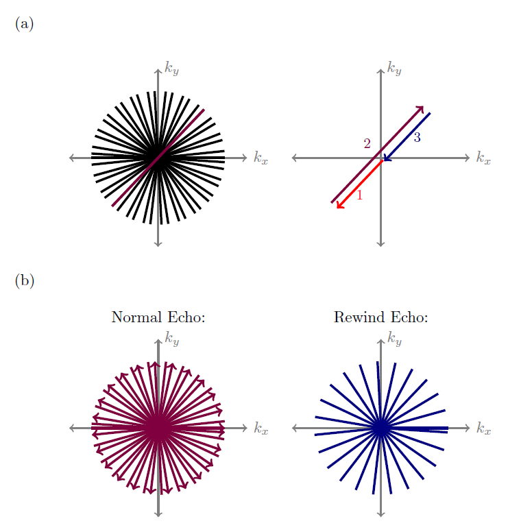
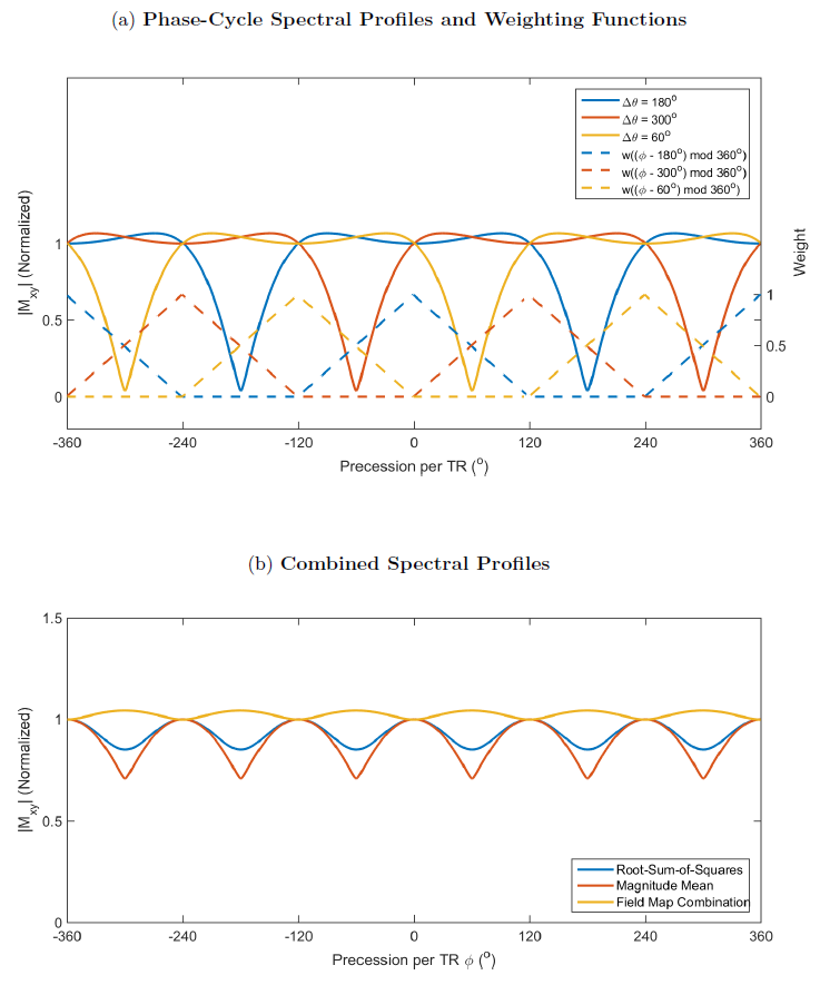
A free-breathing tiny-golden-angle-radial-sparse-parallel (tyGRASP)3 cardiac cine sequence is used to acquire three phase-cycles, which are reconstructed using a framework similar to eXtra-Dimensional-GRASP (XD-GRASP)4. Golden-angle sampling ensures that the rewinds span k-space (Figure 1) and that temporal frames of various lengths are approximately uniformly sampled. A multiscale low-rank (MSLR)5,6 reconstruction is used to reconstruct a low temporal resolution (48 spokes per frame, the length of four cardiac phases) time-series for respiratory-motion estimation, and the raw data is binned accordingly into four respiratory states. MSLR is then used again to reconstruct a higher temporal resolution (41 ms) cine loop at each respiratory position.The rewind images (i.e., the second echo for BMART Bo-map estimation) are reconstructed similarly. The respiratory position estimates from the "normal" data (i.e., data acquired during the readout trapezoid) are used for binning before reconstructing the rewind images with MSLR. A sliding window four cardiac phases long balances the temporal resolution of the Bo map time-series with the SNR of the maps. Finally, the precession per TR $$$\phi$$$ is calculated from the Bo, and a triangular weighting function $$$w(\phi)=tri((\phi-180)/120)$$$ is used to smoothly merge the passbands of the three phase-cycles for each respiratory and cardiac phase. The reconstruction and post-processing code is available at https://github.com/adatta92/PRCine
Data was collected at 1.5T using an eight-channel cardiac coil. Free-breathing cine scans of axial slices were acquired with 1.6 mm in-plane resolution, 36 cm field-of-view, 8-mm slice thickness, 1.3 ms TE, and 3.4 ms TR.
Results
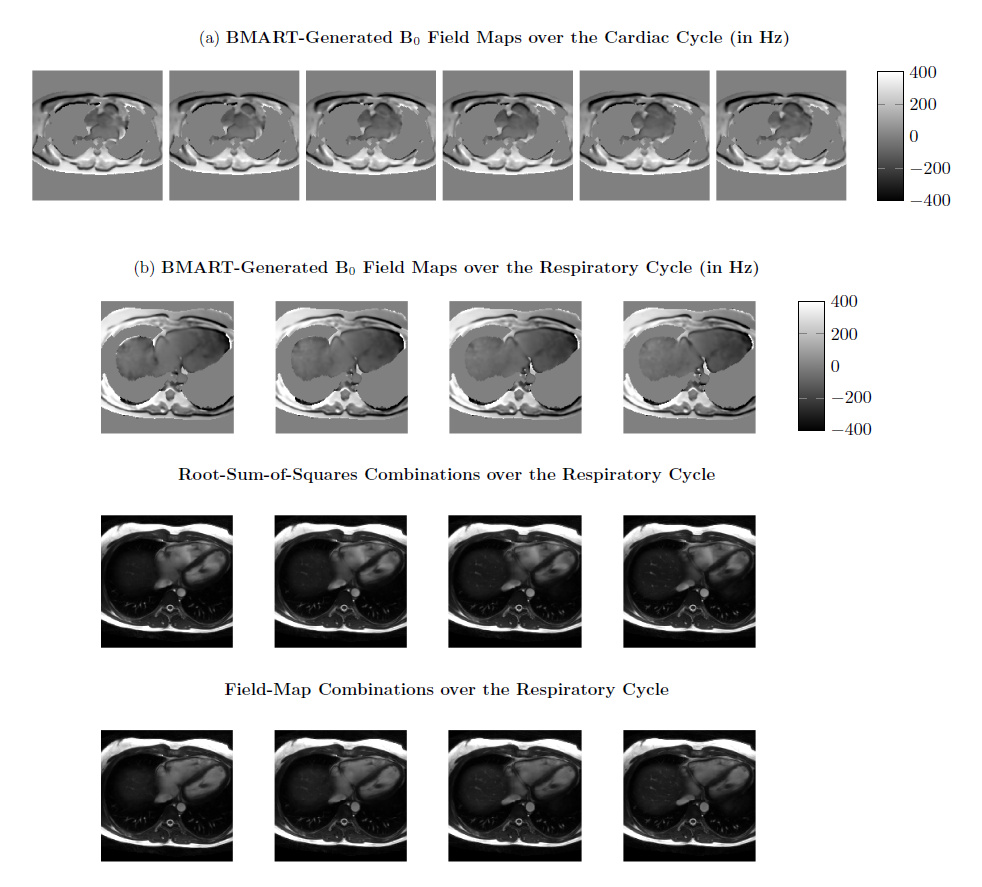
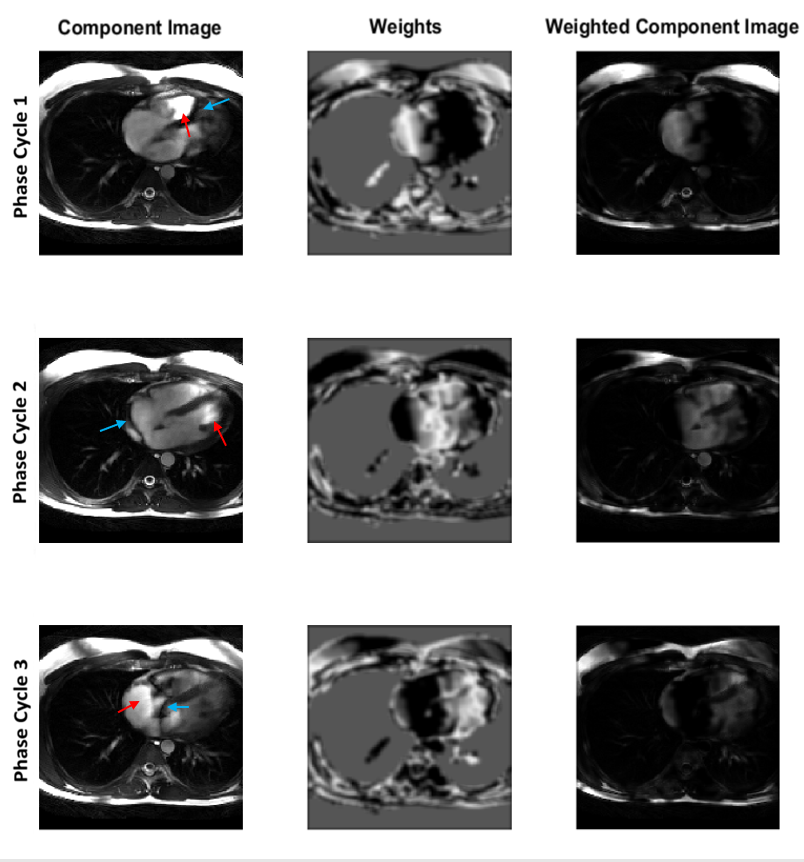
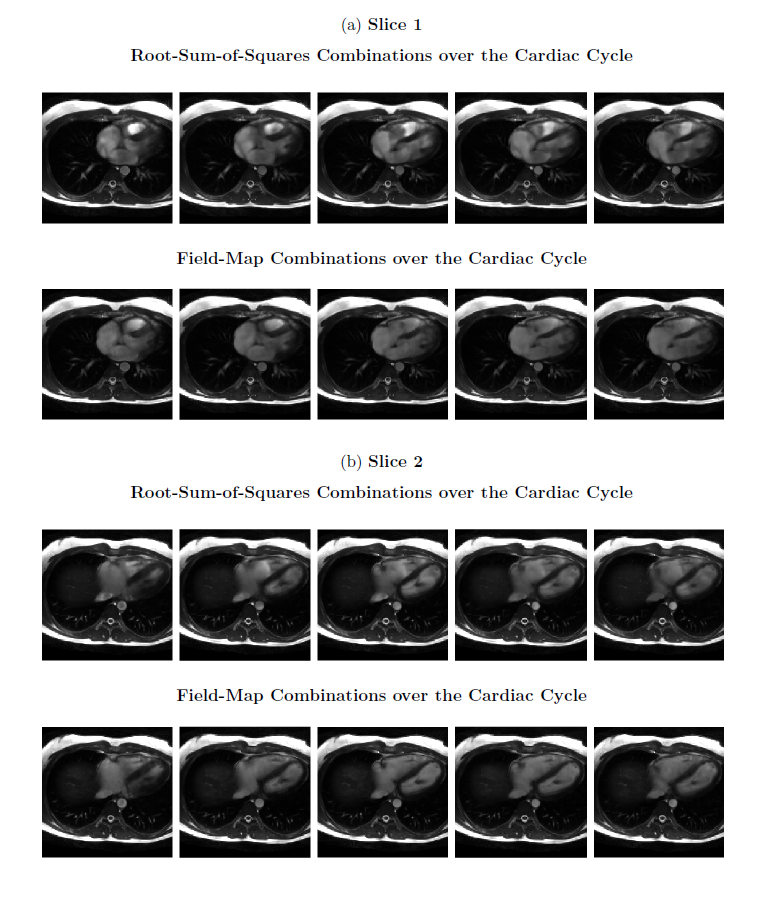
As expected, the Bo maps generated by BMART appear slowly-varying over the image, except in regions with fat (Figure 3a). In addition, the field map changes gradually over the cardiac cycle. The weights derived from the Bo map mask out the banding and near-band flow artifacts in the heart in the component images (Figure 4). As a result, in both slices shown, the field-map combinations have more homogeneous blood pools and substantially reduced hyperintense regions than the RSOS combinations, and less artifactual intensity variations between cardiac phases (Figure 5). The field maps also appear to capture changes over the respiratory cycle (Figure 3b), which are expected due to the susceptibility difference between oxygen and tissue7. Consequently, FMC is effective at suppressing both dark-band artifacts and near-band bright artifacts at all respiratory positions, resulting in substantially flatter blood signal than RSOS.Discussion
We propose a combination method for phase-cycled bSSFP that, unlike traditional methods, is intended to exclude signal outside the pass-band, even if it is bright. Cardiac cine results indicate that using Bo maps to combine phase-cycles greatly suppresses near-band flow artifacts and yields less residual ripple than RSOS. Using BMART enables us to inherently acquire the Bo maps during the scan itself with less than a 5% scan-time increase. Field-map combination should be compatible with any scan protocol that acquires phase-cycles and Bo-field measurements8,9; however, using BMART ensures that the field maps are registered with the bSSFP data and enables changes to the main field over the respiratory and cardiac cycles to be captured, as we have shown here. Although field-map combination is illustrated here in the context of cardiac cine imaging, other applications merit investigation. For example, it may be possible to use FMC to combine the desired signal for either passband or transition-band bSSFP fMRI acquisitions, excluding the other contrast and noise from other parts of the spectral profile.Conclusion
Using a non-Cartesian trajectory for a phase-cycled-bSSFP acquisition facilitates estimation of a Bo-map time series without any additional scans, enabling a field-map-based phase-cycle combination method. By including only passband signal, and excluding stopband and near-band bright signal, the developed combination method results in reduced hyperintense artifacts and less residual ripple than RSOS.Acknowledgements
Thank you to GE Healthcare and NIH R01 HL127039 for their support.References
1. Markl M, Pelc NJ. On flow effects in balanced steady-state free precession imaging: Pictorial description, parameter dependence, and clinical implications. J Magn Reson 2004; 20:697–705.
2. Baron CA, Nishimura DG. B0 mapping using rewinding trajectories. Magn Reson Med 2017, 78: 664-669.
3. Wundrak S, Paul J, Ulrici J, Hell E, Geibel MA, Bernhardt P, Rottbauer W, Rasche V. Golden ratio sparse MRI using tiny golden angles. Magn Reson Med 2016; 75: 2372-2378.
4. Feng L, Axel L, Chandarana H, Block KT, Sodickson DK, Otazo R. XD-GRASP: Golden-angle radial MRI with reconstruction of extra motion-state dimensions using compressed sensing. Magn Reson Med 2016, 75: 775-788.
5. Ong, F, Zhu, X, Cheng, JY, Johnson, KM, Larson PEZ, Vasanawala, SS, Lustig, M. Extreme MRI: Large‐scale volumetric dynamic imaging from continuous non‐gated acquisitions. Magn Reson Med. 2020; 84: 1763– 1780.
6. Datta A, Nishimura DG, Baron CA. Multi-scale low-rank reconstruction for phase-cycled projection-reconstruction bSSFP cardiac cine and BMART-generated B0 maps. In Proceedings of the 2021 ISMRM Annual Meeting, May 2021. p. 1172.
7. Datta A, Ingle RR, Hu B, Nishimura DG. Simulation of respiration-induced B0 shifts in the heart. In Proceedings of the 22nd Annual Meeting of ISMRM, Milan, Italy, May 2014. p. 1624.
8. Datta A, Nishimura DG. Field map combination method for multiple-acquisition bSSFP. In Proceedings of the 25th Annual Meeting of ISMRM, Honolulu, May 2017. p.454.
9. Datta A, Nishimura DG, Baron CA. BMART-enabled field-map combination of phase-cycled projection-reconstruction cardiac cine for banding-free balanced SSFP. In Proceedings of the ISMRM Virtual Conference, August 2020. p. 3407.
Figures