0229
Integrated Multi-Task MR For Abdominal Adaptive Radiation Therapy1Radiology, University of Southern California, Los Angeles, CA, United States, 2Bioengineering, University of California, Los Angeles, Los Angeles, CA, United States, 3Biomedical Imaging Research Institute, Cedars-Sinai Medical Center, Los Angeles, CA, United States, 4Electrical Engineering, University of Southern California, Los Angeles, CA, United States, 5Radiation Oncology, University of Southern California, Los Angeles, CA, United States, 6Cedars-Sinai Medical Center, Los Angeles, CA, United States, 7University of California, Los Angeles, Los Angeles, CA, United States, 8Radiation Oncology, University of California, Los Angeles, Los Angeles, CA, United States, 9Bioengineering, Cedars-Sinai Medical Center, Los Angeles, CA, United States, 10Biomedical Engineering, University of Southern California, Los Angeles, CA, United States
Synopsis
MR applications in radiation therapy has shown great promise for highly conformal treatment. However, MRgRT in the abdomen remains challenging due to motion and the sheer number of organs. In this work, we have demonstrated an integrated multi-task MR platform for adaptive radiation planning and treatment monitoring in the abdomen. In our proposed framework, from a single "pre-beam" scan we are able to generate a volumetric, multi-contrast and motion resolved images for adaptive treatment planning as well as enabling multi-contrast 3D real-time display during treatment. We demonstrate this capability in 3T and 0.55T.
Introduction
In recent years, MR-guided radiation therapy (MRgRT) has gained growing interest since the introduction of MR-Linac as this modality can potentially guide on-board treatment replanning and tracking target movement in real time However, MR sequences commercially available today are limited for these goals. For example, 4D MR sequences for pre-beam motion characterization is based on a fixed contrast weighting that may be suboptimal for resolving different organs-at-risk (OARs) and target, separate sequences to provide multiple contrast weightings are often misaligned because of different motion compensation strategies during acquisition, and real-time imaging during beam-on is based on a 2D acquisition that lacks 3D information and also suffers from the drawback of single contrast weighting1-4. To overcome the above limitations and improve treatment precision, we present an integrated adaptive MRgRT imaging framework, named multi-task MR (MT-MR), technique to generate multi-contrast, volumetric, motion-resolved or real-time images for both adaptive radiation planning pre-beam replanning scan and enabling treatment beam-on real-time monitoring from a single “pre-beam” scan. This MT-MR technique was first developed on a clinically 3T scanner. However, some commercially available MR-Linacs are at lower field strengths. In this work, we also present the initial results and then adapted to a low-field system.Method
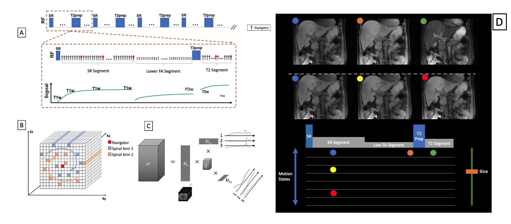
Sequence design:As shown in Fig. 1, each repetition time (TR) of our multi-task (MT-) MR sequence starts with a saturation recovery (SR) pulse followed by a group of FLASH readouts during T1 recovery; Then a lower flip angle (FA) FLASH for signal recovery (Benefits shown in fig. 3); and a T2 Prep followed by additional FLASH readouts. On 0.55T, two gradient echoes are applied between FLASH RF pulses for Dixon-based water-fat separation. K-space is sampled with a Cartesian spiral-in pattern (Fig. 1B) with 10 lines in each spiral arm and the 10th line being the k-space center and used as the navigator. This sampling pattern is used for 1) denser k-space center coverage and 2) reduced eddy-current effects before the center line is sampled 6,7. FAs and length of each segment within TR are optimized for both the 3T and the 0.55T system.
Image reconstruction workflow:
Before treatment, a “pre-beam” scan is conducted to generate the volumetric, multi-contrast motion-resolved images for adaptive planning and retrospective real-time images. Using MR Multitasking5 (MT), In this case,
A = U x Phi
A is the tensor representing a multidimensional image A(r ,tmotion, tcontrast) with r being the physical dimensions [x,y,z], and tmotion, tcontrast being the temporal dimensions. The combined temporal factor Phi is determined from Bloch-constrained low-rank tensor completion and motion binning from the navigator data. In the case of real-time imaging Phi can be extracted directly from SVD of the navigator data.
With a learned U from retrospective real-time application of the “pre-beam” scan, only navigator data (center k-space line) is needed during treatment to generate the real-time Phi and thus the multi-dimensional images. Furthermore, the real-time generated Phi can be projected onto a Bloch simulated subspace to “freeze” the real-time image to a specific contrast while preserving real-time respiratory motion.
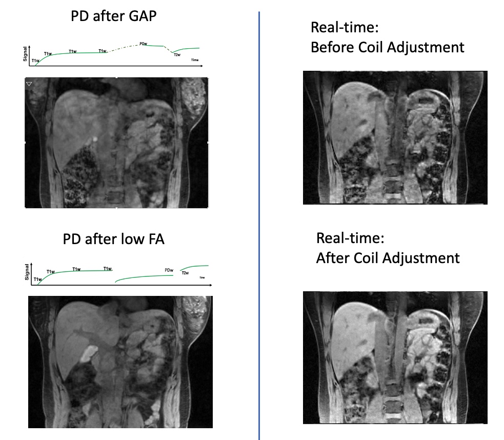
To make the real-time monitoring more robust, we calculate an adjustment matrix R of size (Ncoils x Ncoils) from the time-averaged signal of the “pre-beam scan” and the first 20 seconds of the treatment scan to account for any inter-scan coil settings change and reshimming.
Experiments
Ten healthy volunteers at 3T (MAGNETOM VIDA, Siemens Healthineers) and 3 volunteers at a whole body 0.55T system (prototype Magnetom Aera, Siemens Healthineers, Erlangen, Germany) equipped with high-performance shielded gradients (45 mT/m amplitude, 200 T/m/s slew rate) were included in the study. Following imaging parameters were used: On Both 3T and 0.55T: FOV = 256(SI) x 358(LR) x 256(AP) mm3, resolution = 1.6 x 1.6 x 3.2mm3. On 3T: Water Excitation, TR/TE = 6.0/2.13 ms, total scan time = 10 mins.On 0.55T: TR/(TE1,TE2) = 9.4/(1.54,7,71)ms, total scan time = 14 mins.
Results
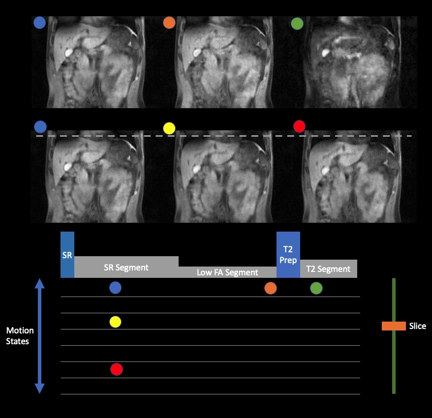
Figure 3 demonstrates the use of low-FA between T1 and T2 acquisition phases is advantageous over the use of gap in providing high-quality images as well as the results of applying the coil adjustment matrix. From 3T, the volumetric, motion-resolved and multi-contrast images are shown in figure 1D, where user can choose to view any contrast, slice and motion states that are available. Real-time monitoring capabilities are demonstrated in figure 2 where not only are we able to generate the real-time images during treatment, we can also freeze at any contrast, keeping only respiratory motion during treatment. Similarly, From 0.55T, the real-time water images are shown in figure 4 and the volumetric, motion-resolved and multi-contrast images are shown in figure Figure5.Discussion
We can demonstrated a solid capability of MT-MR as an integrated platform framework for adaptive abdominal radiation therapy on 3T where multidimensional images for adaptive planning can be generated while enabling volumetric and simultaneous multi-contrast treatment monitoring from a single “pre-beam” scan. We also demonstrated an initial experience of using the MT-MR technique on a low-field scanner. As expected, the results from 0.55T has lower SNR and susceptibility artifacts, they can be overcome by better B0 correction strategies and more advanced acquisition trajectories enabled by the high-performance gradient system on such low-field scanners.Acknowledgements
We acknowledge grant support from the National Science Foundation (#1828736) and research support from Siemens Healthineers.References
1. Huttinga NRF, Berg CAT van den, Luijten PR, Sbrizzi A. MR-MOTUS: model-based non-rigid motion estimation for MR-guided radiotherapy using a reference image and minimal k-space data. Physics in Medicine & Biology. 2020;65(1):015004. doi:10.1088/1361-6560/ab554a
2. Huttinga NRF, Bruijnen T, Berg CAT van den, Sbrizzi A. Nonrigid 3D motion estimation at high temporal resolution from prospectively undersampled k-space data using low-rank MR-MOTUS. Magnetic Resonance in Medicine. [accessed 2020 Dec 13];n/a(n/a). https://onlinelibrary.wiley.com/doi/abs/10.1002/mrm.28562. doi:https://doi.org/10.1002/mrm.28562
3. Rank CM, Heußer T, Buzan MTA, Wetscherek A, Freitag MT, Dinkel J, Kachelrieß M. 4D respiratory motion-compensated image reconstruction of free-breathing radial MR data with very high undersampling. Magnetic Resonance in Medicine. 2017;77(3):1170–1183. doi:10.1002/mrm.26206
4. Stemkens B, Tijssen RHN, Senneville BD de, Lagendijk JJW, Berg CAT van den. Image-driven, model-based 3D abdominal motion estimation for MR-guided radiotherapy. Physics in Medicine and Biology. 2016;61(14):5335–5355. doi:10.1088/0031-9155/61/14/5335
5. Christodoulou AG, Shaw JL, Nguyen C, Yang Q, Xie Y, Wang N, Li D. Magnetic resonance multitasking for motion-resolved quantitative cardiovascular imaging. Nature Biomedical Engineering. 2018;2(4):215–226. doi:10.1038/s41551-018-0217-y
6. Han F, Zhou Z, Han E, Gao Y, Nguyen K-L, Finn JP, Hu P. Self-gated 4D multiphase, steady-state imaging with contrast enhancement (MUSIC) using rotating cartesian K-space (ROCK): Validation in children with congenital heart disease. Magnetic Resonance in Medicine. 2017;78(2):472–483. doi:10.1002/mrm.26376
7. Prieto C, Doneva M, Usman M, Henningsson M, Greil G, Schaeffter T, Botnar RM. Highly efficient respiratory motion compensated free-breathing coronary MRA using golden-step Cartesian acquisition. Journal of magnetic resonance imaging: JMRI. 2015;41(3):738–746. doi:10.1002/jmri.24602
Figures