0220
Improved Agreement of Cerebral Arterial Blood Flow Velocity Measures Using 3D Quantitative QISS MRA
Ioannis Koktzoglou1,2, Rong Huang1, and Robert R Edelman1,3
1Radiology, NorthShore University HealthSystem, Evanston, IL, United States, 2Radiology, Pritzker School of Medicine, University of Chicago, Chicago, IL, United States, 3Radiology, Northwestern University Feinberg School of Medicine, Chicago, IL, United States
1Radiology, NorthShore University HealthSystem, Evanston, IL, United States, 2Radiology, Pritzker School of Medicine, University of Chicago, Chicago, IL, United States, 3Radiology, Northwestern University Feinberg School of Medicine, Chicago, IL, United States
Synopsis
Quantitative time of flight (qTOF) is a recently described 3D magnetic resonance angiography (MRA) technique for simultaneous luminal and hemodynamic evaluation of the intracranial arteries, that provides higher contrast-to-noise ratio efficiency than 3D phase contrast (PC) MRA and reduces in-plane flow displacement artifacts visible on standard 3D TOF MRA. We hypothesized that the use of a quiescent interval slice-selective-based data acquisition strategy (qQISS) that boosts arterial-to-background contrast might improve the quantitation of intracranial arterial flow velocity. Compared to qTOF MRA, we found that qQISS MRA improved agreement with PC MRA for measuring intracranial blood velocity.
Introduction
Quantitative time of flight (qTOF) is a recently described 3D magnetic resonance angiography (MRA) technique for simultaneous luminal and hemodynamic evaluation of the intracranial arteries1. The method provides higher a contrast-to-noise ratio efficiency than 3D phase contrast (PC) MRA for high-resolution morphological evaluation of the intracranial arteries and reduces in-plane flow displacement artifacts visible on standard 3D TOF MRA1. We hypothesized that the use of a quiescent interval slice-selective-based data acquisition strategy (qQISS) that boosts arterial-to-background contrast might improve the quantitation of intracranial arterial flow velocity. The purpose of this study was to test whether qQISS MRA, as compared to qTOF MRA, provides intracranial flow velocities that are in better agreement with 3D phase contrast (PC) MRA.Methods
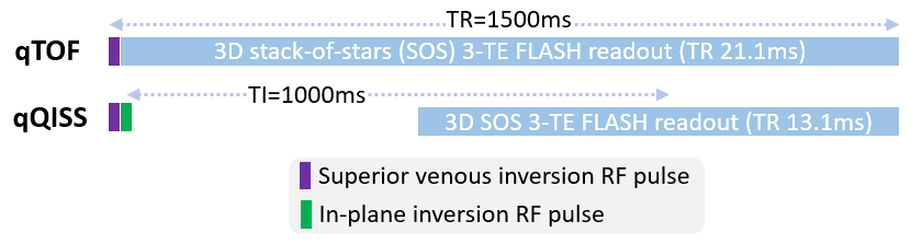
This study was approved by our institutional review board and all subjects provided written informed consent. 8 human subjects (3 males, 5 females, mean age = 38±16 years) were involved in this study. Imaging was done on a 3 Tesla MRI system (MAGNETOM Skyrafit, Siemens Healthineers).Image Acquisition: 3D qTOF, 3D qQISS, and 3D phase contrast MRA (velocity encoding sensitivity=60cm/s) were acquired using ≈4min-3sec-long protocols, with acquired [reconstructed] spatial resolutions of 0.58×0.58×1.00 [0.29×0.29×0.50] mm3 (for qTOF and qQISS) and 0.85×0.85×1.30 [0.43×0.43×0.65] mm3 (for 3D PC). Slightly lower spatial resolution was acquired with 3D PC to maintain similar scan times and adequate signal-to-noise ratio. Imaging parameters for qTOF MRA were as previously described1 (TR=21ms, TEs of 2.9ms and 7.2ms, sequence TR=1500ms, tilted optimized non-saturating RF pulses with central flip angle of 15 degrees, 3 imaging shots acquired per partition, 71 radial views acquired in each imaging shot, 3 overlapping imaging slabs) whereas those for qQISS were similar except for TR=13ms and TI=1000ms. Schematic timing diagrams of the imaging shots used with qTOF and qQISS MRA are shown in Figure 1.
Velocity Quantitation and Data Analysis: Flow velocity quantitation with qTOF and qQISS was done as previously described1, using an image analysis framework leveraging template matching and center-of-mass-based tracking. Time-averaged mean cross-sectional flow velocities were measured every 1.8mm along the bilateral M1, M2, P1, and P2 cerebral arteries. Correlation and agreement of quantitative flow velocity measures were evaluated using Pearson’s correlation coefficient (r), two-way intraclass correlation coefficient for absolute agreement (ICC), and Bland-Altman analysis.
Results
qQISS MRA visually demonstrated lower signal intensity from background brain tissue as compared with qTOF MRA while still portraying the main intracranial arteries (Figure 2). With reference to 3D PC and compared against qTOF, qQISS MRA demonstrated improved correlation (r=0.757 versus 0.689; 95% confidence intervals (CIs) of [0.732,0.781] versus [0.657,0.718]) and agreement (ICC=0.755 versus 0.678; 95% CIs of [0.729,0.779] versus [0.633,0.717]) of velocity measures (Figure 3), as well as reduced Bland-Altman mean bias (-0.36 cm/s versus -1.14 cm/s) and narrower 95% limits of agreement ([-9.46 cm/s,+8.47 cm/s] versus [-11.1 cm/s,+8.84 cm/s]) (Figure 4).Discussion
Compared to qTOF, qQISS MRA of the brain improves the correlation and agreement of quantitative flow velocity measures with respect to 3D PC MRA. Consequently, qQISS MRA may be preferred over qTOF MRA for simultaneous luminal and hemodynamic evaluation of the intracranial arteries when better agreement with PC MRA measures of flow velocity is desirable. We speculate that the improved agreement of qQISS-derived measures is due to improved arterial-to-background contrast, which likely reduces the influence of static non-vascular background signals during the image analysis procedure used for velocity quantitation. Future work will seek to incorporate other image acquisition, reconstruction, and analysis strategies to further improve agreement of flow velocity measures, while further testing and validating the qTOF and qQISS techniques for simultaneous morphologic and hemodynamic evaluation of the intracranial arteries in patients with cerebrovascular disease.Conclusion
Compared to qTOF, qQISS MRA improves the correlation and agreement of intracranial time-averaged mean cross-sectional flow velocity measures with respect to 3D phase contrast.Acknowledgements
NIH NIBIB R01EB027475References
1. Koktzoglou I, Huang R, Edelman RR. Quantitative Time-of-Flight MR Angiography for Simultaneous Luminal and Hemodynamic Evaluation of the Intracranial Arteries. Magn Reson Med. 2021 Aug 10. doi: 10.1002/mrm.28969. Online ahead of print.Figures
Figure
1. Timing diagrams showing one imaging shot for
quantitative time-of-flight (qTOF) MRA (left panel) and quantitative
quiescent-interval slice-selective (qQISS) MRA. These timing structures are
repeated until all radial views, partition encoding steps, and imaging slabs
are collected. FLASH=fast low-angle shot. TE=echo time. TI=inversion time.
RF=radiofrequency.
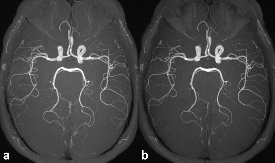
Figure
2. Transversal maximum intensity projection images obtained with
(a) qTOF
MRA and (b) qQISS MRA. Note the improved arterial-to-background contrast
obtained with qQISS.
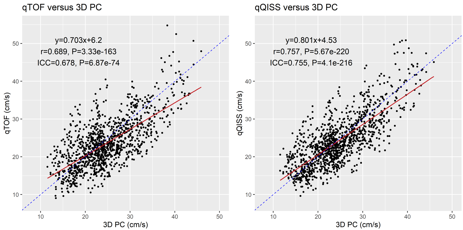
Figure 3. Scatter
plots of time-averaged mean cross-sectional flow velocity measures obtained
with quantitative time-of-flight (qTOF) MRA (left panel) and quantitative
quiescent-interval slice-selective (qQISS) MRA (right panel) with respect to 3D
phase contrast (PC) MRA. Note the improved correlation coefficient (r=0.757
versus 0.689) and intraclass correlation coefficient for agreement (ICC=0.755
versus 0.678) obtained with qQISS MRA.
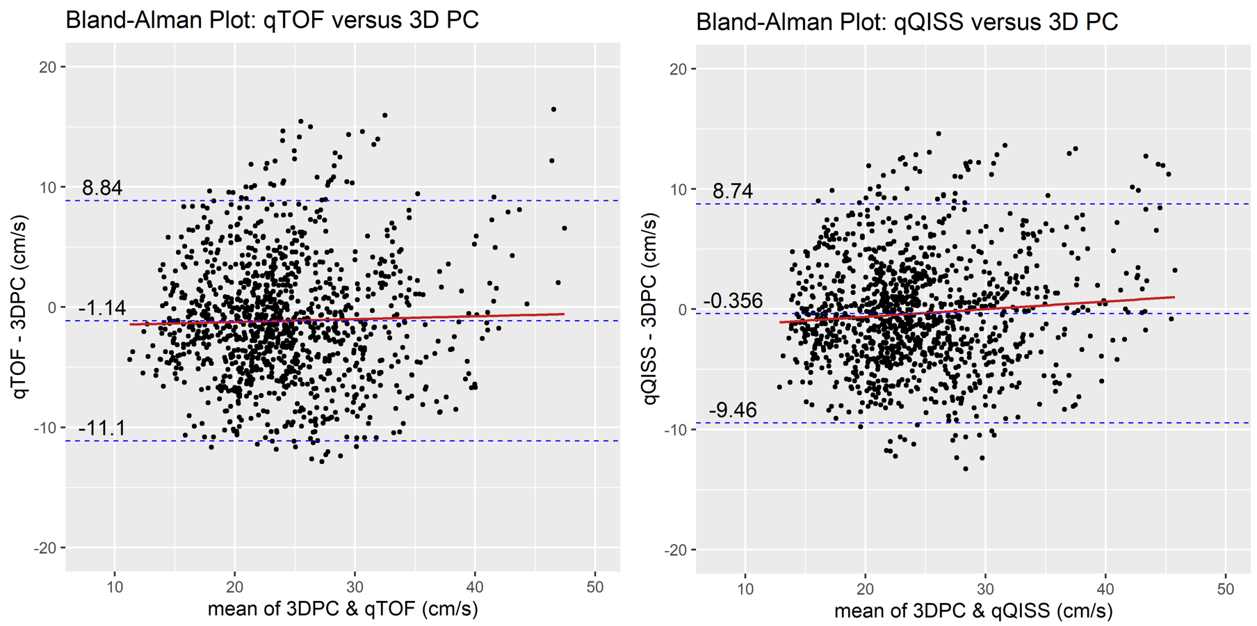
Figure
4. Bland-Altman plots of time-averaged mean
cross-sectional flow velocity measures obtained with qTOF MRA (left panel) and
qQISS MRA (right panel) with respect to 3D PC MRA. Note the reduced mean bias (-0.36
cm/s versus -1.14 cm/s) and narrower 95% limits of agreement ([-9.46 cm/s, +8.74
cm/s] versus [-11.1 cm/s, +8.84 cm/s]) obtained with qQISS MRA.
DOI: https://doi.org/10.58530/2022/0220