0208
Evaluation of a full dynamic density adapted radial 4D-pcASL angiography sequence exploiting B1+-shimming and ramped SPINS excitation at 7T1Institute of Radiology, University Hospital Erlangen, Friedrich-Alexander- Universität Erlangen-Nürnberg (FAU), Erlangen, Germany, 2Physikalisch-Technische Bundesanstalt (PTB), Braunschweig und Berlin, Germany, 3Division of Medical Physics in Radiology, German Cancer Research Center (DKFZ), Heidelberg, Germany, 4Department of Neuroradiology, University Hospital Erlangen, Friedrich-Alexander- Universität Erlangen-Nürnberg (FAU), Erlangen, Germany
Synopsis
In this work, a whole-brain 4D-pcASL angiography sequence exploiting a radial density-adapted gradient echo readout with golden angle sampling was implemented and evaluated on a 7T system. Various parallel transmission (pTx) strategies were compared to the standard circularly polarized mode: To counteract low transmit efficiency in the labeling, B1+-shimming was used in combination with a full spiral-nonselective pTx-optimized readout train. Further, constant flip angle pTx-excitations in the readout were compared to pTx-pulses that were ramped from 3.5° to 6.5° using a linear scaling factor. This ramped readout strategy outperforms constant flip angles and reveals more of the vascular tree
Introduction
To non-invasively image the vascular hemodynamics, non-contrast 4D-angiography MRI techniques, exploiting arterial spin labeling (ASL), have been developed. At low field strengths, balanced steady state free precession readouts are used for 4D-angiography, which are difficult to realize at 7T due to specific absorption rate (SAR) constraints and B0 homogeneity and, therefore, spoiled gradient echo readouts are applied.1 Furthermore, at 7T, pseudo-continuous ASL (pcASL) based 4D-angiography suffers from low B1+ efficiency for the labeling and strong B1+ inhomogeneity across the 3D readout volume. Recently, we could demonstrate that a combination of B1+-shimming, trading B1+ homogeneity and transmit efficiency, applied to pcASL labeling pulses, and a 2-spoke excitation for a turbo flash (TFL) slab-selective readout, significantly improved the 4D-angiography images.2 However, the Cartesian based segmented, 3D slab selective TFL readout does not enable sliding window reconstructions and therefore is inefficient for dynamic imaging (acquisition times>20min). Further, the readout coverage is also limited and no whole-brain coverage is achievable (recently shown slab thickness=5cm). Hence, a whole-brain 4D-pcASL angiography sequence exploiting a radial density-adapted gradient echo readout (DA-3D-GRE-RAD) with golden angle reordering was developed. This enhanced 4D-pcASL sequence was evaluated with regard to the standard circular polarized (CP) mode for labeling and readout as well as B1+-shimmed labeling with a (ramped) spiral-nonselective (SPINS) pTx-optimized readout train.Methods
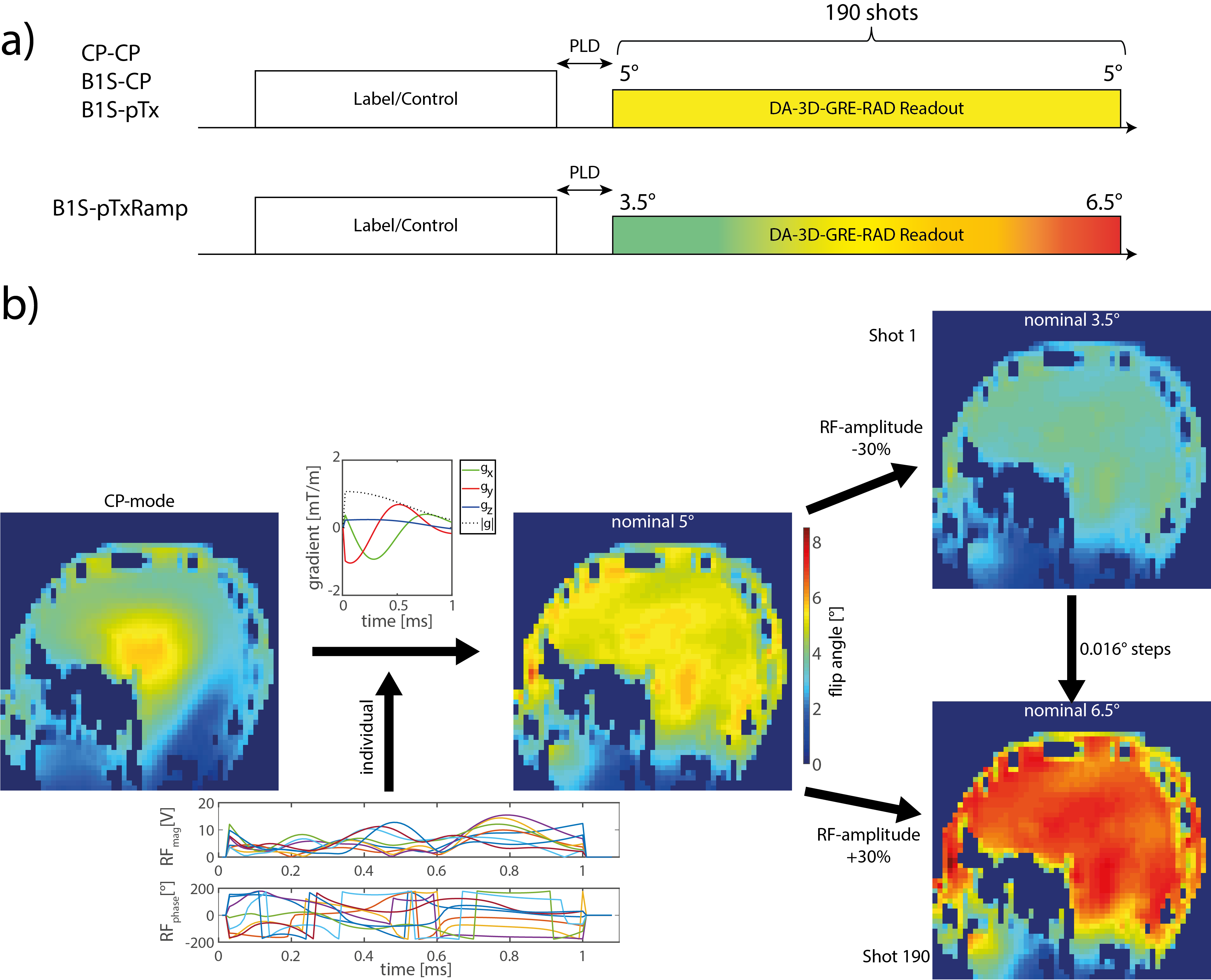
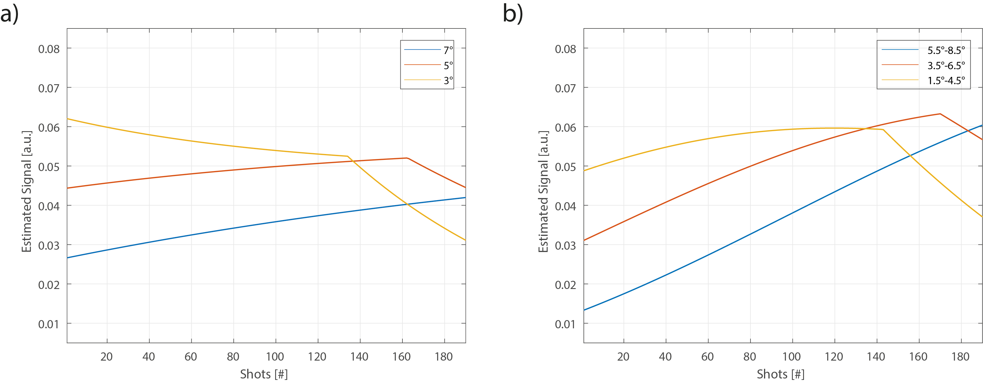
All measurements were performed on a 7T MR system (MAGNETOM Terra, Siemens Healthcare GmbH, Erlangen, Germany) using an 8Tx/32Rx head-coil (Nova Medical, Wilmington, Massachusetts, USA) and carried out in accordance with the institutional guidelines and with approval of the local ethics committee. The unbalanced pcASL labeling scheme using Variable-Rate Selective Excitation (VERSE)3 RF pulses as described4 (Gmax=3.5mT/m, Gave=0.5mT/m, labeling duration=600ms, post labeling delay (PLD)=50ms), was followed by 190 shots/lines of a radial density-adapted5 3D full head gradient echo readout with golden angle reordering6 (Fig.1a). The readout parameters were: FOV=220x220x220mm3, nominal spatial resolution=1.5mm3 (interpolated to 1mm3), TR=4.8ms, TE=2ms, flip angle=5°, radial projections (total)=13300, radial projections/frame=1470 (undersampling factor 23, temporal resolution=100ms).To simulate the signal evolution of the magnitude subtracted angiography signal, a Bloch simulation was carried out, assuming perfect spoiling in the readout. Blood relaxation parameters were T1,blood/T2,blood =2587/43ms7 and a typical labeling efficiency for 7T of α=0.764 was used.
For the parallel transmission (pTx) pulse calculations, B1+ and B0 maps were acquired.8 The B1+-shim trading homogeneity and transmit efficiency for the labeling was used as described2,4. For the readout a SPINS trajectory9 was optimized based on 15 previously acquired B1+/B0-maps with combined optimization values (COV) for 5° pulses.10 The cost function for the COV of the original work10 was normalized to the normalized root mean square error (NRMSE) and the local specific energy dose (SED) of the corresponding averaged rectangular CP excitation pulse (duration=1ms) to ensure low energy deposition:
$$\min\limits_{COV} \sum^{15}_{p=1}\left[ \text{e}^{NRMSE_p - \overline{NRMSE_{CP}}} + \text{e}^{SED_p - \overline{SED_{CP}}} \right] $$
The radio frequency (RF) pulse magnitudes and phases were then optimized individually for a homogenous 5° excitation. In addition, to compensate relaxation processes of the labeling and saturation effects, the 5° homogenous SPINS excitation was ramped up in 0.016° steps for the 190 shots following the labeling, by adjusting the transmit voltage (Fig.1).
In total, 5 healthy subjects (mean age: 25.8±1) were measured with the sequence in CP-mode for labeling/readout (“CP-CP”), with B1+-shimming for labeling/CP-readout (“B1S-CP”), with B1+-shimming for labeling/5° SPINS-readout (“B1S-pTx”) and with B1+-shimming for labeling/ramped 3.5° to 6.5° SPINS-readout (“B1S-pTxRamp”).
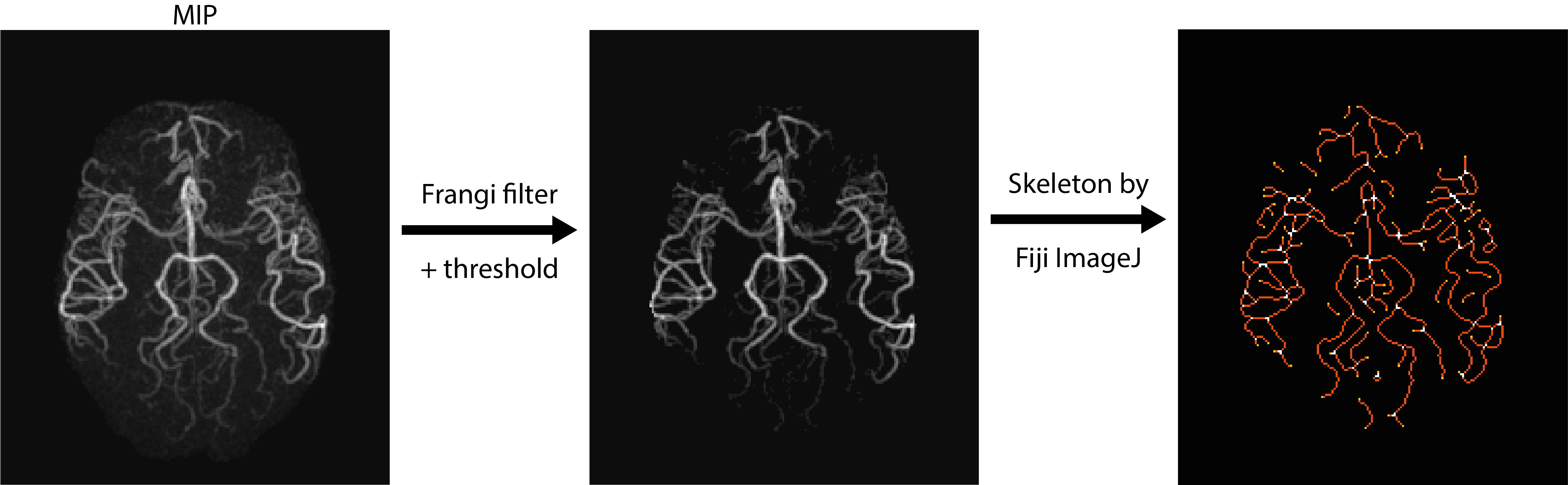
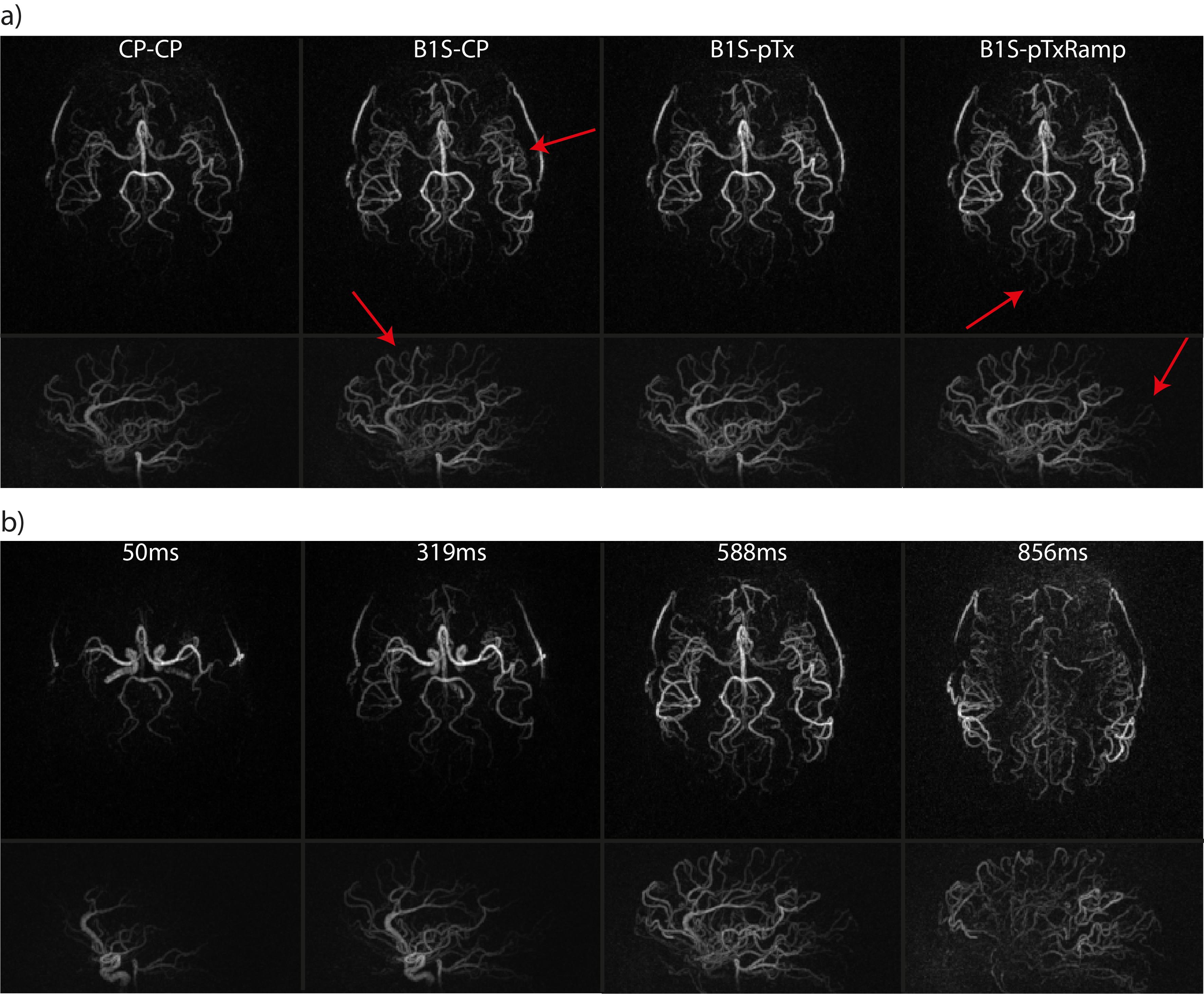
For the 4D-reconstruction, a NUFFT-based reconstruction11 was used on a CPU (Intel i7, 128GB RAM). Afterwards, the 4D-pcASL images were co-registered12, maximum intensity projections (MIPs) were created and filtered by a 2D-Frangi filter13. The vessel length was determined by processing via Fiji ImageJ14 and the results were averaged over four time points (50ms, 319ms, 588ms, 856ms) (Fig.2). Subsequently, the vessel length was normalized to the “CP-CP” acquisition and the data were statistically analyzed via an analysis of variance (ANOVA) test, followed by a Bonferroni corrected multiple comparison test (p<0.05).
Results
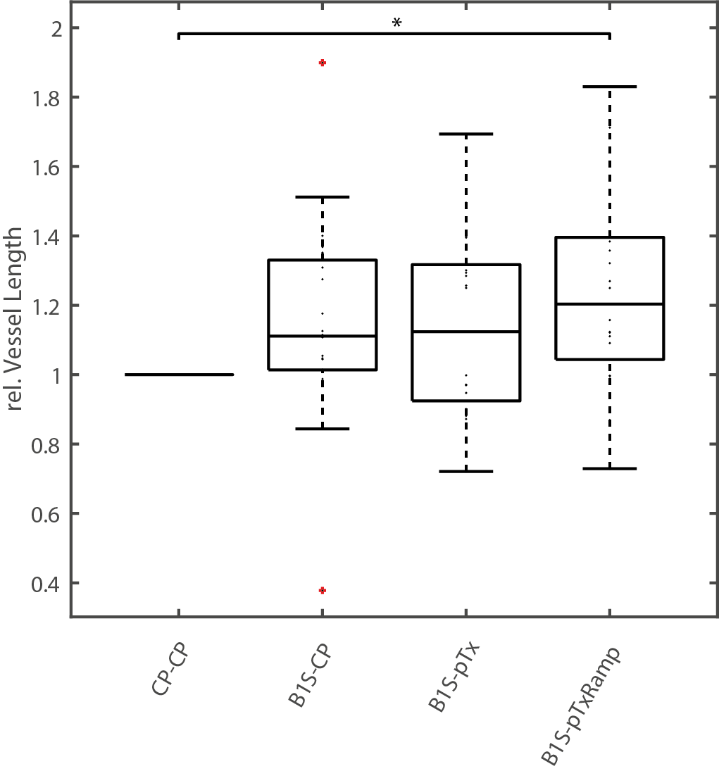
The simulation results are shown in Fig.3. While a constant flip angle in the readout leads to a decreased signal within the last shots, flip angle-ramped excitation pulses could compensate this. This is advantageous to depict small vessels located at the end of the vessel tree.Fig.4 shows the relative vessel lengths of the different measured sequences normalized to “CP-CP” with mean lengths of 1.15±0.29 for “B1S-CP”, 1.14±0.25 for “B1S-pTx” and 1.25±0.29 for “B1S-pTxRamp”. The increase in relative vessel length for “B1S-pTxRamp” is significant. Fig.5 shows MIPs of one representative subject in axial and sagittal orientation. With the hybrid B1+-shimming the overall number of visible vessels increases and with the ramped SPINS pulses, even more vessels can be identified, especially in the posterior part of the brain (Fig.5a). In Fig.5b four time points of the “B1S-pTxRamp” are shown each with a reconstruction time of about 7min.
Discussion and Conclusion
The presented 4D-pcASL angiography sequence with the DA-3D-GRE-RAD readout shows the feasibility of whole-brain 4D-angiography imaging exploiting parallel transmission and highly flexible sliding window reconstruction at 7T. While a constant 5° flip angle SPINS readout did not significantly increase the detected vessel length, the ramped SPINS readout revealed more of the vascular tree by compensating T1-relaxation processes of the labeling and saturation effects.Acknowledgements
No acknowledgement found.References
1. Cong F, Zhuo Y, Yu S, Zhang X, Miao X, An J, et al. Noncontrast-enhanced time-resolved 4D dynamic intracranial MR angiography at 7T: A feasibility study. J Magn Reson Imaging. 2018;48(1):111-20.2. Meixner CR, Schmitter S, Herrler J, Dörfler A, Uder M, Nagel AM. Impact of B1+-Shimming and 2-spoke pTx on 4D Angiography at 7T. Proceedings of the 29th Annual Meeting of ISMRM & SMRT 2021.
3. Conolly S, Nishimura D, Macovski A. Variable-Rate Selective Excitation. Journal of Magnetic Resonance. 1988;78:440-58.
4. Meixner CR, Eisen CK, Schmitter S, Muller M, Herrler J, Hensel B, et al. Hybrid B 1 + -shimming and gradient adaptions for improved pseudo-continuous arterial spin labeling at 7 Tesla. Magn Reson Med. 2021.
5. Nagel AM, Laun FB, Weber MA, Matthies C, Semmler W, Schad LR. Sodium MRI using a density-adapted 3D radial acquisition technique. Magn Reson Med. 2009;62(6):1565-73.
6. Song HK, Yan L, Smith RX, Xue Y, Rapacchi S, Srinivasan S, et al. Noncontrast enhanced four-dimensional dynamic MRA with golden angle radial acquisition and K-space weighted image contrast (KWIC) reconstruction. Magn Reson Med. 2014;72(6):1541-51.
7. Hua J, Jones CK, Qin Q, van Zijl PC. Implementation of vascular-space-occupancy MRI at 7T. Magn Reson Med. 2013;69(4):1003-13.
8. Fautz HP, Vogel M, Gross P, Kerr A, Zhu Y, editors. B1 mapping of coil arrays for parallel transmission. Proceedings of the 22nd Annual Meeting of ISMRM; 2008; Toronto.
9. Malik SJ, Keihaninejad S, Hammers A, Hajnal JV. Tailored excitation in 3D with spiral nonselective (SPINS) RF pulses. Magn Reson Med. 2012;67(5):1303-15.
10. Herrler J, Liebig P, Gumbrecht R, Ritter D, Schmitter S, Maier A, et al. Fast online-customized (FOCUS) parallel transmission pulses: A combination of universal pulses and individual optimization. Magn Reson Med. 2021;85(6):3140-53.
11. BART. Toolbox for Computational Magnetic Resonance Imaging. 2015.
12. Jenkinson M, Beckmann CF, Behrens TE, Woolrich MW, Smith SM. Fsl. NeuroImage. 2012;62(2):782-90.
13. Frangi AF, Niessen WJ, Vincken KL, Viergever MA. Multiscale Vessel Enhancement Filtering. Medical Image Computing and Computer-Assisted Intervention - MICCAI1998. p. 130-7.
14. Schindelin J, Arganda-Carreras I, Frise E, Kaynig V, Longair M, Pietzsch T, et al. Fiji: an open-source platform for biological-image analysis. Nat Methods. 2012;9(7):676-82.
Figures