0185
Bluetooth-enabled reconfigurable metamaterials for MRI1Fraunhofer MEVIS, Bremen, Germany, 2University of Bremen, Bremen, Germany, 3Fraunhofer FHR, Wachtberg, Germany, 4DKFZ, Heidelberg, Germany
Synopsis
Bluetooth (BLE)-controlled, reconfigurable metamaterials (MTMs) for SNR enhancement in MRI are presented. These metasurfaces allow to be wirelessly interfaced and tuned during an MRI scan by means of a digital capacitor (DCAP), which is connected to a low-power microcontroller with BLE capabilities. Two prototypes are manufactured, one of which is a metasurface with adjustable resonance frequency, and the second one is dynamically tunable at the unit cell scale. It includes multiple DCAPS and, thus, is the first wirelessly reconfigurable MTM for MRI that offers field shaping capabilities, adjustable FoV, focal regions, sequence sync., and active detuning.
Introduction
Physiological and technological restrictions are limiting physically feasible improvements in the field of MRI. There are, e.g., higher static background fields needed for signal-to-noise ratio (SNR) improvement and gradient field switching times limit imaging speed. Both aspects come with subtle challenges making it increasingly challenging to innovate.Metamaterial (MTM) technology[4,5] offers promising solutions to advance the efficiency of MRI, yielding sensitivity and speed improvements.[1-3],[6-10] Thin (sub-mm) MTM layers have been shown to locally boost the SNR up to an eightfold improvement,[1] overcoming bulky structures as presented in the previously available literature[2-3],[9-11] to allow for even more applications.
Here we show that, for the first time, such metamaterials can be wirelessly interfaced and dynamically tuned in a digital way. Spatio-temporal reconfiguration is demonstrated, which allows for manifold applications.
The goal is to design MTMs, which i) are smart, i.e., do not influence the excitation by self-detuning in Tx and ensure patient & electronics safety, and ii) allow to be wirelessly interfaced and digitally (fine-)tuned before and during an MRI scan. The tuning can dynamically modify the whole MTM or even local subparts down to the unit cell scale to yield spatio-temporal reconfigurability.
Methods
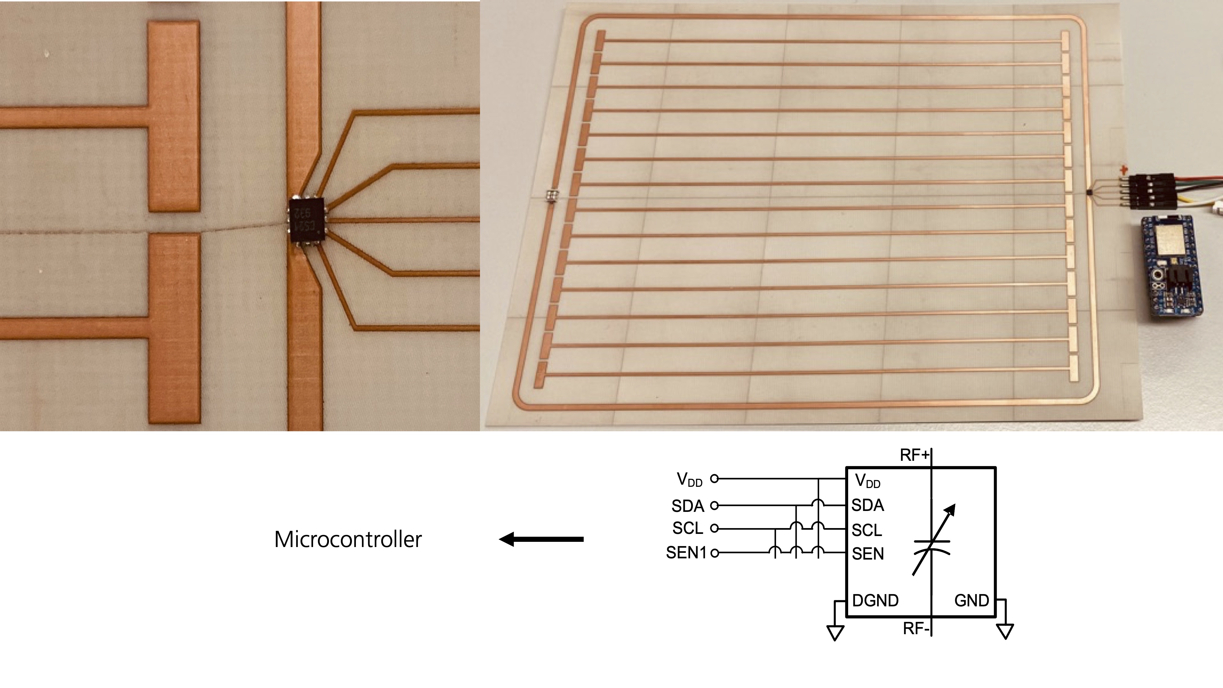
We build on the design of metasurfaces – two-dimensional MTMs – which are manufactured on a single 0.508 mm thin PCB (RO4003C, 17.5 μm-thick Copper layers). The first prototype consists of two inductively coupled structures: 1) a linear metasurface build of flat wire-resonator unit cells, and 2) a limiting diode-loaded loop[1]. The wire-resonators are strongly coupled and are electrically elongated by capacitive end-patches and ground planes on the backside to obtain a resonance frequency at 123.5 MHz. The metasurface can be either seen as an artificial transmission line or an open birdcage coil[12].For the second prototype, the outer detuning loop is not present, but the capacitors used to electrically elongate the wires are digitally-controllable.
For the first prototype, there is a digital capacitor (DCAP) included into the diode-loaded loop. Via an SPI connection, the DCAP is controlled by a Bluetooth-enabled (BLE) microcontroller (MC), which operates at 3.3V and low power, see Fig. 1. The MC is interfaced from a computer via a BLE dongle inserted into the waveguide that leads into the scanner room. Custom-made software allows to control the metasurface at any time and modify its resonance frequency dynamically.
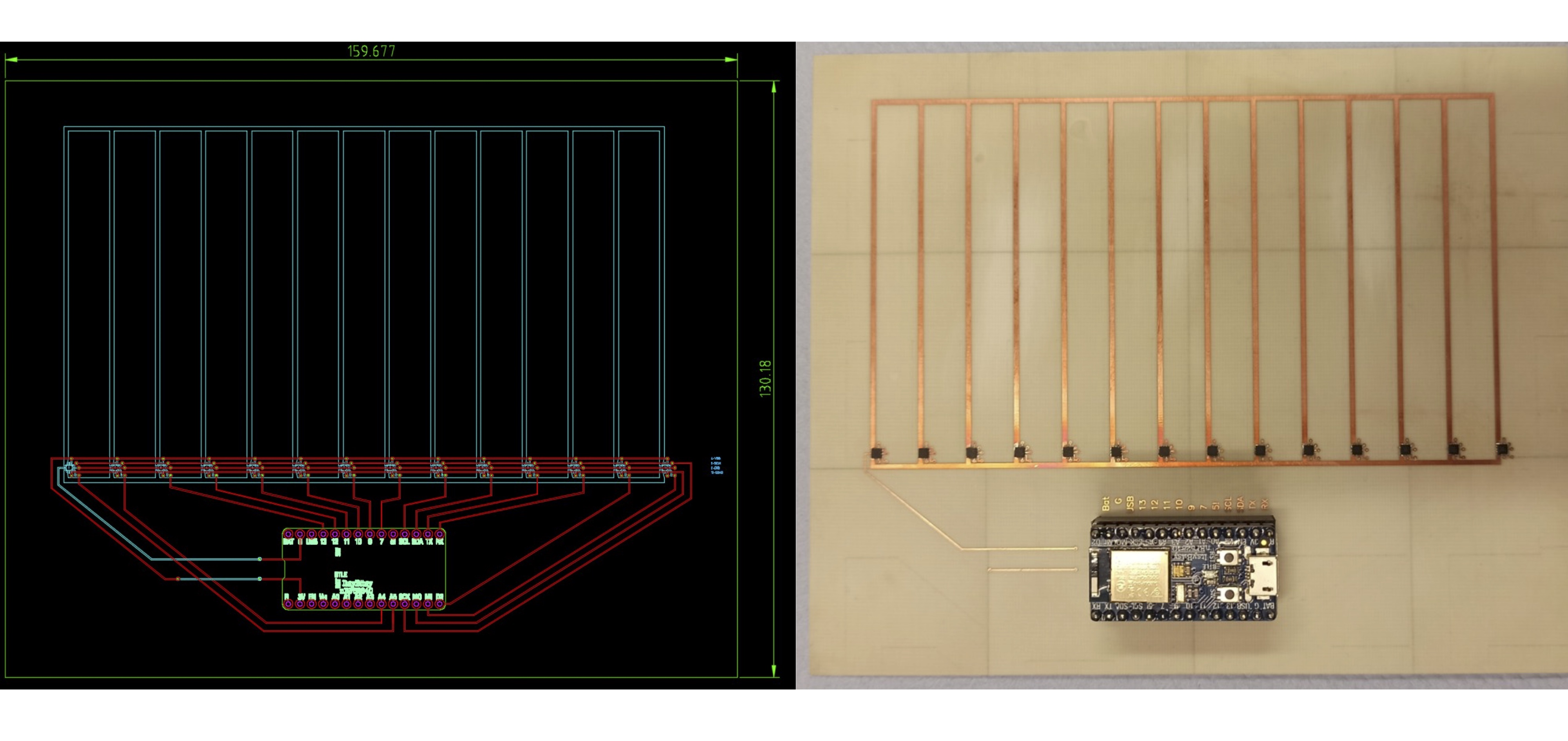
For a second prototype, N = 14 DCAPs are included into all individual wire-resonator unit cells, see Fig. 2. All of them share the SPI interface to the MC but are individually controlled via separate signal-enable lines. Thus, this configuration can be dynamically reconfigured on a local scale. BLE-enabled devices and sensors are in principle compatible with the MRI environment due to the drastically different frequency band[13].
The functionality and wireless control are tested on bench by S-parameter measurements and via MRI experiments. For on-bench measurements, untuned sniffer coils connected to a vector network analyzer are used to characterize the prototypes.
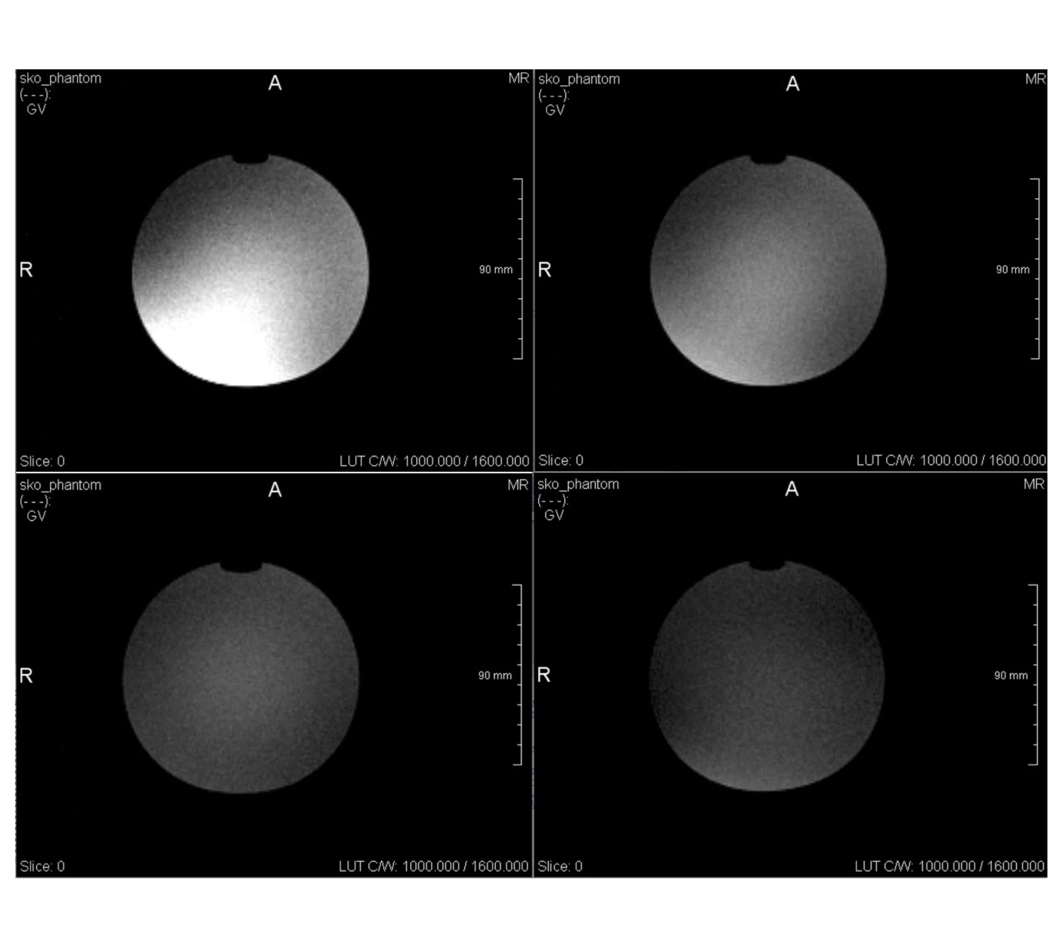
For MRI scans, a gradient echo sequence is used with the following parameters: TR = 100 ms, TE = 5 ms. The imaging is done with a 3 T Magnetom Skyra (Siemens Healthineers, Germany) and i) the scanner’s body coil, ii) the body coil + BLE-MTMs. For all scans, we use the unique, vendor-independent sequence development framework gammaSTAR[14].
Results
The wireless control works under lab and MRI conditions. The first prototype can be tuned in terms of the overall resonance frequency. The second prototype shows spatial reconfigurability and allows to, e.g., shift the first three resonant modes to the scanner’s Larmor frequency. In Fig. 3, exemplary results for MRI tests with the first prototype are shown. The wireless tuning allows to increase the SNR significantly w.r.t. the body coil measurements.Discussion
On-bench measurements of scattering parameters and MRI scans prove the prototypes’ functionality and the wireless tuning mechanism. The electronics works under MRI conditions and the use of non-magnetic components assures device safety. Thus, we have demonstrated the first ever wirelessly interfaced and digitally reconfigurable MTMs for MRI.First MRI experiments with, e.g., higher order resonant modes of the second prototype are ongoing. The successive development will allow for new applications and imaging paradigms to improve, e.g., spatial resolution, imaging speed, and SNR. In particular, locally reconfigurable MTMs allow field shaping capabilities, an adjustable FoV, focal regions, sequence synchronization, and active detuning.
Conclusion
Being a comparatively young research field, MTMs in MRI have a huge potential and are a promising tool to tackle some of the challenges that hamper advancements in the field. With spatial and temporal reconfigurability via fast and low-power wireless interfaces, manifold applications become possible. The field of MTM technology advances from passive structures to dynamical solutions that allow new imaging paradigms and yield entirely new devices for MRI applications.Acknowledgements
This work was supported by the Fraunhofer MAVO project MetaRF. The authors want to thank Peter Erhard for discussions and comments and Michael Jäger for support in the production of prototypes.References
[1] Stoja, E., Konstandin, S., Philipp et al., “Improving magnetic resonance imaging with smart and thin metasurfaces,” Nature Scientific Reports 11, 16179 (2021) DOI: 10.1038/s41598-021-95420-w
[2] Zhao X, et al. Intelligent metamaterials based on nonlinearity for magnetic resonance imaging. Advanced Materials. 2019, Vol. 31, 1905461.
[3] Saha D, et al. A smart switching system to enable automatic tuning and detuning of metamaterial resonators in MRI scans. Sci Rep. 2020, Vol. 10, 10042.
[4] Vesselago V G The electrodynamics of substances with simultaneously negative values of e and m. Soviet Physics Uspekhi. 1968, Vol. 10, 4.
[5] Pendry J B, Schurig D, Smith D R Controlling electromagnetic fields. Science. 2006, Vol. 312, 5781.
[6] Freire M J, et al. On the applications of m = -1 metamaterial lenses for magnetic resonance imaging. Journal of Magnetic Resonance. 2010, Vol. 203.
[7] Duan G, et al. Boosting magnetic resonance imaging signal-to-noise ratio using magnetic metamaterials. Communications Physics. 2019, Vol. 2, 35.
[8] Slobozhanyuk A P, et al. Enhancement of magnetic resonance imaging with metasurfaces. Adv. Mater. 2016, Vol. 28.
[9] Shchelokova A V, et al. Locally enhanced image quality with tunable hybrid metasurfaces. Phys. Rev. Applied. 2018, Vol. 9, 014020.
[10] Kretov E I, Shchelokova A, Slobozhanyuk A P Control of the magnetic near-field pattern inside MRI machine with tunable metasurface. Appl. Phys. Lett. 2019, Vol. 115, 061604.
[11] Alexey P. Slobozhanyuk, Alena V. Shchelokova, Alexander V. Kozachenko, et al. Visualization of Metasurface Eigenmodes with Magnetic Resonance Imaging, Phys. Rev. Applied 16, L021002
[12] Jin, J. (1999). Electromagnetic Analysis and Design in Magnetic Resonance Imaging (1st ed.). Routledge. DOI 10.1201/9780203758731
[13] C. Vogt et al., A wearable bluetooth LE sensor for patient monitoring during MRI scans, 2016 38th Annual International Conference of the IEEE Engineering in Medicine and Biology Society (EMBC), 2016, pp. 4975-4978, DOI: 10.1109/EMBC.2016.7591844.
[14] FraunhoferMEVIS, gammaSTAR, https://gamma-star.mevis.fraunhofer.de
Figures