0183
Low field NMR Relaxometry Studies of Tumours with applications in Intraoperative Tumour Margin Assessment in Breast-Conserving Surgery1Department of Molecular Biotechnology and Health Sciences - Molecular Imaging Center, University of Turin, Torino, Italy, 2Department of Medical Sciences, University of Turin, Torino, Italy, 3Breast Unit, Ospedale Cottolengo, Torino, Italy, 4University of Aberdeen, Aberdeen, United Kingdom, 5IRCCS SDN, Napoli, Italy
Synopsis
A new methodology, based on low field NMR relaxometry, has been employed to characterize breast tumour tissue and to tackle the important clinical need for the assessment of tumour margins during breast-conserving surgery. The method relies on the acquisition of proton longitudinal relaxation rate at low magnetic field strengths which are affected by the changes in the composition of the mammary gland tissue occurring during the development of neoplasia. A sensitivity, specificity, and accuracy of 92%, 85%, and 89%, respectively, were achieved. The information obtained has the potential to support the surgeon in real-time margin assessment.
Introduction
As Breast-Conserving Surgery is routinely applied for treatment of breast cancer (the most common form of cancer for women), the need for new technology to improve intraoperative margin assessment has become increasingly important1. The current gold standard for margin classification is provided by the histopathological evaluation (H&E staining). The method is robust and accurate, but it is time consuming2. About 20–40% of BCS procedures result in margins which are either positive or suspected of having malignant cells at the margins of the resection region and require reoperation3. Several intraoperative margin assessment strategies have been proposed to reduce the need for re-excision, but they all have significant clinical and technical limitations that have hampered widespread adoption4,5. In this study, the potential of fast field-cycling 1H-NMR relaxometry as a new diagnostic tool was evaluated. It is well known that proton longitudinal relaxation rate (R1) values of a given tissue decrease as a function of the applied magnetic field strength, thus providing larger differences among the tissues the lower the applied magnetic field strength is6. It has been shown by our group that the Magnetic Nuclear Relaxation Dispersion (NMRD) profiles can act as a high-sensitivity tool for cancer detection and staging in ex vivo murine breast tissues7 or in breast cancer cell lines8 and in vivo with mice transplanted with murine mammary cancer cells9.Methods
Small freshly excised tissue samples (n=104, with a range of 2.5–8.9 mm Ø) from 41 patients undergoing breast cancer surgery were collected and subjected to R1 measurements at very low magnetic field strengths, then sent to gold standard histopathological evaluation (H&E staining). The acquisition of R1 was performed on a SpinMaster FFC-NMR relaxometer (Stelar S.n.c., Mede, Italy), equipped with a signal-detection microcoil of 10 mm diameter, at 10 °C in the 0.02–1 MHz proton Larmor frequency range (B0 = 0.48 mT–24 mT) pre-polarising the sample at 25 MHz10. The water exchange rate constants across the cell membrane and the extracellular volume fraction were determined by analysing the data of tumour samples using the 2SX model11.Results
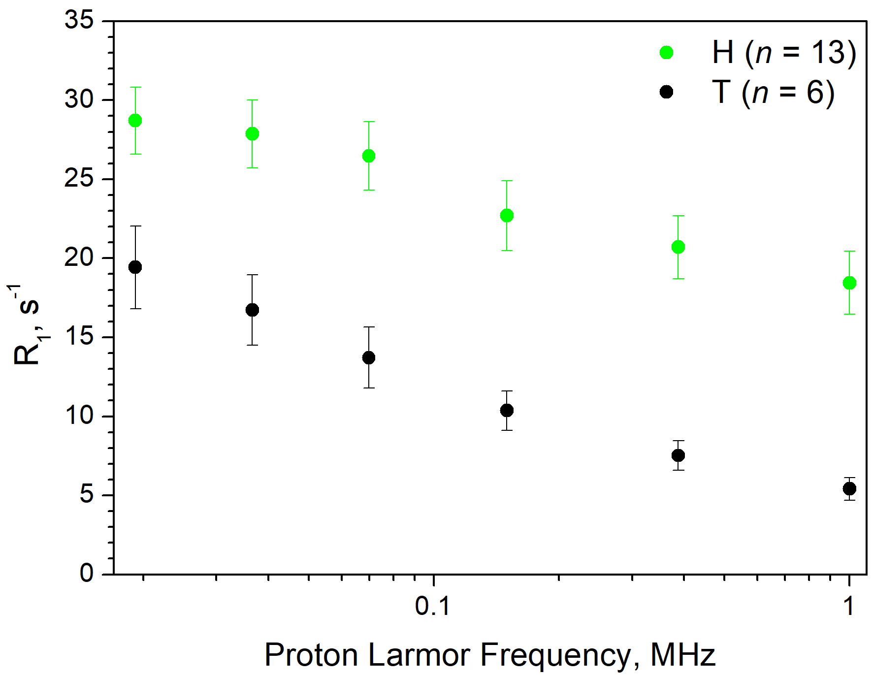
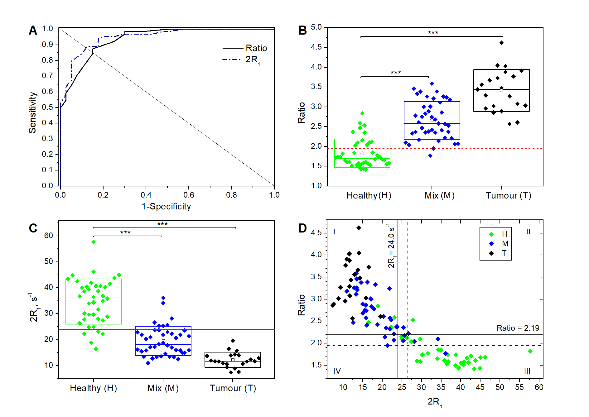
The histological analysis performed by E&H staining allowed the classification of the 104 breast tissue samples investigated as follows: 40 healthy (H), 21 tumours (T), and 43 containing a mixture of both (M). Both H and T tissue relaxation rates increase when the magnetic field strength decreases, but the relative values and slopes of the two curves are significantly different (Fig. 1). Accordingly, we defined two relaxometric quantifiers that captured this behaviour and allowed the assessment of the presence of tumour cells in a breast tissue specimen. The first one was the ratio between the R1 value measured at the lowest (0.02 MHz) and highest (1 MHz) magnetic fields (later referred to as the Ratio). The second one was the sum of the R1 values measured at 0.39 MHz and 1 MHz (later referred to as the 2R1 value). The Receiver Operating Characteristic curve analysis was used to assess the performance of the two quantifiers in the discrimination among negative (H) and positive (M+T) specimens (Fig. 2). The best cutoff values were found to be 2.19 and 24.0 s-1, respectively. The T samples classified as Grade 3 were characterized by high transcytolemmal efflux rate constants.Discussion
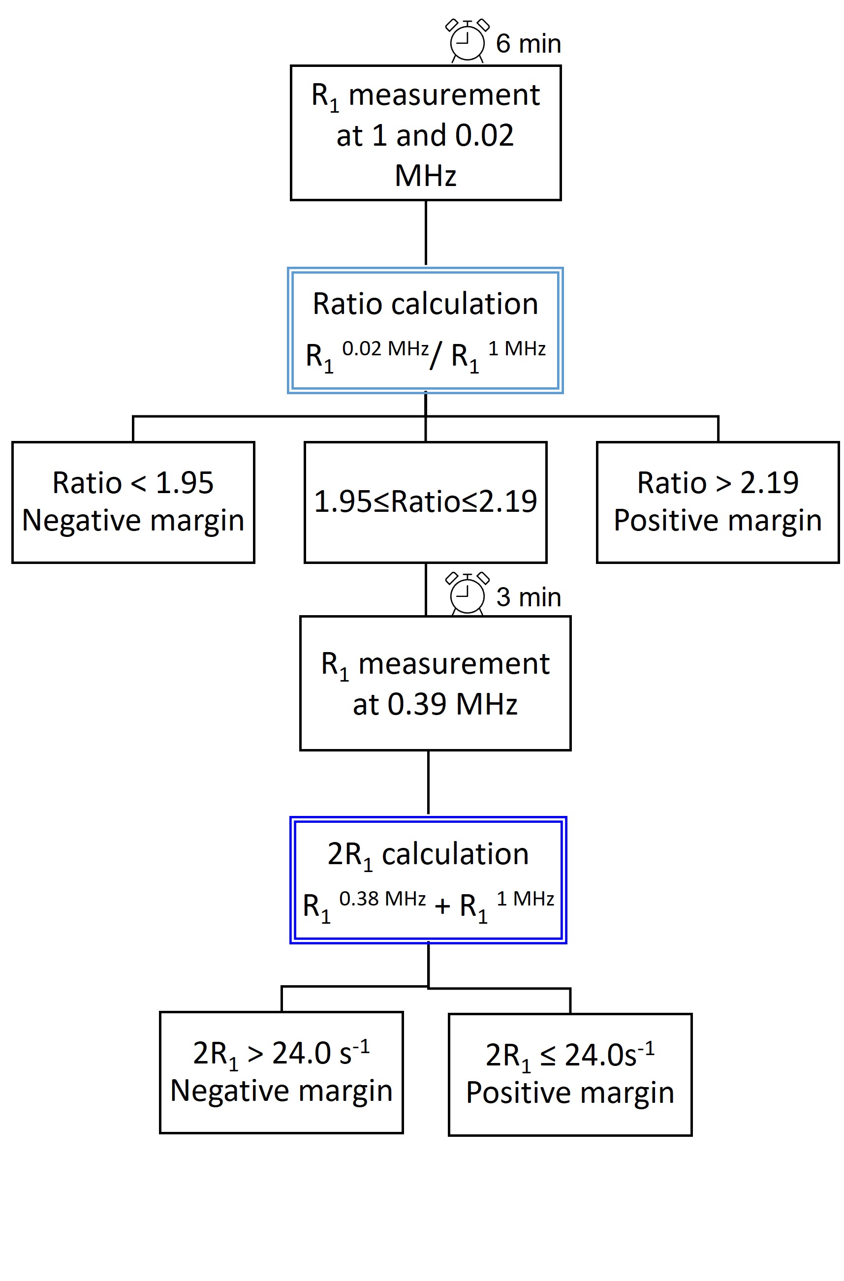
The difference between healthy and malignant tissue appears to be associated with the different water content and water mobility characteristics of the tissues: tumour tissue has a protein/fat/water content that is highly altered with respect to healthy breast tissue, in which adipocytes are dominant and lipids account for up to 70–80% of tissue content. As previously reported7, lipid proton relaxation rates show significatively less dispersion with the magnetic field strength in the range 0.02–10 MHz than water/protein protons. Therefore, relaxation rates of H tissues show higher values and a less pronounced dispersion with the magnetic field with respect to T tissues. Preliminary data indicate that the Ratio and the transcytolemmal efflux rate constants can report on tumour grading. Considering the discriminating ability of the two defined quantifiers, we developed a protocol (Fig. 3) that privileged higher sensitivity over specificity by applying two criteria in a specific order (Ratio first, 2R1 second). The sensitivity (92%) and specificity (85%) attained appear competitive with other methods and well comparable with the best procedures currently proposed for intraoperative tumour margin assessment4,5. The relaxometric method is carried out without requiring any additional treatment of the excised specimen and only needs a reasonable time to complete (currently 6–9 min), which is well compatible within the timeframe of BCS.Conclusion
The proposed relaxometric method has good sensitivity and specificity, is low cost, fast and intraoperatively applicable with minor modifications. We believe that the results reported here clearly indicate that tissue relaxometric data obtained at different magnetic field strengths may provide a useful tool to assess the presence of tumour cells in an excised tissue specimen by a simple quantitative analysis that has the potential to support the surgeon in real-time margin assessment during Breast-Conserving Surgery. Moreover, the water efflux rate constant may be exploited as a specific biomarker giving a fast and specific determination of tumour aggressiveness.Acknowledgements
This work was performed in the frame of the COST Action AC15209 (EURELAX). This project has received funding from the European Union Horizon 2020 research and innovation program under grant agreement No 668119 (project “IDentIFY”) and from AIRC under IG 2019, ID 23267 project (PI Geninatti Crich Simonetta). Maria Rosaria Ruggiero was supported by a “FIRC-AIRC fellowship for Italy”. The authors acknowledge the Italian Ministry of Research for FOE contribution to the Euro-BioImaging MultiModal Molecular Imaging Italian Node (www.mmmi.unito.it).References
1. Schwarz J and Schmidt H. Technology for intraoperative margin assessment in breast Cancer. Ann. Surg. Oncol. 2020;27:2278–2287.
2. Moran MS, Schnitt SJ, Giuliano AE et al. Society of surgical oncology-American society for radiation oncology consensus guideline on margins for breast-conserving surgery with whole-breast irradiation in stages I and II invasive breast cancer. Int. J. Radiat. Oncol. Biol. Phys. 2014;88:553–564.
3. Elmore LC and Margenthaler JA. A tale of two operations: Re-excision as a quality measure. Gland Surg. 2019; 8:593–595.
4. Maloney BW, McClatchy DM, Pogue, BW et al. Review of methods for intraoperative margin detection for breast conserving surgery. J. Biomed. Opt. 2018;23:1–19.
5. Pradipta AR, Tanei T, Morimoto K et al. Emerging Technologies for Real-Time Intraoperative Margin Assessment in Future Breast-Conserving Surgery. Adv. Sci. 2020;7:1901519.
6. Korb JP, Bryant RG. Magnetic field dependence of proton spin-lattice relaxation times. Magn. Reson. Med. 2002;48:21–26.
7. Di Gregorio E, Ferrauto G, Lanzardo S et al. Use of FCC-NMRD relaxometry for early detection and characterization of ex-vivo murine breast cancer. Sci. Rep. 2019;9:1–8.
8. Ruggiero MR, Baroni S, Aime S, et al. Relaxometric investigations addressing the determination of intracellular water lifetime: A novel tumour biomarker of general applicability. Mol. Phys. 2018;117: 968–974.
9. Ruggiero MR, Baroni S, Pezzana S et al. Evidence for the Role of Intracellular Water Lifetime as a Tumour Biomarker Obtained by In Vivo Field-Cycling Relaxometry. Angew. Chemie Int. Ed. 2018;57:7468–7472.
10. Ferrante G and Sykora S. Technical aspects of Fast Field Cycling. Adv. Inorg. Chem. 2005;57:405–470.
11. Springer CS, Li X, Tudorica LA et al. Intratumor mapping of intracellular water lifetime: metabolic images of breast cancer? NMR Biomed. 2014;27:760–773.
Figures