0122
Early tumour response to radiotherapy assessed by magnetic resonance elastography1Division of Radiotherapy & Imaging, The Institute of Cancer Research, Sutton, United Kingdom, 2The Institute of Cancer Research, Sutton, United Kingdom, 3The Royal Marsden NHS Foundation Trust, Sutton, United Kingdom, 4School of Biomedical Engineering and Imaging Sciences, King's College London, London, United Kingdom
Synopsis
Radiotherapy is widely used to treat cancer. In this study, we used MR elastography to evaluate early tumour response to radiation therapy. A significant increase in stiffness |G*| and elasticity Gd were determined 3 days after orthotopic 4T1 breast tumours were irradiated with a single 8Gy dose, compared to untreated control tumour-bearing mice. The increase in tumour stiffness was associated with an increased macromolecule pool measured by magnetisation transfer, and increased collagen deposition observed histologically.
Introduction
In addition to its direct effects on malignant cells, there is increasing appreciation of the role of the tumour microenvironment in influencing response to radiotherapy.1 Numerous tumour types, including breast cancer, are characterized by excessive desmoplastic stromal reaction and aberrant tensional homeostasis. Non-invasive imaging techniques for the early assessment of tumour radiotherapeutic response would enable personalised adaptation of treatment for improved patient outcomes. Magnetic resonance elastography (MRE) is being exploited to image and quantify the viscoelastic properties of tumours in vivo and revealed the markedly stiff nature of orthotopically-propagated breast tumour models, consistent with a dense extracellular matrix (ECM).2,3Radiotherapy can elicit dynamic effects on tumour ECM remodelling.4 In this pre-clinical study, the hypothesis that MRE can inform on early breast tumour response to radiotherapy was tested.
Methods
All experiments were performed in accordance with the UK animal (Scientific Procedures) Act 1996. Murine 4T1 breast tumour cells (1x10⁵) were implanted orthotopically into the third right mammary fat pad of female BALB/c mice (n=7). Around ~11 days later MRI was performed on anaesthetized tumour-bearing mice (~300mm3) using a 7T Biospec 70/20 horizontal system (Bruker, Ettlingen, Germany). Following localization of the tumour using T2-weighted RARE images, MRE was performed by applying a 1,000 Hz mechanical vibration to the tumour and acquiring a spin echo sequence with the following parameters: TE/TR=30/504ms, 2 averages, FOV=19.2x19.2mm, matrix=64x64, slice thickness=0.3mm, 9 transverse slices, scan duration=12min, 4 phase time-points. MT was acquired from two RARE scans with TE/TR=19.22/1500ms, 4 averages, RARE factor=8, FOV=30x30mm, matrix=128x128, slice thickness=1mm, 1 slice, saturation=100kHz (no saturation) or 7.5kHz (macromolecule pool saturation), scan duration=1min36s each. Tumours (n=4) were then treated with a single 8Gy radiation dose administered using a small animal radiation research platform (SARRP, XSTRAHL Ltd), and MRI repeated 3 days later.Data analyses were performed on regions-of-interest covering the whole tumour. The complex shear modulus |G*| (stiffness), Gd (elasticity) and Gl (viscosity) (kPa) were reconstructed from the central three 0.3 mm consecutive slices. The MT ratio (MTR) was derived from the signal depletion from the macromolecule-saturated scan using in-house software (in IDL). Voxel-based histograms were constructed using Matlab (MathWorks). All data plots and statistical analysis were done in Prism 9 (GraphPad).
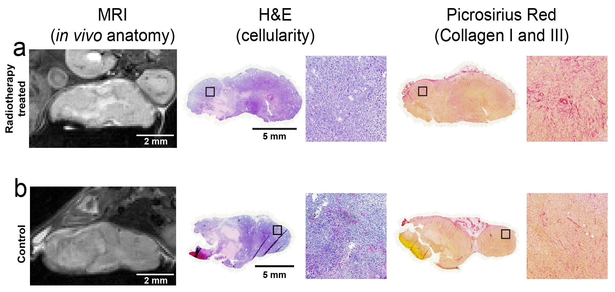
Following post-treatment MRI, tumours were carefully excised, and formalin-fixed paraffin-embedded sections (5µm) aligned with the MRI imaging plane were tinctorially stained with either haematoxylin and eosin (H&E), or picrosirius red, to evaluate cellularity, and collagen I & III, respectively.
Results
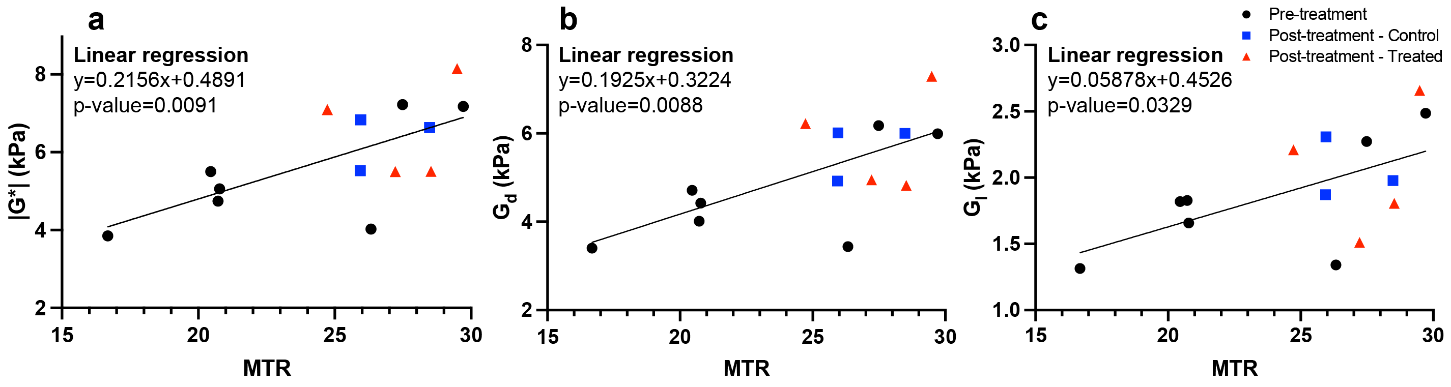
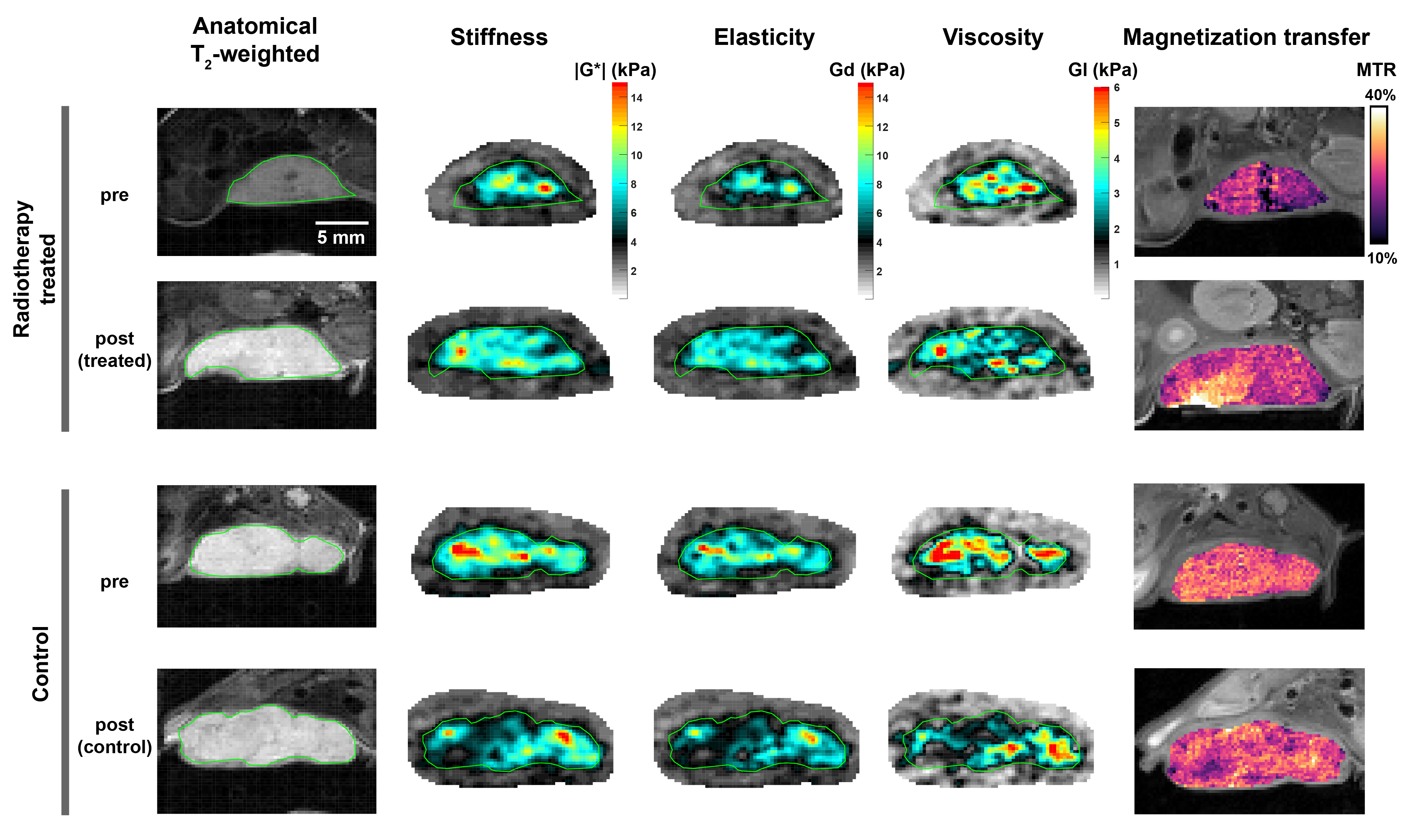
Representative tumour MRE parametric maps along with matching anatomical scans and MTR maps from irradiated and control tumours are illustrated in Figure 1. Voxel values from all control and treated tumours prior to and following radiation are plotted in grouped histograms in Figure 2. The median pre-treatment |G*| for all the tumours was ~5kPa. Radiation-treated tumours showed a marked increase in |G*| after treatment compared to control.To evaluate the change in viscoelasticity induced by the radiation treatment, the median values of all parameters were extracted from each tumour region-of-interest and plotted in Figure 3. A significant (p=0.013) ~47% increase in |G*|was determined in the irradiated group, compared to a ~2% decrease in the control group (Figure 3a). Similar patterns of response were seen for Gd (p=0.014) and Gl (p=0.096). A non-significant ~34% increase in MTR was observed in the irradiated group, compared to a ~6% increase in the control cohort (p=0.2). Irrespective of treatment, a linear relationship was found between the MRE measurements |G*|, Gd and Gl, and MTR (Figure 4).
Histological staining with picrosirius red revealed higher collagen deposition in viable regions of the irradiated tumours (Figure 5).
Discussion and Conclusion
MRE revealed a heterogeneous distribution of viscoelastic properties across the 4T1 tumours, consistent with previous measurements in this model.3 Radiotherapy elicited marked tumour stiffening just 3 days after treatment, with increases in tissue elasticity being the main determinant of this response.The chronology of ECM-remodelling following radiotherapy suggests that an increase in both collagen content and cross-linking occur within days following treatment with an intermediate (4 to 10Gy) radiation dose.4 MRE has been shown to provide sensitive imaging biomarkers of tumour collagen deposition and its therapeutic modulation in a range of preclinical tumour models including breast cancer xenografts.2 The increase in stiffness seen herein may thus reflect radiation-induced changes in collagen organization and reflected in the picrosirius red staining.
Positive linear relationships of MRE-derived |G*| and elasticity Gd with MTR measured by magnetisation transfer suggest an association of elevated tumour stiffness with increased extracellular macromolecule content (e.g. collagen).
In conclusion, early radiation-induced ECM remodelling was detected in murine breast tumours in vivo by MRE. The increase in stiffness and elasticity is consistent with a rearrangement of collagen days after treatment with an intermediate 8Gy dose.
Acknowledgements
We acknowledge support from Cancer Research UK grant C16412/A27725, the Cancer Research UK Centre at the ICR, Children with Cancer UK and the European Union Horizon 2020 Research and Innovation Programme (Grant #668039).
References
1. Barker, H.E., et al., The tumour microenvironment after radiotherapy: mechanisms of resistance and recurrence. Nature reviews. Cancer, 2015. 15(7): p. 409-425.2. Li, J., et al., Investigating the contribution of collagen to the tumor biomechanical phenotype with noninvasive magnetic resonance elastography. Cancer research, 2019. 79(22): p. 5874-5883.
3. Reeves, E.L., et al., MR elastography reveals a marked increased in breast cancer viscoleasticity in vivo following hyaluronan degradation by PEGPH20, in Proc. Intl. Soc. Mag. Reson. 2021.
4. Martinez-Zubiaurre, I., A.J. Chalmers, and T. Hellevik, Radiation-induced transformation of immunoregulatory networks in the tumor stroma. Frontiers in immunology, 2018. 9: p. 1679.
Figures
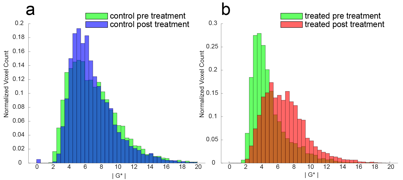
Normalized grouped histograms of the tumour complex shear modulus |G*| in the (a) control and (b) irradiated cohorts.
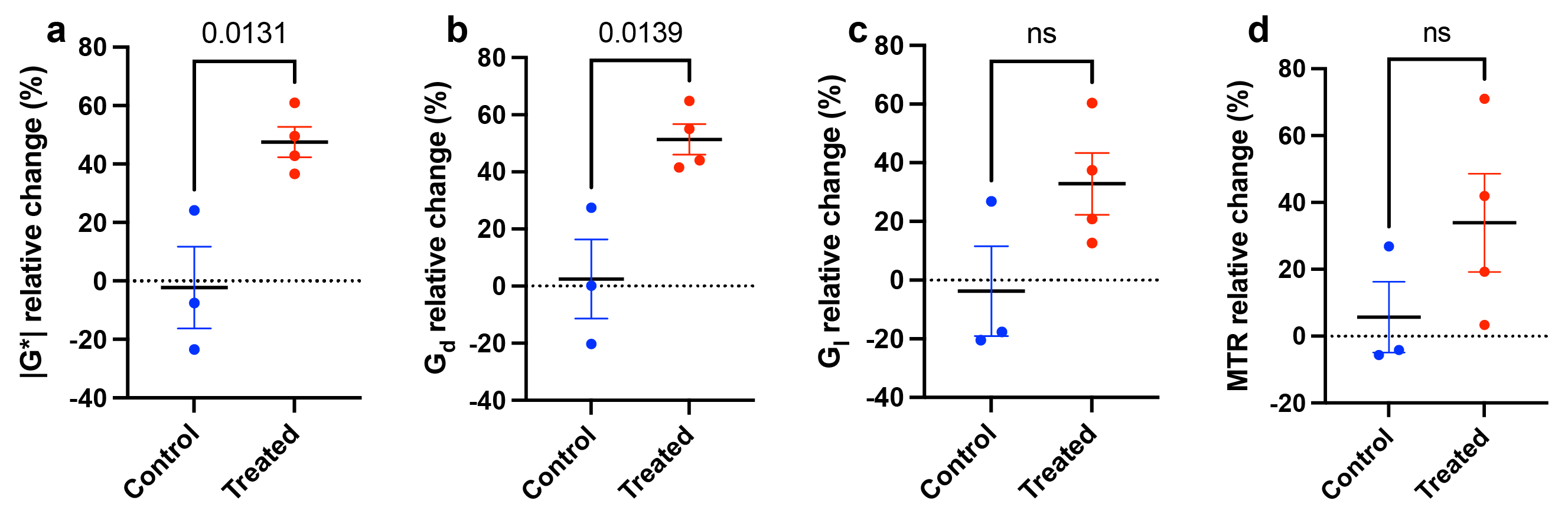
Relative change in tumour (a) complex shear modulus |G*|, (b) elasticity Gd, (c) viscosity Gl and (d) magnetization transfer ratio, determined prior to and after treatment with radiotherapy. Both |G*| and Gd significantly increased in the irradiated group (p<0.05, Student’s unpaired t-test).