0105
Simultaneous 3D T1- and T2-weighted imaging using RF phase-modulated GRE with retrospective contrast adjustment1Radiology, University of Wisconsin-Madison, Madison, WI, United States, 2Biomedical Engineering, University of Wisconsin-Madison, Madison, WI, United States, 3Medical Physics, University of Wisconsin-Madison, Madison, WI, United States, 4Emergency Medicine, University of Wisconsin-Madison, Madison, WI, United States, 5Medicine, University of Wisconsin-Madison, Madison, WI, United States
Synopsis
A novel method to obtain simultaneous 3D T1- and T2-weighted imaging with retrospective contrast adjustment was developed. Acquisitions are performed using a 3D RF phase-modulated GRE acquisition with a small RF phase increment. After phase correction, real and imaginary components are separated into T2- and T1-weighted images, respectively. Image contrast can also be modified by modulating additional phase into the signals retrospectively. Simulation and in vivo examples are shown. The results suggested the proposed method successfully provides arbitrary image contrasts with clinically acceptable quality and short acquisition time.
INTRODUCTION
T1- and T2-weighted acquisitions are widely accepted for clinical MRI exams. Fast spin-echo (FSE) is most common approach for T1w and T2w imaging. FSE-based imaging has the limitation of long acquisition time, despite the use of parallel imaging (1,2) and compressed sensing (3).We propose a method to obtain 3D T1w and T2w images using RF phase modulated gradient echo (GRE), which enables faster acquisition compared to conventional FSE-based imaging (4). However, depending on the underlying T1 and T2 of the tissue, the image contrast may be limited.
In this study, we proposed a new approach to obtain simultaneous T1w and T2w images with retrospective contrast adjustment by modulating signal phase into acquired signals. The results suggested that the proposed method enables improvement of T1w and T2w image contrast.
THEORY
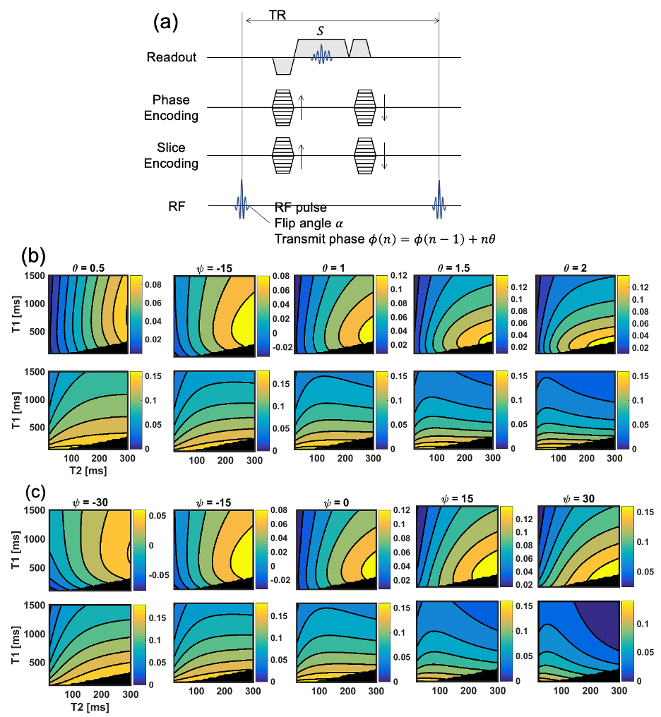
The RF phase-modulated GRE signal can be explain using Sobol's approach(5). The pulse sequence assumed in this study is shown in Figure 1a. The RF phase modulation was performed by incrementing the transmit RF phase (φ) quadratically such that φ(n)=φ(n-1)+nθ, where θ is the RF phase increment. The signal with RF pulse with flip angle (FA) of α, RF phase of φ, and a repetition time of TR can be expressed as$$Re(S) = \beta \eta e^{-\frac{TR}{T2}} \quad [1]$$
$$Im(S) = \beta [\eta^2 - \epsilon (e^{-\frac{TR}{T2}} -\epsilon)] \quad [2]$$
with
$$\beta = \frac{(1-e^{-\frac{TR}{T1}} M_0 sin \alpha) ) }{ (e^{-\frac{TR}{T2}}-\epsilon) [ e^{-\frac{TR}{T2}}(cos \alpha - e^{-\frac{TR}{T1}}) + \epsilon (1-e^{-\frac{TR}{T1}} cos \alpha) ] - \epsilon^2 (1-e^{-\frac{TR}{T1}} cos \alpha) } \quad [3]$$
where $$$M_0$$$ is the proton density, and ε and η are real coefficients, determined by recursive calculation(5). Simulated signal intensities of real and imaginary components shown in Figure 1b shows these components have different response against T1 and T2.
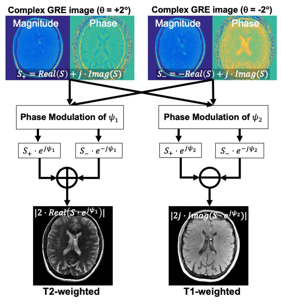
Since two different contrasts are encoded into real and imaginary components separately, it can be adjusted by modulating the signal phase. The signal equations with phase modulation can be expressed as
$$S_1 = Re(S \cdot e^{j \phi_1}) \quad [4]$$
$$S_2 = Im(S \cdot e^{j \phi_2}) \quad [5]$$
where S1 and S2 are real and imaginary components of the phase modulated signal, and Ψ1 and Ψ2 are the modulated phase for S1 and S2 , respectively. Ψ1 and Ψ2 can be determined independently to modulate the T2 and T1 contrast. The real and imaginary signals with varying phase modulation, shown in Figure 1c, suggested that adjustment of image contrast can be achieved.
Two-pass imaging was adopted to remove a background phase, achieved by using acquisition with the positive and negative RF phase increments (Figure 2). T1w and T2w images can be reconstructed using an appropriate value for Ψ1 and Ψ2, chosen to optimize image contrast.
METHODS
All data acquisitions were performed using a clinical 3.0T MRI system (GE Healthcare, Waukesha, WI) with a 48-channel head coil. Parameters for the acquisitions are shown in Table 1.Bloch equation simulation and in vivo studies were performed to discuss the image contrast dependency on Ψ. MR images of a digital brain phantom were calculated using varying Ψ. Also, a volunteer brain, recruited from IRB approved database, was acquired and reconstructed with varying Ψ.
To demonstrate the feasibility of the proposed method, pre- and post-contrast MRI of patients, who have gadolinium enhanced exams, were performed using both standard of care 2D FSE and the proposed method. A total of 8 patients were recruited after obtaining IRB approval.
T1/T2 contrast, and contrast enhancement of the acquired images were evaluated with four-point Likert scale (1=poor, 2=acceptable, 3=good, 4=excellent) by two board-certified radiologists. Additionally, the presence and severity of motion artifacts (1=severe, 2=substantial apparent artifact, 3=mild artifact, 4=no apparent artifact) was evaluated.
RESULTS
Modulating Ψ also affects image contrast, as shown in Figure 3a. Positive phase modulation weakens T2w contrast from the real component, whereas a negative phase provides stronger T2w. In contrast to the real component, T1w contrast becomes stronger as increases. Similar tendency was observed in the results of in vivo study shown in Figure 3b.Figure 4 shows an example pre- and post-T1w/T2w images of a patient, compared to FSE images. The evaluation by the radiologist suggested the contrast (Reader 1: 3.0±0.0, Reader 2: 3.75±0.71) and enhancement quality (Reader 1: 3.6±0.53, Reader 2: 3.5±1.1) are good to excellent. Also, minor motion artifacts were observed (Reader 1: 2.6±0.52, Reader 2: 2.6±0.52).
DISCUSSION
In this work, we have proposed and demonstrated the feasibility of a novel 3D RF phase-modulated GRE imaging technique to obtain simultaneous T1- and T2-weighted images with retrospective contrast adjustment. Simulation and in vivo studies demonstrated the feasibility of the proposed method.The proposed strategy may offer an alternative approach to the acquisition of acquisition of sequentially acquired T1w and T2w imaging. Further, the short acquisition time may be advantageous for rapid or focused MRI protocols that require both contrast mechanisms as part of the standard examination.
There are several limitations to this work that should be addressed, such as the presence of inter-pass motion, the effects of short T2*, and a limited cohort size of clinical evaluation.
In conclusion, we have successfully developed RF phase-modulated GRE method for simultaneous 3D T1w and T2w imaging with a retrospective reconstruction to provide enhanced T1w and T2w image contrasts.
Acknowledgements
The authors acknowledge support from UW ICTR through CTSA grant UL1TR002373 from NIH/NCATS. Further, the authors acknowledge GE Healthcare who provides research support to the University of Wisconsin-Madison. Finally, Dr. Reeder is a Romnes Faculty Fellow, and has received an award provided by the University of Wisconsin-Madison Office of the Vice Chancellor for Research and Graduate Education with funding from the Wisconsin Alumni Research Foundation.References
1. Pruessmann KP, Weiger M, Scheidegger MB, Boesiger P. SENSE: sensitivity encoding for fast MRI. Magnetic Resonance in Medicine: An Official Journal of the International Society for Magnetic Resonance in Medicine 1999;42(5):952-962.
2. Griswold MA, Jakob PM, Heidemann RM, Nittka M, Jellus V, Wang J, Kiefer B, Haase A. Generalized autocalibrating partially parallel acquisitions (GRAPPA). Magnetic Resonance in Medicine: An Official Journal of the International Society for Magnetic Resonance in Medicine 2002;47(6):1202-1210.
3. Lustig M, Donoho D, Pauly JM. Sparse MRI: The application of compressed sensing for rapid MR imaging. 2007;58(6):1182-1195.
4. Tamada D, Field AS, Reeder SB. Simultaneous T1- and T2-Weighted 3D MRI Using RF Phase-Modulated Gradient Echo Imaging. Magnetic Resonance in Medicine 2021;Accepted on October 25, 2021.
5. Sobol WT, Gauntt DM. On the stationary states in gradient echo imaging. Journal of Magnetic Resonance Imaging 1996;6(2):384-398.
Figures