0069
MIITRA atlas: T1w and DTI templates of the older adult brain in a common space with 0.5mm resolution1Biomedical Engineering, Illinois Institute of Technology, Chicago, IL, United States, 2Rush Alzheimer's Disease Center, Rush University Medical Center, Chicago, IL, United States
Synopsis
As part of the Multichannel Illinois Institute of Technology & Rush university Aging (MIITRA) atlas project, the present work (A) developed high quality 0.5mm isotropic resolution T1-weighted (T1w) and diffusion tensor imaging (DTI) templates in common space using data from a large, diverse, community cohort of non-demented older adults, and (B) compared the new templates to existing templates of the older and younger adult brain. The new templates exhibited higher image quality, were more representative of the older adult brain, and allowed higher spatial normalization accuracy across subjects and across modalities compared to other templates.
Introduction
High quality multimodal MRI templates of the older adult brain that are located in a common space and are of high spatial resolution have the potential to enhance the sensitivity and accuracy of template-based neuroimaging studies of older adults. The Multichannel Illinois Institute of Technology & Rush university Aging (MIITRA) atlas aims to accomplish this goal. The present work: (A) developed high quality 0.5mm isotropic resolution T1-weighted (T1w) and diffusion tensor imaging (DTI) templates in common space using data from a large, diverse, community cohort of non-demented older adults, and (B) compared the new templates to existing templates of the older and younger adult brain in terms of image quality and accuracy in spatial normalization of older adult data.Methods
DataT1w (1x1x1 mm3) and DTI (2x2x2 mm3) data were collected using two 3T MRI scanners on 400 non-demented older adults (50% male; 64.9-98.9 years of age; 54% white, 43% black) participating in longitudinal cohort studies of aging1,2.
Multimodal template construction
The template construction process combined recently introduced methods for the development of unbiased, detail-preserving templates3, high quality multimodal templates in common space4, and high spatial resolution templates based on principles of super-resolution5,6.
Step 1: T1w data were center-aligned and groupwise normalized by pairwise affine registration to create an unbiased initial T1w template3.
Step 2: ANTs7 registration was driven by T1w data, and the resulting transformations were also applied to the DTI data.
Step 3: DR-TAMAS8 registration was driven by DTI data, and the resulting transformations were also applied to the T1w data.
Steps 4, 5: Steps 2 and 3 were repeated.
Steps 2-5 followed iterative template building9,10 with higher amount of warping allowed with more iterations. The transformations from each step were combined to minimize interpolations. To account for residual misregistration, a patch-based tissue-guided sparse-representation approach was employed instead of simple averaging when computing the template3.
Step 6: The final transformations were used to forward map signals from raw space to exact physical locations in template space, eliminating interpolations that occur in conventional template building methods5,6. The transformed images were averaged using the sparse-representation approach3. The resulting templates are referred to in the following as MIITRA_0.5mm T1w and DTI templates.
Evaluation
T1w and DTI data from 202 ADNI311 non-demented older adults (50% male, 65-93.2 years) were used for evaluation.
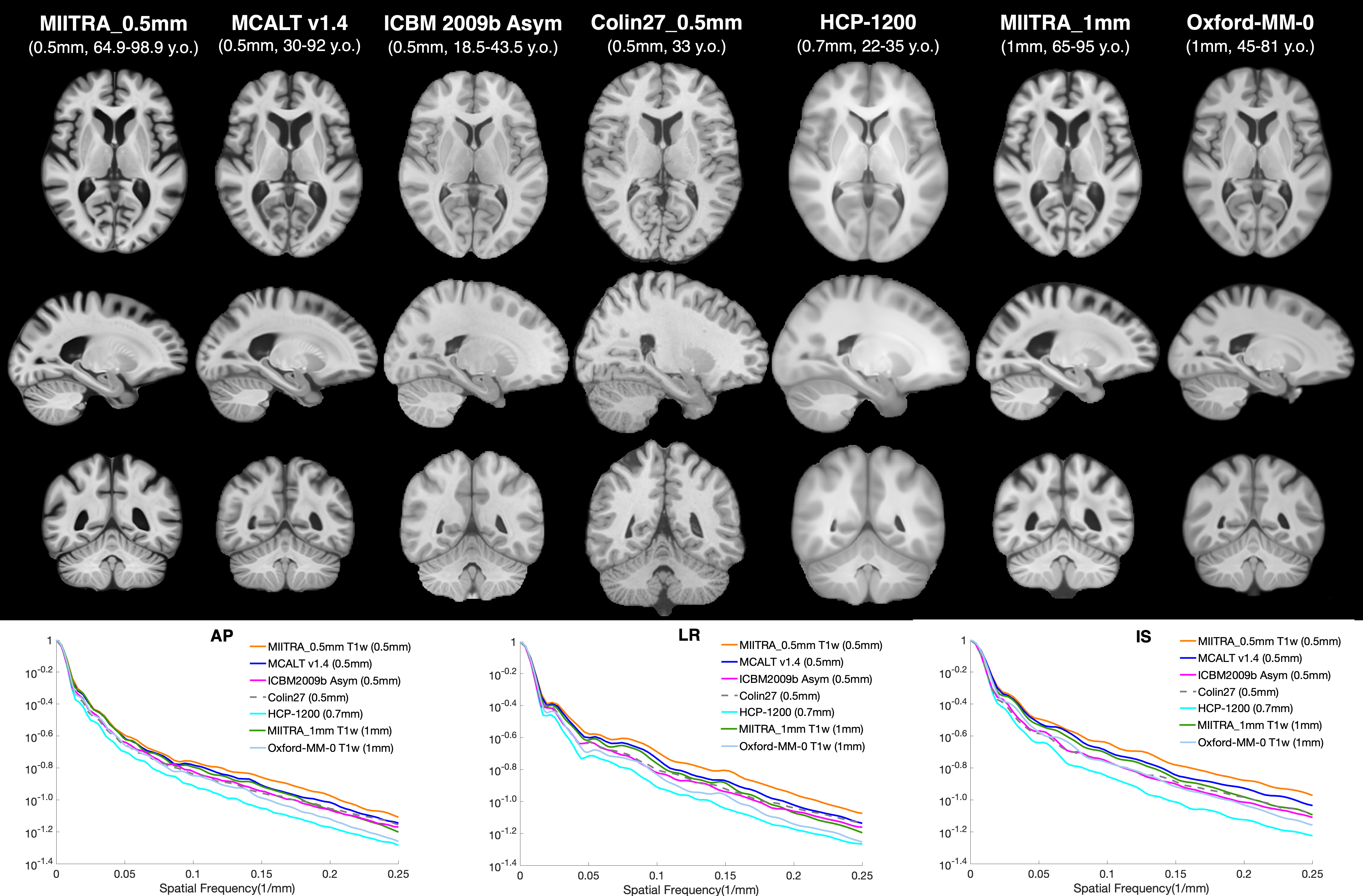
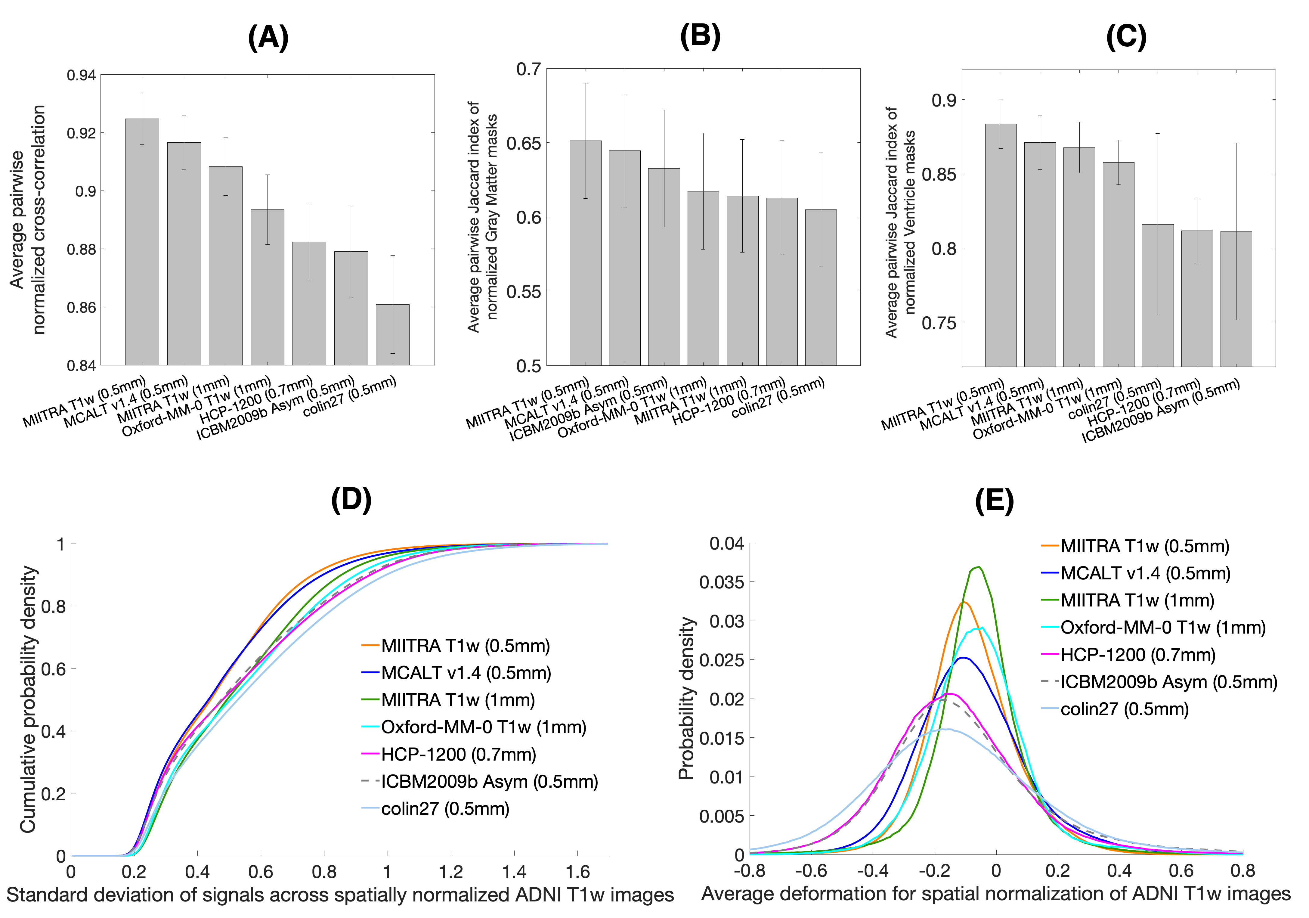
The MIITRA_0.5mm T1w template was compared to other high resolution templates (MCALTv1.4_0.5mm12, ICBM2009b_Asym_0.5mm9,13, colin27_0.5mm14, HCP1200_0.7mm15), as well as 1mm resolution templates (MIITRA_1mm10, Oxford-MM-0 T1w16). Only the MIITRA templates were constructed exclusively from data on older adults (Fig.1). The templates were compared in terms of image sharpness and inter-subject spatial normalization accuracy of ADNI3 T1w data (ANTs7 registration). Normalization accuracy was assessed by the average pairwise normalized cross correlation (APNCC) and average pairwise Jaccard index (APJI) of gray matter and ventricle masks across normalized T1w images. The average log-Jacobian determinant of the deformations was also calculated.
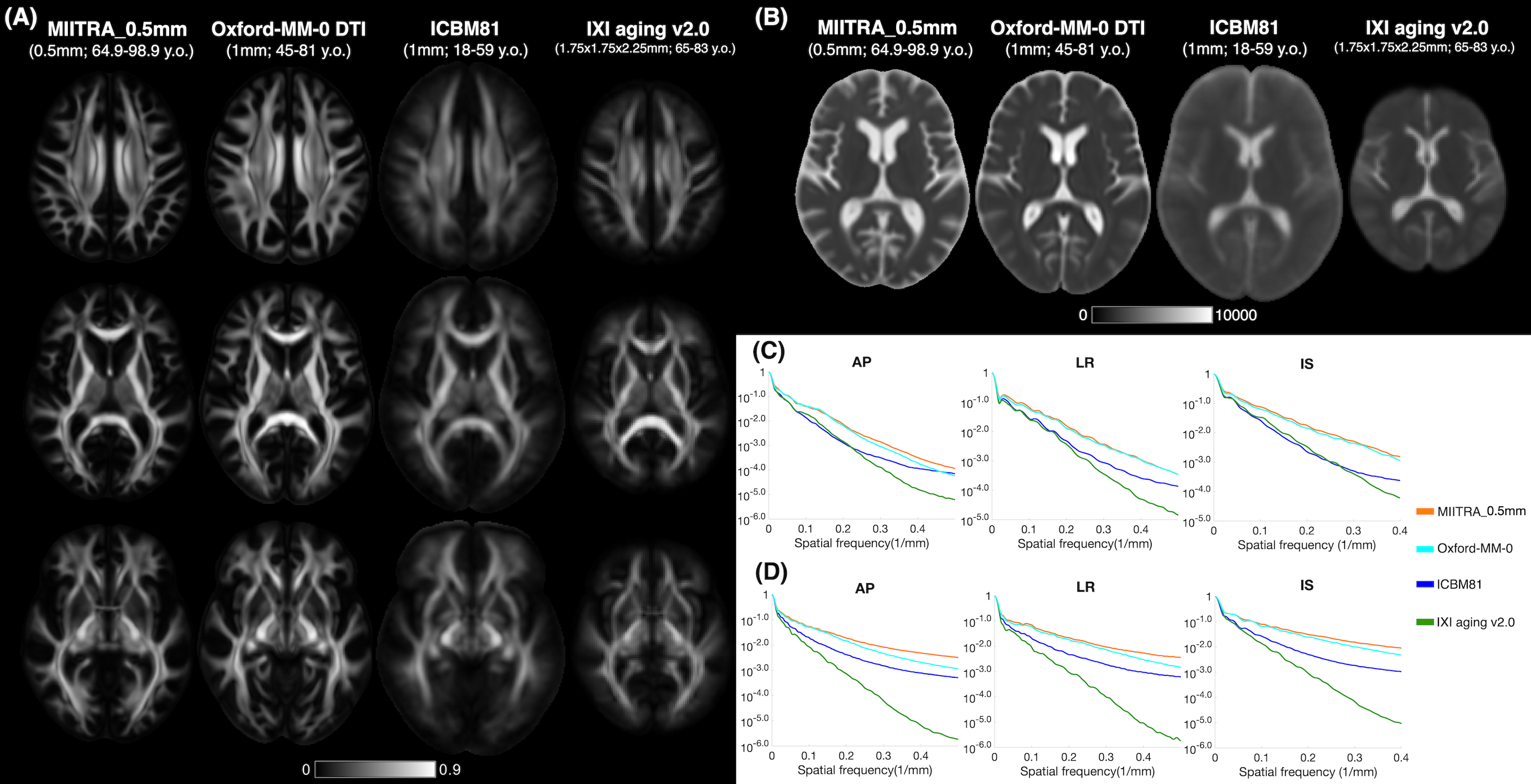
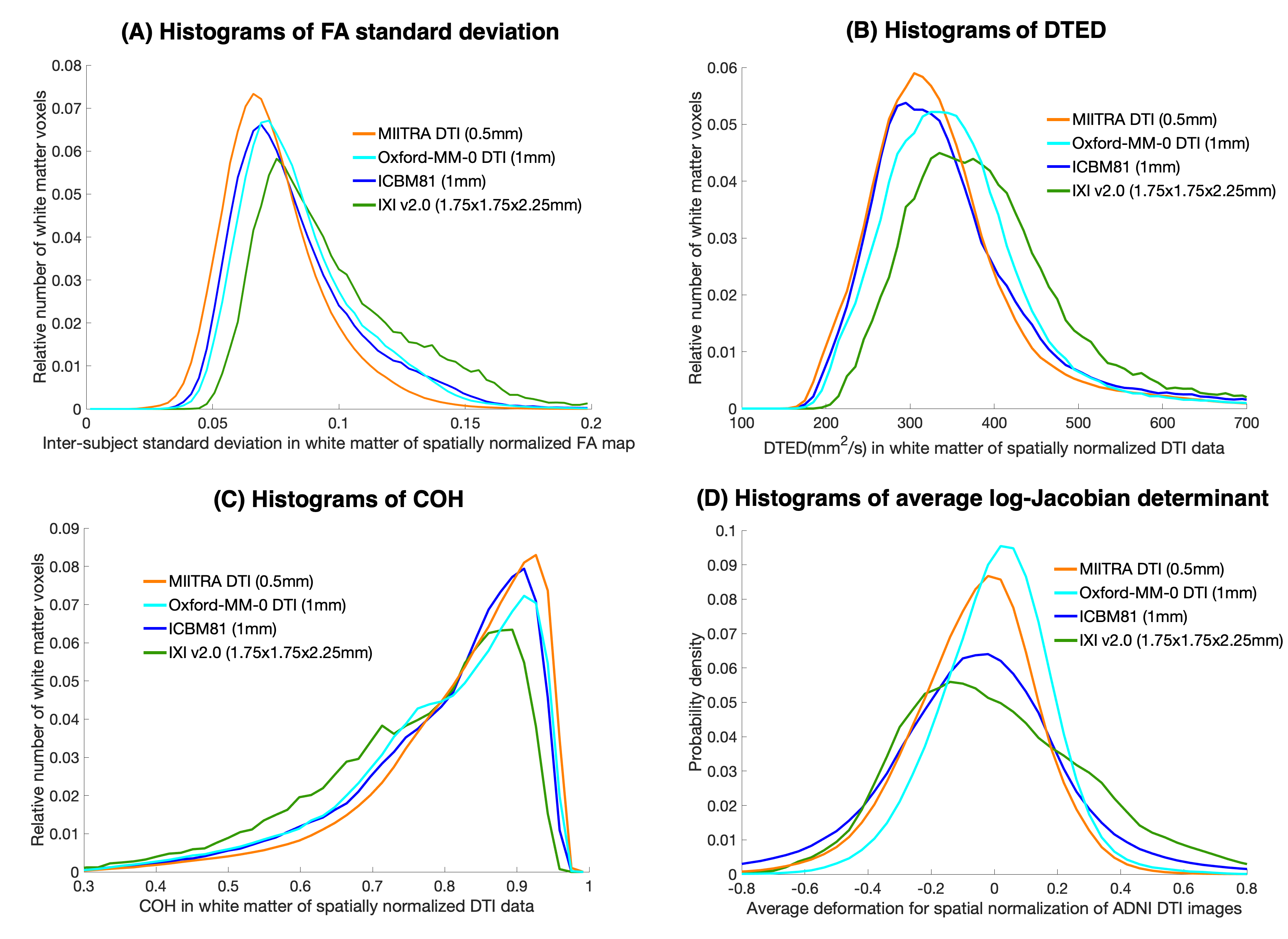
The MIITRA_0.5mm DTI template was compared to other DTI templates (Oxford-MM-0 DTI16, IXI v2.017, ICBM8118) none of which, however, was of high resolution, and only IXI v2.0 was also constructed exclusively from data on older adults. DTI templates were compared in terms of image sharpness of FA and trace maps and inter-subject spatial normalization accuracy of ADNI3 DTI data (DR-TAMAS8 registration). Normalization accuracy was assessed by the standard deviation of FA maps, average pair-wise Euclidean distance (DTED)19 and coherence of primary eigenvectors (COH)19 across normalized DTI data. The average log-Jacobian determinant of the deformations was also calculated.
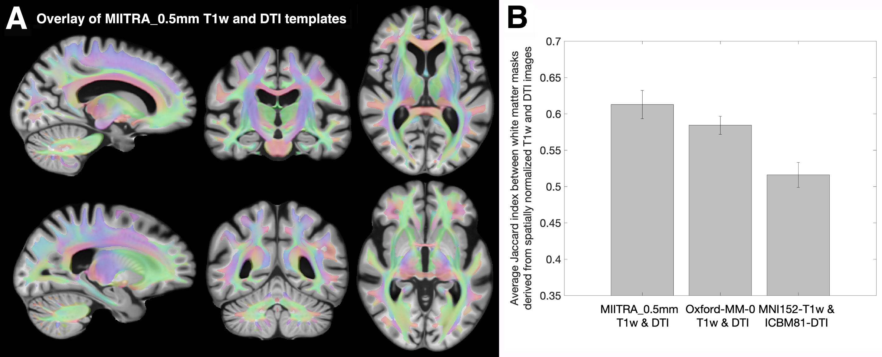
The inter-modality spatial matching achieved with the MIITRA_0.5mm templates was compared to that achieved with other multimodal templates (Oxford-MM-016; MNI152-T1w20and ICBM81-DTI18) by means of the average Jaccard index between white matter masks generated from T1w images and FA maps of spatially normalized ADNI3 data.
Results
MIITRA_0.5mm T1w (Fig.1) and DTI (Fig.2) templates exhibited higher image sharpness compared to other templates as demonstrated by larger high spatial frequency content in the normalized power spectra. MIITRA_0.5mm T1w allowed higher APNCC, higher APJI and lower standard deviation for spatial normalization of ADNI3 data compared to other T1w templates (Fig.3A,B,C,D). MIITRA_0.5mm, MIITRA_1mm and Oxford-MM-0 T1w required less deformation than other T1w templates (Fig.3E). The MIITRA_0.5mm DTI template allowed lower standard deviation of FA maps, lower DTED and higher COH across spatially normalized ADNI3 data compared to other DTI templates (Fig.4A,B,C). MIITRA_0.5mm and Oxford-MM-0 DTI required less deformation than other DTI templates (Fig.4D). It should be noted that 1mm templates require on average less deformation than high resolution templates because the larger voxels limit local deformations. Finally, the MIITRA_0.5mm T1w and DTI templates exhibited high spatial matching to each other and allowed higher inter-modality spatial matching of normalized older adult data (Fig.5).Discussion
This work developed high resolution T1w and DTI templates of the older adult brain in common space for the MIITRA atlas using data from a large, diverse, community cohort of non-demented older adults. The new templates exhibited higher image quality, were more representative of the older adult brain, and allowed higher spatial normalization accuracy across subjects and across modalities compared to other templates. The new templates will be made available for download at www.nitrc.org/projects/miitra.Acknowledgements
This study was supported by:
National Institute on Aging (NIA) R01AG052200
National Institute on Aging (NIA) P30AG010161
National Institute on Aging (NIA) P30AG072975
National Institute on Aging (NIA) R01AG017917
National Institute on Aging (NIA) RF1AG022018
National Institute on Aging (NIA) R01AG056405
References
1. Bennett DA, Buchman AS, Boyle PA, et al. Religious Orders Study and Rush Memory and Aging Project. J Alzheimers Dis. 2018;64(s1):S161-S189.
2. Barnes LL, Shah RC, Aggarwal NT, et al. The Minority Aging Research Study: Ongoing Efforts to Obtain Brain Donation in African Americans without Dementia. Curr Alzheimer Res. 2012;9(6):734-745.
3. Ridwan AR, Wu Y, Niaz MR, et al. Construction of an unbiased high resolution and detail-preserving structural T1-weighted template for use in studies on older adults. Proc. Intl. Soc. Mag. Reson. Med. 2021; 29.
4. Wu Y, Ridwan AR, Qi X, et al. Development of High Quality T1w and DTI Templates of the Older Adult Brain in a Common Space. Proc. Intl. Soc. Mag. Reson. Med. 2019;27.
5. Niaz MR, Ridwan AR, Qi X, et al. Development and evaluation of a 0.5mm isotropic resolution structural template of the older adult brain. Proc. Intl. Soc. Mag. Reson. Med. 2019;27.
6. Niaz MR, Wu Y, Ridwan AR, et al. Development and evaluation of a high spatial resolution diffusion tensor template of the older adult human brain. Proc. Intl. Soc. Mag. Reson. Med. 2020;28.
7. Avants et al. ANTS: Open-Source Tools for Normalization And Neuroanatomy.
8. Irfanoglu MO, Nayak A, Jenkins J, et al. DR-TAMAS: diffeomorphic registration for tensor accurate alignment of anatomical structures. Neuroimage. 2016;132:439–454.
9. Fonov V, Evans AC, Botteron K, et al. Unbiased average age-appropriate atlases for pediatric studies. Neuroimage. 2011;54(1):313-27
10. Ridwan AR, Niaz MR, Wu Y, et al. Development and evaluation of a high performance T1-weighted brain template for use in studies on older adults. Hum Brain Mapp. 2021;42:1758-1776.
11. http://adni.loni.usc.edu/
12. Schwarz CG, et al. THE MAYO CLINIC ADULT LIFE SPAN TEMPLATE: BETTER QUANTIFICATION ACROSS THE LIFE SPAN. Alzheimer's & Dementia: The Journal of the Alzheimer's Association. 2017;13(7):93-4.
13. Fonov VS, Evans AC, McKinstry RC, et al. Unbiased nonlinear average age-appropriate brain templates from birth to adulthood. NeuroImage. 2009(47):S102.
14. Aubert-Broche B, EvansAC, Collins DL, A new improved version of the realistic digital brain phantom. NeuroImage. 2006;32(1):138–45.
15. Glasser MF, Sotiropoulos SN, Wilson JA, et al. The minimal preprocessing pipelines for the Human Connectome Project. Neuroimage. 2013;80:105-124.
16. Arthofer C, Smith SM, Jenkinson M, Andersson J, Lange F. Multimodal MRI template construction from UK Biobank: Oxford-MM-0. Organisation for Human Brain Mapping (OHBM). 2021.
17. Zhang H, et al. A computational white matter atlas for aging with surface-based representation of fasciculi. Biomedical Image Registration. WBIR 2010. Lecture Notes in Computer Science, vol 6204.
18. Mori S, et al. Stereotaxic white matter atlas based on diffusion tensor imaging in an ICBM template. Neuroimage 2008;40(2):570-582.
19. Zhang S., et al. Enhanced ICBM diffusion tensor template of the human brain. Neuroimage. 2011;54(2):974-84.
20. Grabner G,Janke AL, Budge MM, et al. Symmetric atlasing and model based segmentation: an application to the hippocampus in older adults. Med Image Comput Comput Assist Interv. 2006;9:58–66.
Figures