0045
Perfusion Observations using Spherical Slice-shuffled Ultra-high-field MRI (POSSUM)
Jana Hutter1,2, Raphael Tomi-Tricot3, Thomas A Wilkinson1,2, Philippa A Bridgen1,2, Enrico DeVita2, Shaihan A Malik1,2, and Joseph V Hajnal1,2
1London Collaborative Ultra high field System (LoCUS), London, United Kingdom, 2Centre for Medical Engineering, King's College London, London, United Kingdom, 3Siemens Healthcare Limited, Frimley, United Kingdom
1London Collaborative Ultra high field System (LoCUS), London, United Kingdom, 2Centre for Medical Engineering, King's College London, London, United Kingdom, 3Siemens Healthcare Limited, Frimley, United Kingdom
Synopsis
Spin tagging perfusion MR imaging allows the movement of blood in human tissue to be visualized and quantified non-invasively and contrast agent-free. Velicity-selection Arterial Spin Labeling (VSASL) relies on the velocity instead of the location for tagging. It is less dependent on the anatomy and coil coverage but includes directional dependence and can suffer from reduced SNR. POSSUM expands VSASL to spherical, directional-independant encoding and slice shuffling to enable more robust perfusion imaging at 7T.
Introduction
Spin-tagging perfusion MR imaging allows the movement of blood in tissue to be visualized and quantified non-invasively. Commonly, a preparation module is followed by the imaging module. While most preparations tag blood in a labelling location and then observe its inflow into the imaging region, velocity-selective arterial spin labeling (VSASL) labels based on velocity and not position [1,2]. Spins flowing in a chosen direction $d$ above a velocity $v_c$ are labeled globally to focus on blood entering small arteries and capillary beds resulting in reduced anatomy and transit time dependency. Furthermore, it facilitates perfusion imaging on 7T, where the use of local transmit coils inherently raises challenges for distal tagging such as in the carotid arteries. It does, however, impose an SNR penalty as it crushes rather than inverts the magnetization, as well as a dependency on the chosen labelling direction $d$. Another common challenge for spin tagging perfusion methods is the background suppression (BGS) using inversion pulses. The continuous recovery during the imaging module leads to inhomogeneity between slices and limits the number slices that can be sampled.Moving to higher field strengths allows the SNR challenge to be addressed through both an advantage of longer blood T1, and the increase in intrinsic sensitivity [3]. This work presents “Perfusion Observations using Spherical Slice-shuffled Ultra-high-field MRI (POSSUM)” designed to address the other two VASL challenges:(1) Directional dependency: We make tagging direction free by introducing tensor-valued encoding gradients, as proposed and employed in diffusion MRI [4,5]. (2) Coverage and slice signal variation: We Include slice-shuffling to acquire every slice at every time point during the post BGS recovery curve.Methods
A Siemens Terra product ASL sequence using single-shot Echo Planar Imaging was modified to include the above described VSASL and BGS modules (see Figure 1a). The VSASL preparation consists of two 900 and two 1800 RF pulses and the velocity sensitising gradients parametrized by their duration $\delta$, the distance $\Delta$ (see Figure 1b) and the chosen maximal gradient amplitude $G_{max}$. The control acquisition has no gradients included in the first labelling module. The BGS module consists of two background suppression pulses. In the following a dynamic pair will mean two successive volumes acquired with control/tagging modules. To achieve isotropic labelling, the conventional single axis simple gradient pulses (1b) were replaced by continuous free waveform gradients on all three axes (Figure 1c). Next, the sequence was modified to enable dynamic modification of the used perfusion preparation (shape, direction and $G_max$) for each dynamic, facilitating motion-robust acquisition. Finally, the slice order was modified to allow dynamic shuffling for each volume by a parameterizable length (see Figure 1d).Two volunteers were scanned on a 7T scanner (MAGNETOM Terra, Siemens Healthcare, Erlangen, Germany) with an 8-ch transmit/32-ch receive head coil (Nova Medical, Wilmington MA, USA) using the modified POSSUM sequence. The following parameters were employed for the data shown below: FOV=240x240mm, resolution=2.7x2.7mm, slice thickness=3mm, 20 slices, TE=59ms, TR=4s, BGS pulses at 1000ms/2000ms, VSASL parameters were $\delta$=10ms, $\Delta$=40ms, PLD=3s. 20 dynamics were acquired with linear encoding on d1=(1,0,0), d2=(0,1,0) and d3=(0,0,1) and 20 dynamics with spherical encoding (using previously generated waveforms [4]) in a total acquisition time of 4:52 (1:14 for the spherical encoded data alone). Basic perfusion analysis was performed to obtain perfusion-weighted maps by averaging the difference between subsequent pairs of control and tag dynamics.Results and discussion
The POSSUM sequence was successfully acquired and perfusion weighted maps calculated individually for the three directions d1, d2 and d3 with linear encoding, for their combination (pseudo isotropic) and with spherical encoding (Fig 2). The dynamics acquired with linear encoding applied in different directions display clear directional dependencies, the spherical encoded perfusion weighted map on the right shows a more homogeneous pattern throughout the brain.The proposed POSSUM sequence opens an additional dimension for VSASL imaging, adding the shape of the encoding tensor as an additional degree of freedom and thus increasing robustness to varying vascular anatomy and transit times. For the present study, only one free waveform corresponding to spherical tensor encoding was evaluated but the framework developed allows exploration of further combinations as previously successfully shown in dMRI [5]. Similarly, while the data acquired at different T1 times due to the slice shuffling was averaged for this study, ongoing work focuses on extracting additional T1 information by including the slice-level varying TI and PLD times into the analysis.Acknowledgements
This work was supported by the NIH Human Placenta Project grant 1U01HD087202-01 (Placenta Imaging Project (PIP)), Wellcome Trust Collaboration in Science grant [WT201526/Z/16/Z], a Sir Henry Wellcome Fellowship, a UKRI FL fellowship and by core funding from the Wellcome/EPSRC Centre for Medical Engineering [WT203148/Z/16/Z] and by the National Institute for Health Research (NIHR) Biomedical Research Centre based at Guy’s and St Thomas’ NHS Foundation Trust and King’s College London and/or the NIHR Clinical Research Facility. The views expressed are those of the author(s) and not necessarily those of the NHS, the NIHR or the Department of Health and Social Care.References
[1] Wong, E.C., Cronin, M., Wu, W.-C., Inglis, B., Frank, L.R. and Liu, T.T. (2006), Velocity-selective arterial spin labeling. Magn. Reson. Med., 55: 1334-1341[2] van Osch MJ, Teeuwisse WM, Chen Z, Suzuki Y, Helle M, Schmid S. Advances in arterial spin labelling MRI methods for measuring perfusion and collateral flow. J Cereb Blood Flow Metab. 2018 Sep;38(9):1461-1480[3] X. Zhang,E. T. Petersen, E. Ghariq,J. B. De Vis,A. G. Webb,W. M. Teeuwisse,J. Hendrikse,M. J. P. van Osch, In vivo blood T1 measurements at 1.5 T, 3 T, and 7 T, Magn. Reson. Med., 70: 1082-1086.[4] Nilsson, M, Szczepankiewicz, F, Brabec, J, et al. Tensor-valued diffusion MRI in under 3 minutes: an initial survey of microscopic anisotropy and tissue heterogeneity in intracranial tumors. Magn Reson Med. 2020; 83: 608– 620[5] Szczepankiewicz F, Westin C-F, Nilsson M, Gradient waveform design for tensor-valued encoding in diffusion MRI, Journal of Neuroscience Methods, Volume 348, 2021Figures

Figure 1: Schemata of the POSSUM sequence. (a) schemata of a pair of dynamics displaying the tag and control VSASL modules, the BGS module and the acquisition. (b) Detailed view of the VSASL module displaying both the conventional encoding, here in direction $d=(1,0,0)$ and the spherical tensor encoding. (c) Schematic representation of the slice-shuffling displayed for 5 slices and slice shuffle length 1.
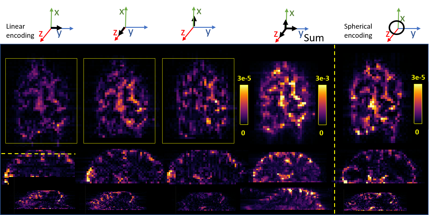
Figure 2: Perfusion weighted images obtained from (from left to right) the linear single-axis encoding in directions (1,0,0), (0,1,0), (0,0,1), the sum over these directions and (right) from the spherical encoding. The first row shows a transverse plane, the second a coronal cut and the bottom row a sagittal cut.
DOI: https://doi.org/10.58530/2022/0045