0019
Single-breathhold multiband cine DENSE MRI using novel phase modulation and a self-calibrated slice-low-rank reconstruction
Changyu Sun1, Yu Wang1, and Frederick H. Epstein1,2
1Biomedical Engineering, University of Virginia, Charlottesville, VA, United States, 2Radiology, University of Virginia, Charlottesville, VA, United States
1Biomedical Engineering, University of Virginia, Charlottesville, VA, United States, 2Radiology, University of Virginia, Charlottesville, VA, United States
Synopsis
Multiband (MB) excitation of spiral cine DENSE MRI has the potential to provide three-slice coverage in single breathhold with 2D displacement encoding. To acquire three slices in one breathhold of 14 heartbeats, we develop MB3+ phase modulation, and use golden-angle rotation, outer volume suppression and variable flip angle. We extend our recent Cartesian slice-L+S method to develop a non-Cartesian slice-low-rank reconstruction model. DENSE data with MB=3 were acquired in 4 volunteers. The MB3+ DENSE method shows fewer artifacts than CAIPIRINHA and Hadamard encoding phase modulations. The slice-low-rank method with MB+ significantly improves slice separation providing fewer artifacts and signal decay.
Purpose
Accurate and reproducible whole-slice and segmental circumferential strain assessed rapidly in three short-axis slices would provide valuable diagnostic and prognostic information and facilitate an efficient clinical workflow. For spiral cine DENSE, a well-established strain MRI method, three slices are typically acquired with each slice requiring one breathhold (14 heartbeats)1,2,3. We have previously demonstrated multiband (MB) DENSE, acquiring three slices in two breathholds; however, slice separation artifacts and low signal-to-noise ratio presented challenges for further acceleration. Here we present improved results by modifying the MB phase modulation method and utilizing an improved iterative MB reconstruction model.Methods
We implemented a modified spiral cine DENSE sequence with variable flip angle MB excitation, outer volume suppression3, and golden angle-rotation (Figure 1A). To acquire three slices in a single breathhold with temporal resolution of 30ms, the acquisition protocol uses 4 spiral interleaves3. However, when the number of interleaves is not a multiple of the MB factor, slice separation is challenging (for example, when MB=3 and number of interleaves = 4, as in Figure 2A). To overcome this challenge, we employed CAIPIRINHA phase modulation corresponding to MB=4 to simultaneously excite 3 slices using a 4 interleave acquisition (termed MB3+, Figure 1A). Experimentally, we compared CAIPIRINHA MB=3 phase modulation, Hadamard encoding phase modulation4 and MB3+. These methods were compared using a non-Cartesian slice GRAPPA (NCSG) reconstruction5. Single-band DENSE images at matched slice locations served as reference standards for comparison.To improve the reconstruction, we developed a self-calibration method to fit NCSG kernels5 and coil sensitivity maps for each slice (Figure 1B). Specifically, the artifact-generating T1-relaxation echo was removed, followed by phase demodulation, NUFFT and temporal averaging of images to generate kernels and sensitivity maps without calibration scans. Next, the reconstruction employed a non-cartesian slice-low-rank approach based on a recently-developed Cartesian slice-L+S method6, which enforces MB data consistency, uses in- and through-plane coil information, exploits golden-angle rotation and enforces temporal low rank of multiple slices to reduce slice separation artifacts (Figure 1C). The slice-low-rank reconstructed images were compared to non-iterative NCSG reconstructed images5 and reference single-band DENSE images at matched slice locations. We evaluated the single-breathhold MB DENSE method in 4 human volunteers, where 3 slices with 2D displacement encoding were simultaneously acquired in a single breathhold (14 heartbeats). For one volunteer, datasets were acquired using three different phase modulation methods. MRI was performed on a 3T system (Prisma, Siemens) using 24-30 RF receiver channels.
Results
Figure 2A compares NCSG and slice-low-rank, using MB3 phase modulation in the acquisition. Slice-low-rank shows better image quality and is more similar to reference images than NCSG. Figure 2B illustrates the comparison of different MB phase modulation strategies, where NCSG-reconstructed images using MB3, Hadamard, MB3+ and reference single-band images are shown. MB3+ shows higher SNR of the myocardium than MB3 and Hadamard (arrows). Figure 3 illustrates example results from one volunteer, where MB3 phase modulation was used along with the NCSG reconstruction, and these images are compared to ones using MB3+ phase modulation and the slice-low-rank reconstruction. MB3+ combined with slice-low-rank shows significant improvement of image quality compared to MB3 with NCSG, demonstrating fewer artifacts and better SNR. Figure 4 illustrates example slice-low-rank and MB3+ results for 3 slices imaged simultaneously, and, adjacently, single-band reference images. The MB images show image quality similar to single-band images at basal, mid-ventricular and apical locations for both magnitude and phase. Example circumferential strain maps (Ecc) and segmental strain-time curves also show a close correspondence between the single-band and optimized MB methods (Figure 5A). Correlation and Bland-Altman plots in Figure 5B, and intraclass correlation of 0.98 quantitatively show excellent agreement between single-breathhold optimized MB DENSE and single-band DENSE for all segments and all three slices of four volunteers.Discussion
The proposed MB spiral cine DENSE method with MB3+ phase modulation, self-calibration, golden-angle rotation, and a slice-low-rank reconstruction provides single-breathhold strain imaging of the left ventricle with excellent agreement compared to single-band cine DENSE strain. When combined with fully-automated deep-learning strain analysis7, these methods can provide accurate and reproducible global and segmental strain imaging with an efficient clinical workflow, requiring the addition of just a single breathhold to any 3T cardiac MRI protocol.Acknowledgements
This work was supported by R01HL147104.References
[1] Zhong X, Spottiswoode BS, Meyer CH, Kramer CM, Epstein FH. Imaging three-dimensional myocardial mechanics using navigator-gated volumetric spiral cine DENSE MRI. Magn Reson Med 2010;64(4):1089-1097. [2] Chen X, Yang Y, Cai X, Auger D, Meyer CH, Salerno M, Epstein FH. Accelerated two-dimensional cine DENSE cardiovascular magnetic resonance using compressed sensing and parallel imaging. J Cardiovasc Magn Reson 2016;18(1):38. [3] Tayal U, Wage R, Ferreira PF, Nielles-Vallespin S, Epstein FH, Auger D, Zhong X, Pennell DJ, Firmin DN, Scott AD, Prasad SK. The feasibility of a novel limited field of view spiral cine DENSE sequence to assess myocardial strain in dilated cardiomyopathy. Magnetic Resonance Materials in Physics, Biology and Medicine. 2019 Jun;32(3):317-29. [4] Yang Y, Meyer CH, Salerno M, Kramer MK, Epstein FH. Whole-heart spiral simultaneous multi-slice first-pass myocardial perfusion imaging. Magnetic Resonance in Medicine, 81, 852-862,2019. [5] Sun C, Yang Y, Cai X, Salerno M, Meyer CH, Weller D, Epstein FH. Non-Cartesian slice-GRAPPA and slice-SPIRiT reconstruction methods for multiband spiral cardiac MRI, Magnetic Resonance in Medicine, 83, 1235-1249, 2020. [6] Sun C, Robinson A, Schumann C, Weller D, Salerno M, Epstein FH. Multiband first-pass myocardial perfusion MRI using a slice-low-rank plus sparse model. ISMRM 28th Annual Meeting, Virtual meeting, 2020, p.1097. [7] Ghadimi S, Auger DA, Feng X, Sun C, Meyer CH, Bilchick KC, Cao JJ, Scott AD, Oshinski JN, Ennis DB, Epstein FH. Fully-automated global and segmental strain analysis of DENSE cardiovascular magnetic resonance using deep learning for segmentation and phase unwrapping. J Cardiovasc Magn Reson 2021;23(1):20.Figures

Figure
1: Schematic diagram of the proposed multiband DENSE acquisition and
reconstruction. (A) A single breathhold MB spiral cine DENSE acquisition method
using MB3+, (B) the spiral cine DENSE self-calibration method and (C) the
reconstruction method.
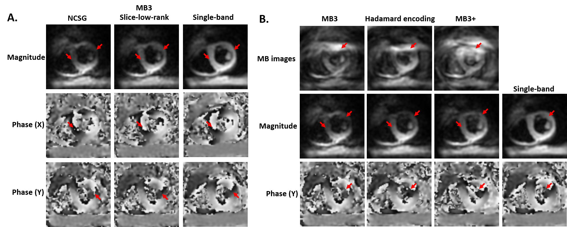
Figure 2: (A) Comparison of NCSG MB,
slice-low-rank MB, and single-band cine DENSE images at a basal location using
CAIPIRINHA MB3 phase modulation. Red
arrows indicate the artifacts. The slice-low-rank method provides fewer
aritfacts and less signal decay. (B) Comparison of NCSG reconstructed images using
MB3, Hadamard encoding, MB3+ phase modulations in the MB acquisition, and reference
single-band cine DENSE images at a basal location. Red arrows indicate the
artifacts. Using the same NCSG reconstruction, MB3+ shows fewer aritfacts and better
SNR.
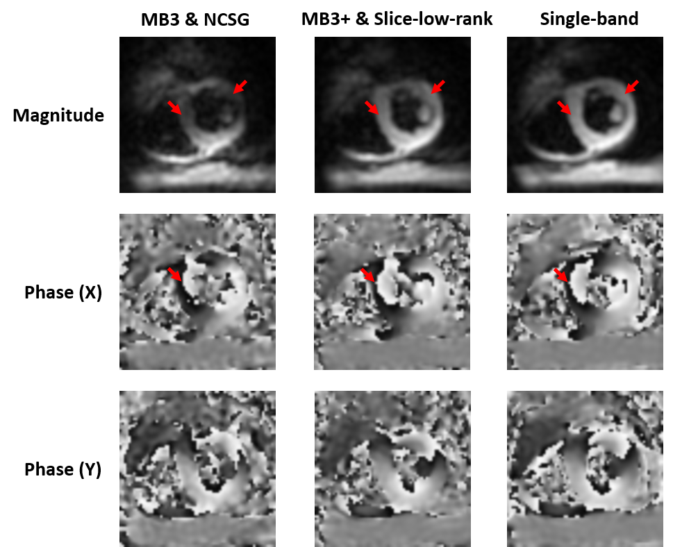
Figure 3: Comparison of NCSG
reconstructed images using CAIPIRINHA MB3 phase modulation, slice-low-rank
reconstructed images using MB3+ phase modulation, and single-band cine DENSE
images. Red arrows indicate artifacts. The
MB3+ & slice-low-rank provides similar image quality to the single-band
image and provides fewer artifacts and better SNR.
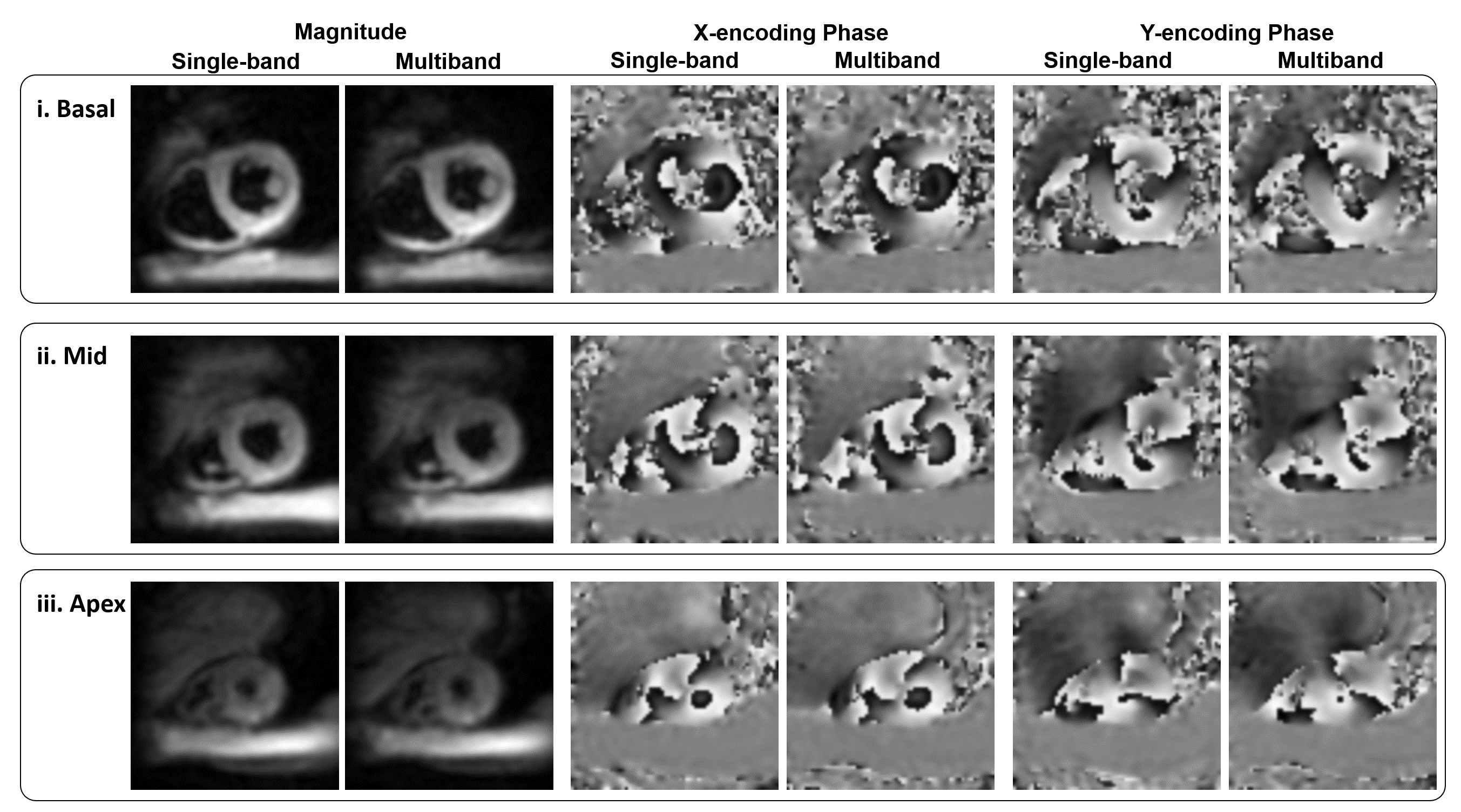
Figure
4: Example images of slice-low-rank reconstructed images using MB3+ and
single-band images of three short-axis slices at basal, mid-ventricular, and
apical locations. Magnitude and phase images show similar image quality.
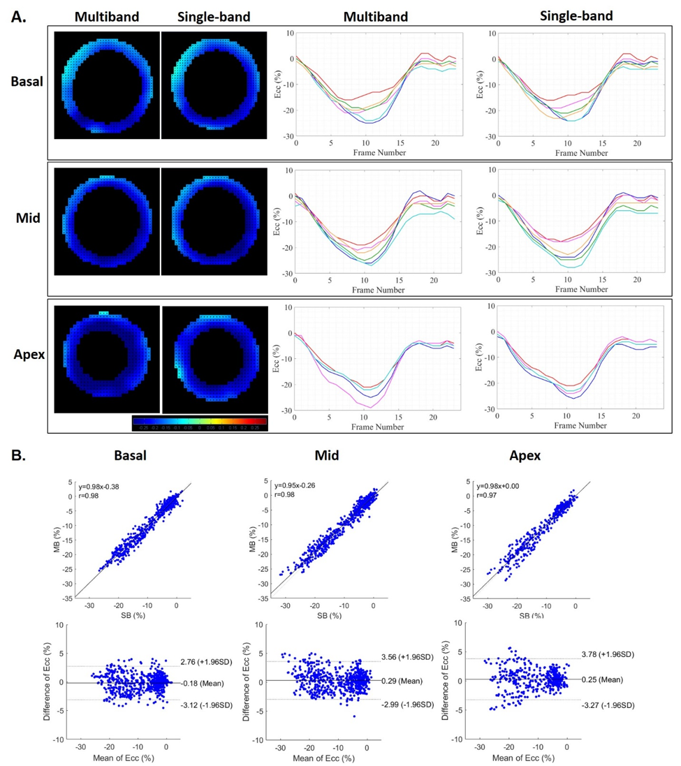
Figure
5: (A) Comparison of strain maps and strain-time curves of three short-axis slices at basal, mid-ventricular, and apical
locations using single-breathhold optimized multiband and single-band
acquisitions. (B) Correlation plots and Bland-Altman plots comparing strain (Ecc)
computed from optimized multiband and single-band acquisitions for all segments
and all three slices of four volunteers.
DOI: https://doi.org/10.58530/2022/0019