0018
Fetal cardiac 4D imaging from multi-planar balanced SSFP MRI1Translational Medicine, Hospital for Sick Children, Toronto, ON, Canada, 2Cardiology, Hospital for Sick Children, Toronto, ON, Canada, 3Hospital for Sick Children, Toronto, ON, Canada, 4Diagnostic Imaging, Hospital for Sick Children, Toronto, ON, Canada, 5Medical Biophysics, University of Toronto, Toronto, ON, Canada
Synopsis
This work explores the feasibility of 4D fetal whole-heart image reconstruction from high resolution 2D multiplanar radial bSSFP acquisitions. High quality 4D reconstructions (1 mm isotropic spatial resolution, ~20 ms temporal resolution) are demonstrated in 3 congenital heart disease cases with distinct cardiac abnormalities. These dynamic, highly detailed visualizations of the fetal heart facilitate prenatal CHD diagnosis and planning for delivery and surgical intervention.
Introduction
With an estimated prevalence of ~8 in 1000 live births, congenital heart disease (CHD) is the most common birth defect. Prenatal detection of CHD has been shown to impact neonatal survival and morbidity and neurocognitive outcomes, likely due to planned delivery and immediate interventions, which prevent circulatory failure after birth [1].Fetal magnetic resonance imaging (MRI) is now an established adjunct modality for fetal cardiovascular imaging when ultrasound visualization is difficult or more detailed vascular imaging is required. Recently, several groups have recently demonstrated four dimensional (i.e., 3D volumetric + cardiac phase) MRI depictions of fetal cardiovascular anatomy [2,3]. 4D, whole-heart data permits dynamic visualizations of the heart and great vessels in arbitrary, oblique planes. This flexibility is very helpful in the detection of small and potentially complex cardiac abnormalities. It also shortens scan time by eliminating the need for careful prescription of multiple 2D scans to visualize the suspected abnormality.
To gain these benefits, we explore retrospective reconstruction of 4D fetal cardiac data from accelerated 2D multi-slice radial balanced SSFP (bSSFP) acquisitions. Given the high spatio-temporal resolution of these 2D data, the resulting 4D reconstructions promise to push the boundaries of fetal cardiac MRI by providing dynamic, highly detailed visualizations of the fetal heart.
Methods
MRI AcquisitionLate-gestation (>34 weeks) pregnant subjects, with a known fetal CHD diagnosis were scanned on a 1.5 T clinical MRI system (AvantoFit, Siemens Healthineers Germany). Three multi-slice, tiny golden angle (θGA=23.58°) radial scans were prescribed in roughly axial, sagittal, and coronal fetal views. Scans were performed under maternal free-breathing conditions with the following acquisition parameters: flip angle=70°, 2000 radial spokes/slice, 256 samples/spoke, FOV=260 x 260 mm2, slice thickness=4 mm, TR/TE=4.06/2.03 ms, and a scan duration of ~8.5 seconds/slice. 12 axial, 16 coronal, and 16 sagittal slices were acquired for complete coverage of the fetal chest in roughly 6 minutes.
2D Reconstruction
2D cine reconstructions were performed using our previously published pipeline [4]. Briefly, the pipeline included preliminary reconstruction of a real-time image series, which was used for rejection of data corrupted by through-slice motion, estimation of in-plane translational motion, and extraction of a fetal cardiac gating signal via metric optimized gating [5]. A final cine reconstruction was then performed on motion corrected (by phase modulation) k-space data, which was re-sorted into 20 frames based on the extracted cardiac signal. As described in [4], both reconstruction steps relied on compressed sensing (CS) to suppress artifacts from radial undersampling.
2D ‘static’ reconstructions were also performed by Fourier transformation of all 2000 acquired spokes from each slice (without CS). These “time-average” images were required for the initial static registration and reconstruction steps in the 4D reconstruction pipeline (see below).
4D Reconstruction
4D cine reconstructions were performed at 1 mm isotropic resolution from 2D cine data via the recently published framework developed by van Amerom et al [2], which was implemented using the open-source slice to volume reconstruction toolkit (SVRTK) from King’s College London (https://github.com/SVRTK/SVRTK). Briefly, the multi-stage algorithm consists of an initial registration of the 3 time-average multi-slice stacks, followed by a 3D time-average reconstruction, and concluding with the interleaved registration and 4D reconstruction of the dynamic cine data.
Results
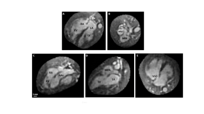
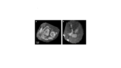
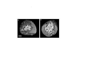
4D reconstructions were performed on 3 cases with distinct congenital abnormalities. Case 1 was a fetus with a rare form of transposition of the great arteries with a posteriorly positioned aorta. Case 2 was a fetus with a normal heart, but a vascular anomaly, whereby the umbilical vein bypassed the liver and fed directly into the right atrium of the heart. Case 3 had a hypoplastic right heart, along with a double-inlet left ventricle (DILV) fed by both the right and left atria. Anatomical features of each case are summarized in Figures 1-3, which show a series of oblique slices through the 4D data that highlight the specific cardiac abnormalities.Discussion
This work demonstrates the feasibility of 4D fetal whole-heart visualization reconstruction from 2D multiplanar radial bSSFP acquisitions. While the duration of the 2D acquisitions is similar to those reported in the original work of van Amerom et al [2], the spatial and temporal resolution are significantly higher (1x1x4 mm3 and ~20 ms compared to 2x2x6 mm3 and 76 ms). The successful 4D reconstructions of the 2D radial cine data demonstrates the robustness of the algorithm, which was originally developed for real-time reconstructions of Cartesian k-t SENSE data.Modifications to the reconstruction pipeline may bring even further improvements to the quality of 4D data. In particular, the reconstruction assumed a 3D Gaussian point spread function (PSF) with full width at half maximum equal to the slice-thickness in the through-plane direction and 1.2 times the voxel-size in-plane. A good approximation of the PSF is essential for high quality reconstruction [6]. The accuracy of this Gaussian model for the current radial acquisition scheme should therefore be investigated.
Acknowledgements
No acknowledgement found.References
[1] Waern et al. BMC Pregnancy and Childbirth. 2021;21:579
[2] van Amerom et al. Magn Reson Med. 2019;82:1055–1072
[3] Roberts et al. Nature Communications. 2020;11:4992
[4] Roy et al. Journal of Cardiovascular Magnetic Resonance. 2017;19:29
[5] Roy et al. Magn Reson Med. 2013;70:1598–1607
[6] Kuklisova‐Murgasova et al. Med Image Anal. 2012;16:1550–1564.
Figures


