S60
Osteoporotic vertebral fracture analysis with intravoxel incoherent motion: a preliminary study1Division of Radiology and Nuclear Medicine, Sapporo Medical University Hospital, Sapporo, Japan, 2Department of Orthopaedic Surgery, Sapporo Medical University School of Medicine, Sapporo, Japan, 3Department of Orthopaedic Surgery, Sapporo Maruyama Orthopedic Hospital, Sapporo, Japan
Synopsis
The parameters of vertebral fracture (VF) and without fracture using intravoxel incoherent motion (IVIM) were analyzed. In addition, the usefulness of using IVIM for VF was investigated. The subjects of this study were 52 patients with VF in the acute phase. As a result, the ADC, D, and f of VF were significantly higher than without fracture. Meanwhile, the D* of VF were significantly lower than without fracture. In the future, new findings may be obtained by analyzing the IVIM parameters and VF prognosis.
Background and Purpose
Recent studies of osteoporotic vertebral fractures (OVF) have revealed that some patients develop delayed bone union or nonunion, leading to unfavorable outcomes, residual pain, and decreased activities of daily living or quality of life.1 It will be possible to perform therapeutic interventions such as vertebroplasty and surgical treatment from an early stage if the occurrence of such delayed union or nonunion and the progression of vertebroplasty can be predicted at the initial treatment stage. Meanwhile, some reports exist that the blood flow in the fractured part was evaluated by using a contrast medium because the disorder of intramedullary perfusion in the vertebral fracture delays the bone union process.2 IVIM has recently been demonstrated to be an attractive approach for the assessment of tissue water diffusivity and microcapillary perfusion.3 However, few reports evaluating OVF using IVIM are available but it is unclear whether IVIM parameters reflect intramedullary perfusion at VF. This study aimed to analyze VF and without fracture using IVIM. Moreover, the usefulness of using IVIM for VF was investigated.Methods
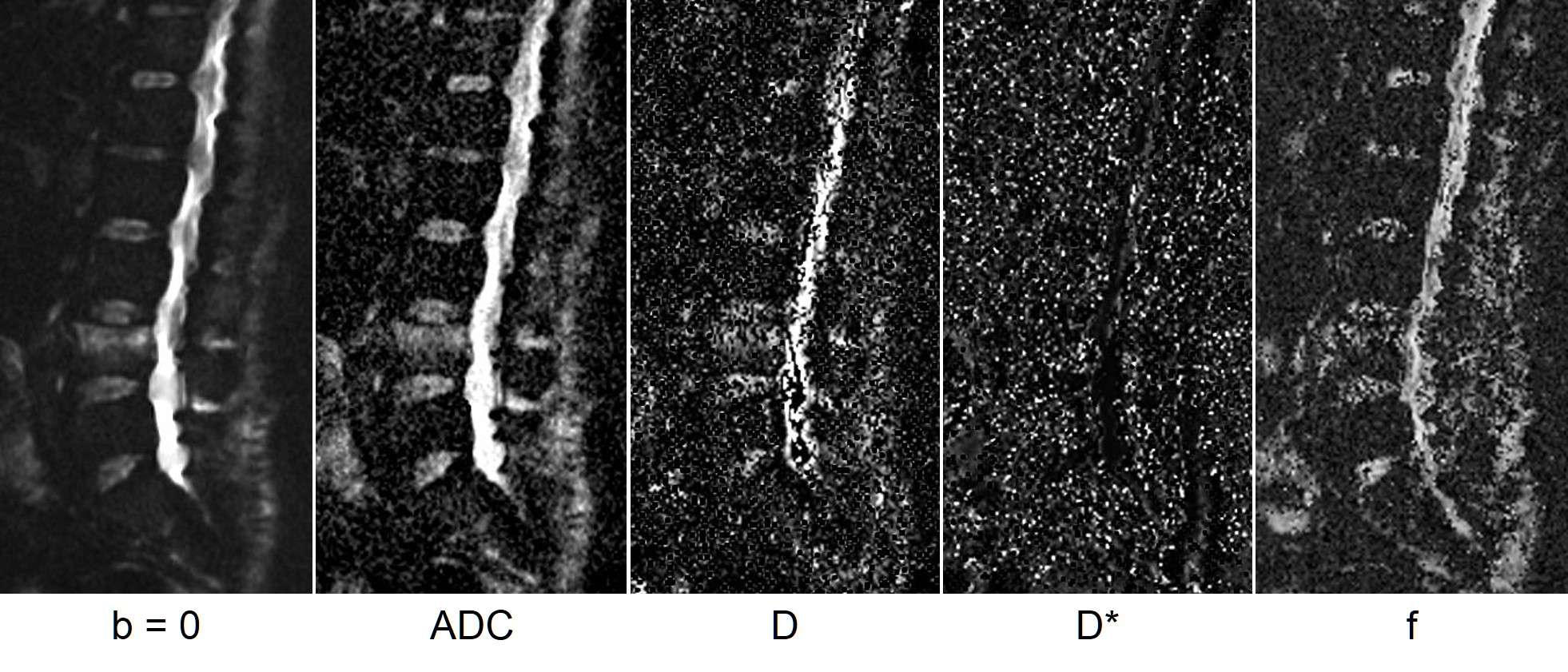
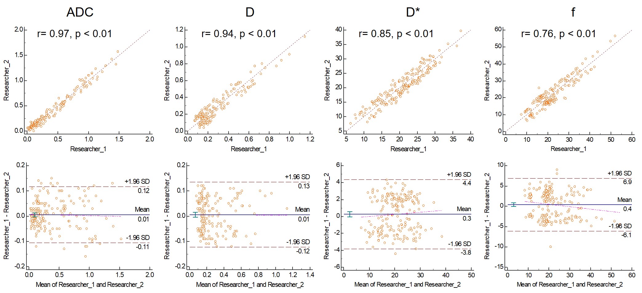
The subjects of this study were patients who underwent X-ray and MR examination of the thoracolumbar spine because of back pain and were diagnosed with VF. Among them, 52 patients (37 females, 15 males, 76.8 ± 12.5 years old) with VF were enrolled indicating high signal change on STIR or T2-weighted sagittal image. MRIs were performed using a GE Signa Creator 1.5T (GE Healthcare, USA). In addition, IVIM parameters (ADC, D, D*, and f maps) were obtained from IVIM data (TR, 3,300 ms; TE, 85 ms; b value = 0, 10, 20, 30, 50, 80, 120, 200, 300, 500, and 800 s/mm2; voxel size, 0.45 × 0.55 × 5 mm; Fig. 1). Two researchers measured the IVIM parameters on VF and without fracture, and the IVIM parameters were compared. However, interclass correlation coefficients (ICCs) and Bland–Altman (BA) analysis were calculated as the inter-rater reliability between the two researchers.Results
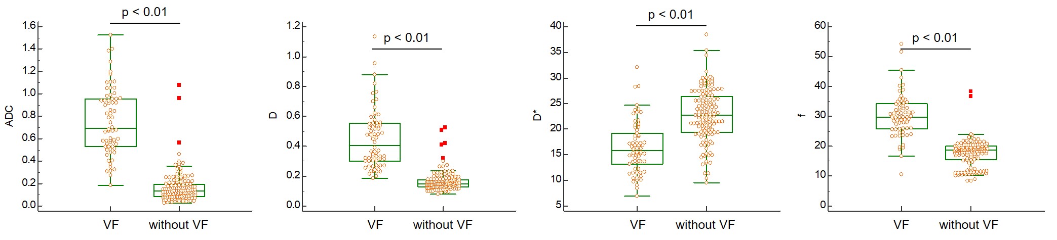
Finally, the analyzed VF and without VF were 66 and 134, respectively. ICCs were calculated between the two researchers for the ADC (ICC, 0.97; 95% CI, 0.9544–0.9737; p < 0.01), D (ICC, 0.94; 95% CI, 0.9254–0.9566; p < 0.01), D* (ICC, 0.85; 95% CI, 0.8038–0.8829; p < 0.01), and f (ICC, 0.76; 95% CI, 0.6964–0.8143; p < 0.01), respectively. Both ICCs and BA analyses and inter-rater reliability tended to decrease on D* and f (Fig. 2). Moreover, the ADC, D, and f of VF were significantly higher than those of without VF (p < 0.05). Meanwhile, D* of VF were significantly lower than those without VF (p < 0.05; Fig. 3).Conclusion
IVIM distinguished between VF and without fracture. This study provides the basic data for analyzing VF using IVIIM, and new findings may be obtained by analyzing the IVIM parameters and VF prognosis in the future.Acknowledgements
I am grateful to MR room staff, especially Mitsuhiro Nakanishi and Nobuyasu Yoshinaka for useful discussions and their help regarding patient treatment. This study is supported by grant of Japan osteoporosis foundation.References
1. Hoshino M, Nakamura H, Terai H. et al. Factors affecting neurological deficits and intractable back pain in patients with insufficient bone union following osteoporotic vertebral fracture. Eur Spine J. 2009; 18 (9): 1279-1286.
2. Kanchiku T, Taguchi T, Toyoda K. et al. Dynamic contrast-enhanced magnetic resonance imaging of osteoporotic vertebral fracture. Spine (Phila Pa 1976). 2003; 28 (22): 2522-2526.
3. Le Bihan D. Intravoxel incoherent motion perfusion MR imaging: a wake-up call. Radiology. 2008; 249 (3); 748-752.
Figures