S30
Depicting capsular injury and disc tears of the sternoclavicular joint with the use of direct MRI arthrography1MRI, Cambridge University Hospitals, Cambridge, United Kingdom
Synopsis
Advances in technology in MRI have allowed for the improved spatial resolution of small joints like the sternoclavicular joint (SCJ). We have shown that through the use of arthrography and MRI we can provide better visualisation of SCJ anatomy, disc tears and other pathologies. Helping to increase knowledge and greatly improve treatment and surgical planning options for the patient.
This presentation aims to understand the techniques for MRI arthrography of the SCJ, review anatomy demonstrated on SCJ arthrography and demonstrate common pathologies of the SCJ.
Background
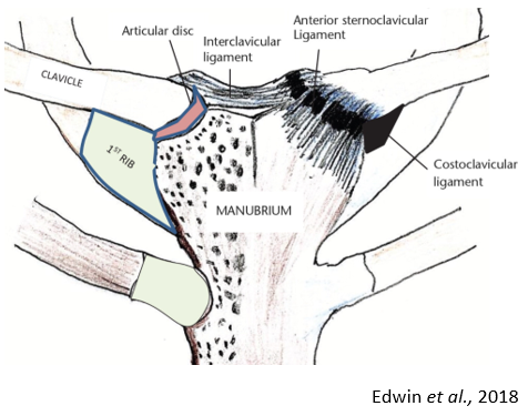
The sternoclavicular joint (SCJ) is formed by the sternal end of the clavicle, the clavicle notch of the manubrium and the first rib cartilage (Figure 1 – sternoclavicular anatomy). It is the only point of articulation between the upper limb and axial skeleton. Although only a small amount of movement occurs at the joint, it can move in 3 axes (forward and backward, up and down, and rotation) – this is essential for the function of the rest of the shoulder girdle. SCJ stability is provided by strong anterior and posterior ligaments, and interclavicular and costoclavicular ligaments. The ligaments and tendons around the SCJ are subjected to large forces whilst maintaining joint stability (Purcho et al., 2014 & Tytherleigh-Strong G., 2020). The relative well reinforced SCJ requires substantial forces to disrupt it and SCJ injuries are uncommon, with injuries at the acromioclavicular and gleno-humeral articulations occurring more frequently. The SCJ contains a fibrocartilaginous disc between the articular surfaces which functions as important shock absorber (Kiel et al., 2020). It is the most frequently injured structure (Benitez et al., 2004). SCJ disc tears usually do not settle without operative management. The most common problems seen at the SCJ relate to osteoarthritis and chronic disc tears or instability (dislocations, acute disc tears) (Tytherleigh-Strong G., 2020). Because SCJ disorders are relatively uncommon clinicians experience in treating them can be limited (Purcho et al., 2014).The evaluation and subsequent management of patients with SCJ region pain can be clinically challenging (Purcho et al., 2014). Routine radiographs with CT or MRI follow-up are usually used to visualise the SCJ (Edwin et al., 2018 & Johnson et al., 2009). Standard radiographs are difficult to interpret with overlapping vertebrae and ribs. Digital tomograms have helped with this (Tytherleigh-Strong et al., 2019). MRI is a more useful technique for detailed and specific identification of soft tissue injuries, usually in the context of investigating a potential acute disc tear (Benitez et al., 2004 & Tytherleigh-Strong et al., 2019). The ability to describe and visualise relevant anatomy can aid an arthroscopic approach or an aspiration or injection of the joint in any treatment plan (Wijeratna et al., 2012).
Magnetic Resonance Imaging (MRI) arthrography involves direct intra-articular injection of contrast under ultrasound guidance before MR imaging. MRI arthrography has shown significant increased sensitivity for the detection of fibrocartilage and ligamentous injury in the shoulder, hip and wrist (triangular fibrocartilage) (Grainger et al., 1999 & Magee T., 2009). The technique is recognised as improving sensitivity to internal derangements, including labral and triangular fibrocartilage tears and ligamentous injury. SCJ arthrography allows excellent visualisation of the fibrocartilage discs and capsuloligamentous structures providing diagnostic information to guide surgical intervention and leading to greater understanding of joint anatomy and internal derangement.
Teaching Points
- To understand the techniques for MRI arthrography of the SCJ
- To review anatomy demonstrated on SCJ arthrography
- To demonstrate common pathologies shown with SCJ arthrography
Conclusion
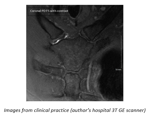
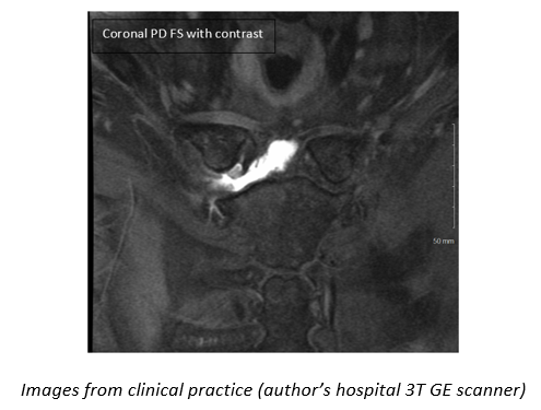
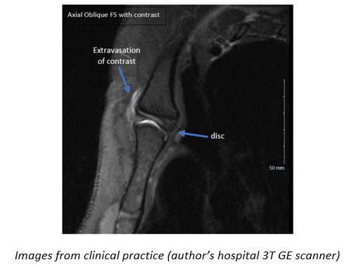
Advances in technology in MRI have allowed for faster imaging and improved spatial resolution of smaller joints like the SCJ. We have shown that through using arthrography and MRI imaging of the SCJ we can provide better visualisation of SCJ anatomy, disc tears and other pathologies (Figure 2, 3, 4 – images from clinical practice). Helping to increase knowledge, and greatly improving treatment and surgical planning options for the patient.Acknowledgements
Thank you for the help and support of...
- Dr Andrew J Grainger FRCR, Musculoskeletal Consultant Radiologist at Cambridge University Hospitals Trust
- Mr Rhys Slough MSc, MRI Manager at Cambridge University Hospitals Trust
References
Benitez CL, Mintz DN, Potter HG (2004) MR Imaging of the Sternoclavicular joint following trauma Journal of Clinical Imaging 28 p.59-63
Edwin J, Ahmed S, Verma S, Tytherleigh-Strong G, Karuppaiah K, Sinha J (Aug 2018) Swellings of the sternoclavicular joint: review of traumatic and non-traumatic pathologies EFFORT Open Reviews 3 p.471-484
Grainger AJ, Elliott JM, Campbell RSD, Tirman PFJ, Steinbach LS, Genant HK (Aug 1999) Direct MR Arthrography: A Review of Current Use Clinical Radiology 55 p.163-176
Johnson MC, Jacobson JA, Fessell DP, Kim SM, Brandon C, Caoili E (Oct 2009) The sternoclavicular joint: can imaging differentiate infection from degenerative change? Skeletal Radiology 39 p.551-558
Kiel J, Ponnarasu S, Kaiser K (Sep 2020) Sternoclavicular Joint Injury, Stat Pearls Publishing
Kusak AS, Podgórski MT, Grzelak P, Kwapisz A (Dec 2018) The advantages of a spine coil over a torso coil in magnetic resonance imaging examination of the sternoclavicular joints Polish Journal of Radiology 83 p.e514-e518
Magee T (Jan 2009) 3-T MRI of the Shoulder: Is MR Arthrography Necessary Musculoskeletal Imaging 192 p.86-92
Purcho AM, Sellon JL, Smith J (Jun 2014) Sonigraphically Guided Sternoclavicular Joint Injection Journal Ultrasound Medicine 34 p.325-331
Tytherleigh-Strong G, Rashid A, Lawrence C, Morrissey D (Jun 2017) Arthroscopic Intra-articular Disk Excision of the Sternoclavicular Joint Arthroscopy Techniques 6 (3) p.e599-e605
Tytherleigh-Strong G, Mulligan A, Babu S, See A, Al-Hadithy N (Apr 2019) Digital Tomography is an effective investigation for sternoclavicular joint pathology European Journal of Orthopaedic Surgery and Traumatology 29 p.1217-1221
Tytherleigh-Strong G (2020) Sternoclavicular Joint [online] https://cambridgeshoulder.co.uk/sternoclavicular/ (Accessed: 08/12/2020)
Wijeratna MD, Turmezei TD, Tytherleigh-Strong G (Aug 2012) Novel assessment of the sternoclavicular joint with computed tomography for planning interventional approach Skeletal Radiology 42 p.473-478
Figures